Full Length Research Paper
ABSTRACT
West African Dwarf (WAD) goats are important source of animal protein, ubiquitous in rural Nigerian households and undergo necessary surgical procedures. Induction of general anaesthesia in goats is associated with resultant severe systemic side effects due to position-induced stress. This study investigated cardiopulmonary and stress responses of WAD goats to three sedatives in different body positioning. Six adult WAD bucks, weighing 11±2.0 kg were randomly selected for three separate experiments using of xylazine (0.05 mg/kg), acepromazine (0.2 mg/kg) or midazolam (0.3 mg/kg) intramuscularly in five clinical trials lasting five days each. Goats were restrained in standing (control), Right Lateral (RL), Left Lateral (LL), supine and prone positions. Venous blood (5 ml) via jugular venipuncture was collected before and after each sedation to determine selected stress biomarkers [blood glucose (mg/dL), plasma cortisol (mmol/L) and lactate dehydrogenase LDH (U/L)]. Heart Rate-HR (beats/min), Respiratory Rate-RR (breaths/min), Mean Arterial Pressure-MAP (mmHg), Oxygen-haemoglobin Saturation-SpO2 (%) and Rectal Temperature -RT (°C) were measured at intervals of 10 min for 1 h. Bucks were rested for 14 days in between clinical trials. Data were analysed using descriptive statistics and ANOVA at p<0.05. In LL, supine and prone body positions, peak blood glucose levels and plasma cortisol, respectively were significantly increased with xylazine, acepromazine and midazolam compared to control. Plasma levels of LDH were significantly decreased compared to control for the RL, LL, supine and prone positions with xylazine and acepromazine sedation. Acepromazine and Midazolam had highest HR with supine position. Xylazine sedation resulted in severe hypotension and hyperglycaemia with LL and supine body positions whereas acepromazine and midazolam sedation resulted in mild hypotension and increased heart rate with LL and prone body positions. The LL and prone positioning should be used cautiously in caprine sedation. Acepromazine and midazolam were found to be safer alternatives to xylazine.
Key words: Body positioning, cardiopulmonary stress responses, sedation, West African Dwarf goats.
INTRODUCTION
The estimated population of goats in Nigeria is put at about 34.5 million (RIM, 1992) of which West African Dwarf (WAD) breed are the commonest (about 45%) of this population (Chiejina et al., 2009). They are reared for different reasons at different locations (Ajala et al., 2008; Olatunji-Akioye and Adeyemo, 2009). Goats are very important to the increasing population as source of animal protein and regular income especially for the rural populace (Chiejina et al., 2015; Fasae et al., 2015). Their compatibility with rural communities is exemplified by their ability to graze and utilize poor quality forage, walk long distances, short generation interval, high reproductive rate, and low risk on investments (Lebbie, 2004). An ever increasing population in Nigeria places a greater demand on goat supply and this also leads to a steady rise in the population of goats reared in the country (Okpeku et al., 2011). The common management system practice of rearing goats in Nigeria is semi-intensive, this method exposes them to various harsh weather condition and health challenges including rumen foreign body impaction, laceration from sharp twigs and grasses, which consequently necessitate handling, medical, diagnostic or surgical intervention (Abu et al., 2013).
Most of several unique complications from sedation and anaesthesia in ruminants arise from the interaction of alimentary and respiration systems (Adetunji et al., 1985; Clarke et al., 2014). Loss of eructation usually accompanies sedation and general anaesthesia with continued fermentation (Seddighi and Doherty, 2016). In the absence of eructation, gas accumulation in the rumen relates directly to the rate of gas produced from fermentation. Gas distension of the rumen may depress respiratory function by reducing the lung volume and venous return (Musewe, 1979; Valverde and Doherty, 2008). A proper body position ideally should provide an unrestricted access to the operative site, should not interfere with peripheral tissue perfusion, ventilation, and oxygenation and should not put undue pressure on neuromuscular system and joints, otherwise this is stressful for the animal. In the goat, various surgical procedures have been done with right-lateral position, including surgeries of forestomach (rumenotomy and rumenostomy), oesophagus (oesophagotomy) and gravid uterus (Caesarean section) while the left-lateral position is indicated for the procedures on the abomasum and intestine (Fubini and Ducharme, 2014). The supine position is indicated in the goats for procedures on the udder, umbilicus, urinary bladder, abomasal displacement and genital organs such as preparation of teaser buck while prone position is indicated for cerebrospinal fluid (CSF) tap, myelography and spinal procedures (Fubini and Ducharme, 2014).
In veterinary practice, goats are often presented for diagnostic, medical and surgical procedures necessitating sedation, anesthesia, restrain and body positioning. The commonly used classes of sedatives in caprine practice include alpha2-agonists (e.g. xylazine, medetomidine, detomidine and dexmedetomidine), phenothiazine derivative (e.g. acepromazine) and benzodiazepines (e.g. diazepam, midazolam and zolazepam) (Taylor, 1991; Udegbunam and Adetunji, 2007). Reports have shown that a number of variables, including handling and animal behaviour can elicit stress in animals (Hemsworth et al., 2011).
Stress is an adaptive event of neuroendocrine, metabolic, haematological, immune and behavioural changes following anxiety, pain, trauma or injury designed towards the restoration or maintenance of homeostasis (Desborough, 2000; Allen et al., 2014). A stressor is capable of disrupting homeostasis as it is normally accompanied by series of predictable events such as biochemical, physiological, cognitive and behavioural changes (Taylor, 2008). The neuroendocrine response to stress includes elevated circulating catecholamines and cortisol (Finnerty et al., 2013; Saidu et al., 2016). Behavioural changes in response to stress in animals include flight and fight, immobility (freezing stance), panting, sweating, piloerection and facial expression (Breazile, 1987). Hyperglycaemia has also been reported as one of the markers of stress via the activation of various glucose pathways (Armario et al., 1996).
Naturally, all ruminants voluntarily assume either the standing or sternal position when ruminating, resting or sleeping. Any other body positions would appear to be unphysiological. Indeed, awake or sedated cattle placed in either lateral or supine position showed significant cardiopulmonary disturbances and intense stress (Adetunji et al., 1984; Wagner et al., 1990; Tagawa et al., 1994). They are generally not used to handling, thus when presented for clinical procedures, there is the need for physical restraint, sedation, or anaesthesia, which in turn initiates stress response especially in long term recumbency. Also, they are considered poor subjects for general anaesthesia because of the associated complications such as inadequate ventilation, prolonged recumbency, ruminal tympany, regurgitation and excessive salivation with potential for aspiration into the lungs (Adetunji et al., 1984; Clarke et al., 2014). Consequently, sedation and loco-regional anaesthesia are usually employed in ruminants for clinical procedures.
Most clinical procedures in large ruminants such as cattle are performed on the standing animal with the aid of sedation under loco-regional analgesia and/or restraining chute (Fubini and Ducharme, 2004). However, standing procedures are not practical for small ruminants such as goat and sheep owing to their small body size and they have to be made recumbent for this purpose (Clarke et al., 2014). Considering that goats differ considerably from cattle with regard to species, body size and body weight, it would seem inappropriate to extrapolate large ruminant data to the goat as a basis for providing supportive care.
Body positions most commonly used in the goats are Right-Lateral (RL), Left-Lateral (LL), Supine (SUP) and Prone (PP) positions (Fubini and Ducharme, 2004; Ames, 2014). Physiological effects of these positions are yet to be reported in sedated sheep and goat. Therefore, the responses of sedated goat or sheep to different body positions are not known at the present time. The aim of this research was to determine and compare cardiopulmonary and stress responses of sedated goats placed in different body positions.
MATERIALS AND METHODS
This study was approved by the Animal Care and Use Research Ethics Committee (ACUREC), University of Ibadan (UI-ACUREC/18/0029). Six healthy bucks, aged 1.5 to 2 years and having a mean body weight of 11±2.0 kg were used for the research. They were bought from a local goat market in Ibadan and selection was based on signalment and normal physical examination findings. The selected goats were housed at the experimental unit of the Faculty of Veterinary Medicine in a communal goat pen with a concrete floor bedded with wood shavings. They were fed a basal diet of star grass (Cyanodon alaniferensis) and cassava peels, additional cereal concentrate ration at a rate of 50 g/head/day ad libitum. Salt lick and fresh, clean water were provided ad libitum. A period of one month was allowed for the goats to acclimatize to their new housing, feeding regimen and handling. Just before the start of the trials, the health status was evaluated by complete blood cell count, haematocrit, plasma proteins and electrolytes assessments.
Drugs used in the research consisted; Xylazine hydrochloride (Xylased ® Bioveta Laboratory, Ivanovice na Hane Czech Republic) supplied as a 20 mg/ml solution for injection in a 50ml multidose vial. Acepromazine maleate (ACP injection, Novartis Animal Health UK Ltd) marketed as a 20 mg/ml solution for injection in a 20 ml multidose vial. Midazolam hydrochloride (Midazolam Hamelin Pharm. Ltd Glaucester, UK) supplied as a 50 mg/ml solution for injection in a 2 ml glass ampoule.
Research design
The research design used comprised of prospective, randomized crossover experimental trials, carried out on sedated goats. A crossover design is a repeated measurement in which the (entire) whole sample size is used (and moved) from one treatment to another during the course of the trial using an appropriate washout effect to eliminate the impact of carryover effect (Siyasinghe and Sooriyarachi, 2011). Based on the three classes of sedative drugs employed, three main studies were carried out each consisting of five trials. In each trial, each of the six bucks was sedated and positioned for an hour in appropriate positions based on the trial in use. During this sedation, vital parameters were taken using the patient monitor at 10 min interval for an hour. Blood samples were also collected before and after positioning and sent to the laboratory for analysis. Each trial took a week as a buck went through the experiment per day. Thereafter, the bucks were rested for 14 days in between clinical trials to allow for the wash-out of the sedative drugs. This was repeated for each study till all the trials were carried out.
Experimental procedure
At the start of each trial, the body weight of each goat was determined using bathroom scale for calculating drug dosage. A total of 5 ml whole blood each was collected from the jugular vein into lithium heparin sample tube to determine baseline values of Cortisol and LDH. Another 5 ml of blood was collected into sodium fluorine EDTA sample tube to determine baseline values of glucose. Another sample was taken at the end of an hour for determination of cortisol, LDH and glucose. The goat was administered sedative agent at recommended clinical doses of 0.05 mg per kg xylazine, 0.2 mg/kg acepromazine or 0.3 mg/kg midazolam as appropriate (Clarke et al., 2014). All drugs were injected intramuscularly in the femoral muscle of the goats. The sedated goat was then restrained as appropriate in a chosen body position, which included; standing, supine and prone positions with the aid of constructed specialized chute that also stabilized the heads. Right and left lateral positions were achieved with aid of an operating table. The standing position was used as control. The goat was maintained in a body position for one hour.
The restrained sedated goat was attached to a veterinary multiparameter patient monitor (Cardell® 9500HD) for measurement of physiological variables. Heart rate (HR) in beats/minutes, respiratory rate (RR) in breath/min, mean arterial pressure (MAP) in mmHg, and oxygen saturation (SpO2) in percentage (%) were recorded. Rectal temperature (RT) in °C was measured using an electronic thermometer. All measurements were taken immediately and during maintenance of body position at 10 min intervals over a period of one hour.
It is usual to take preanaesthetic baseline cardiopulmonary parameters but this was not done in this study for several reasons; first, such preanaesthetic values are easily obtained in companion animals that are used to human handling but difficult in farm animals that will tend to resist handling and instrumentation. Secondly, such physiological parameters are usually quoted in ranges rather than as absolute values. Therefore, it will be difficult to establish such baseline ranges with only six experimental animals. Thirdly, this research makes use of trends rather than absolute values in the physiological parameters.
Furthermore, trends rather than absolute blood pressure readings were needed. Normal blood pressure varies throughout the cardiac cycle as the ventricles contract and relaxes. Systolic blood pressure is produced by the contraction of the left ventricle as it propels blood through the systemic arteries. Diastolic blood pressure is the pressure that remains in the arteries when the heart is in its resting phase between contractions. The MAP is the average pressure through the cardiac cycle and is the most important value because it represents actual tissue perfusion of the internal organs. For these reasons, only the MAP was considered in this study. A multiparameter electronic monitor with ability to display data on the screen was employed to facilitate measurement of numerous cardiopulmonary variables at short intervals.
Blood samples collected in tubes were sent to a specific laboratory at the University College Hospital, Ibadan on ice pack for the analysis for three stress biomarkers; glucose, cortisol and lactate dehydrogenase (LDH).
The Glucose HK Gen. 3 Kit (GLU3, Roche Diagnostics GMbH Sandfofer Strasse 116 D-68305 Manheim, Germany) was used for plasma glucose analysis. It uses the UV test, which is enzymatic reference method with hexokinase (Hexokinase catalyzes the phosphorylation of glucose to glucose-6-phosphate by ATP). Glucose + ATP G-6-P + ADP. Glucose -6-Phosphate dehydrogenase oxidizes glucose-6-phosphate in the presence of NADP to gluconate-6-phosphate. No other carbohydrate was oxidized. The rate of NADPH formation during the reaction was directly proportional to the glucose concentration and was measured photometrically. The equipment used was cobas c311 by Roche and it was done on the cobas auto analyser.
The ARCHITECT Cortisol Reagent Kit (Abbott) Laboratories Diagnostics Division by Fisher Diagnostics, a division of Fisher Scientific Company LLC, 8365 Valley Pike, Middle town, VA 22645-1965, USA) was used to determine the plasma cortisol activity. This was done on Abbott i1000SR (auto analyses). The ARCHITECT Cortisol assay is a delayed one-step immunoassay for the quantitative determination of cortisol in serum or plasma using CMIA technology with flexible assay protocol referred to as Chemiflex. The samples collected were combined with anti-cortisol paramagnetic micro particles. The cortisol present in the sample binds to the anticortisol coated microparticles. After incubation, cortisol acridinium-labeled conjugate was added to the mixture. Following a second incubation, the micro particles were washed and pre-trigger and trigger solutions were added to the reaction mixture. The resulting chemiluminescent reaction was measured as relative light unit (RLUS). There was an inverse relationship between the amount of cortisol in the sample and the RLUS detected by the ARCHITECT iSystem optics.
Lactate dehydrogenase in the sample was analysed using Lactate HK Gen3 (LDH3, Roche diagnostics GmbH Sandhofor Strasse 116 D-68305 Mannheim, Germany). The principle of LDH estimation is optimized standard method according to the Deutsche Gesellscaft fur Klinische Chemie (DGKC). LDH catalyzes the reaction between pyruvate and NADH to form L-lactate and NAD+; Pyruvate + NADH + H+LDH L-lactate + NAD+. The initial rate of the NADH oxidation is directly proportional to the catalytic LDH activity. It was determined by measuring the decrease in absorbance at 340 nm. The LDH analysis was auto analysed using cobas c311 by Roche using the aforementioned principle.
Statistical analysis
The data generated were analysed statistically using the PRISM Software package (version 5.0 Graph pad Software Inc, San Diego CA, USA). Where applicable, all data were expressed as mean ± standard deviation (SD). The physiological variables (HR, RR, MAP, SPO2, RT) were compared using analysis of variance (ANOVA) and student’s t-test was used to assess statistical significance for paired data (Stress markers; Glucose, Cortisol and LDH). Values of probability less than 5% (p<0.05) were considered significant for all data.
RESULTS
Responses of xylazine-sedated goats
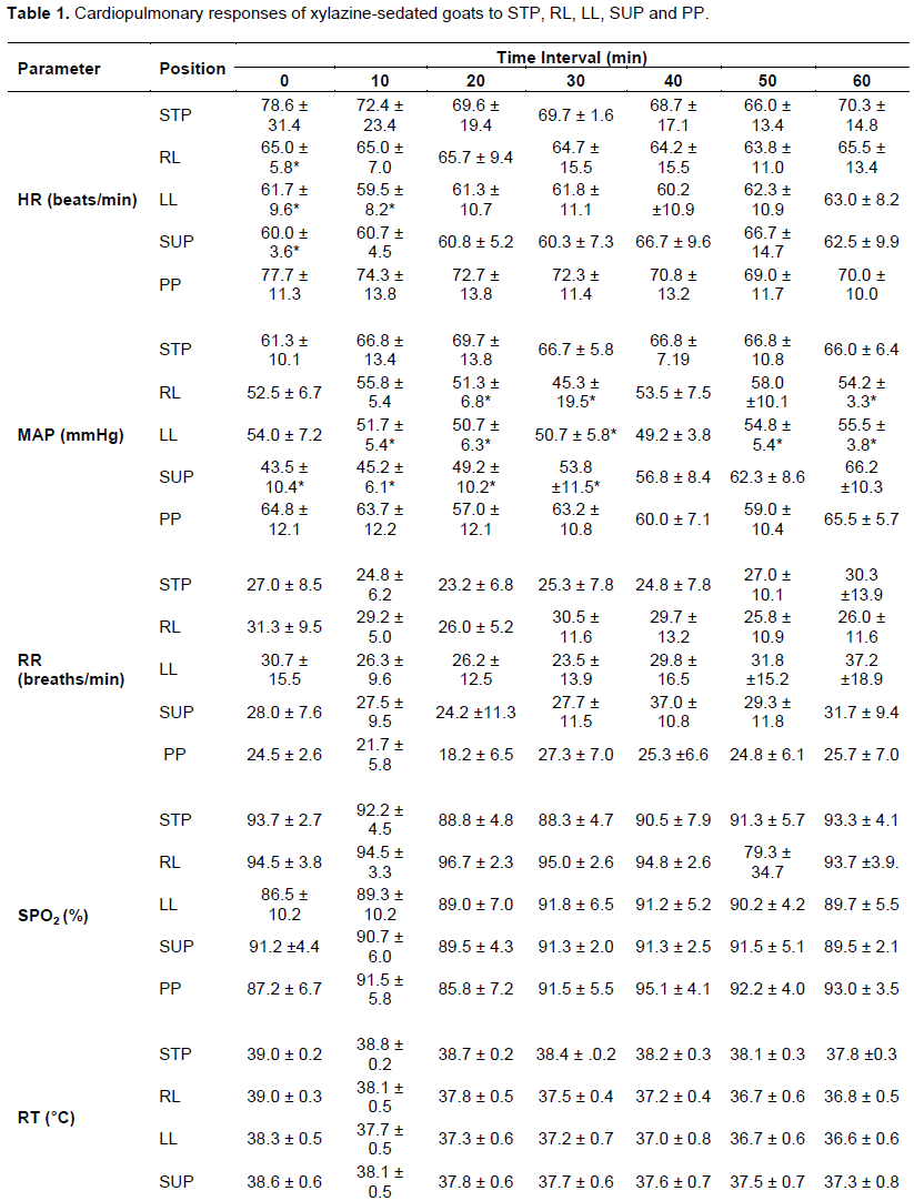
In all the five trials involving xylazine, the sedative agent produced cooperative animals that did not resist head and rope restrain. The sedated goats salivated for the first 10 minutes. All laterally recumbent sedated goats developed transient ruminal tympany due to their inability to eructate. Xylazine induced good muscle relaxation, which facilitated body positioning of the experimental goats. Cardiopulmonary responses of xylazine-sedated goats to STP, RL, LL, SUP and PP are shown in Table 1
Stress responses
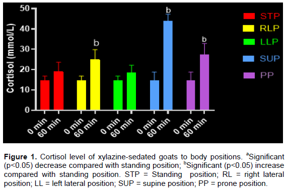
Plasma cortisol responses of xylazine-sedated goat to all the studied body positions are as shown in Figure 1 Mean plasma cortisol levels at 60 min rose above corresponding baseline levels for STP, RL, LL, SUP, and PP. After 60 min, mean plasma cortisol levels were significantly (p<0.05) higher in RL, SUP and PP than in STP.
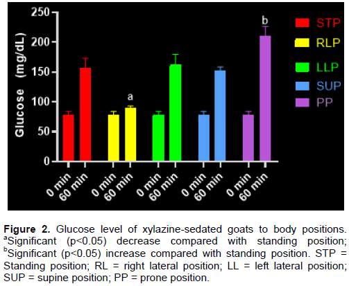
Mean blood glucose responses of xylazine-sedated goats to all the studied body positions are as shown in Figure 2 at 60 min, mean blood glucose levels rose above the corresponding baseline levels for STP, RL, LL, SUP and PP. Mean blood glucose levels were significantly (p<0.05) higher in RL and PP than STP. Mean values for LL and SUP were not significantly (p>0.05) different than the STP value.
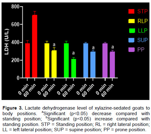
The mean plasma LDH values of xylazine-sedated goats to all the studied body positions are as shown in Figure 3. Except for STP, mean plasma LDH levels for RL, LL, SUP and PP were lower than the corresponding baseline values and the corresponding baseline values and also significantly (p<0.05) lower than in STP.
Responses of goats to acepromazine sedation
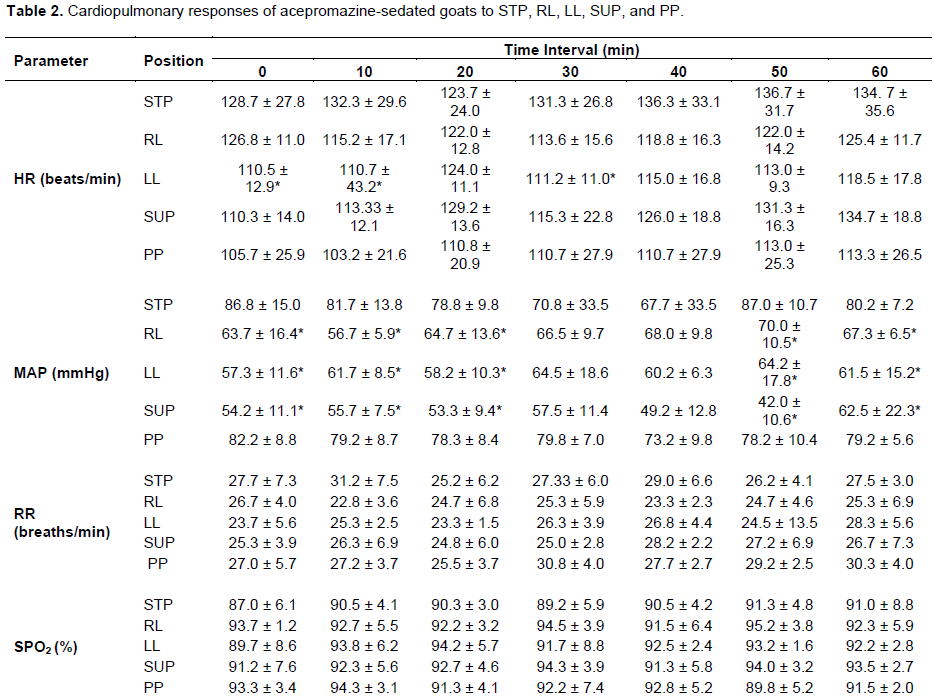
In all the five trials involving acepromazine, the sedative agent produced pliable animal that tolerated head and rope restrain. Sedation produced only mild muscle relaxation with penile prolapse and no ruminal tympany or salivation was observed. In addition, no oesophageal reflux occurred. Cardiopulmonary responses of acepromazine-sedated goats to STP, RL, LL, SUP and PP are shown in Table 2.
Stress responses of goats to acepromazine sedation
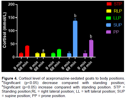
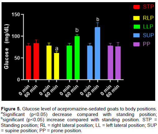
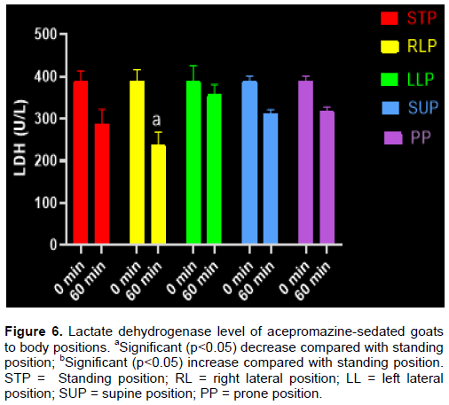
Plasma cortisol responses of acepromazine-sedated goats to all body positions studied are as shown in Figure 4. Mean plasma cortisol levels at 60 min rose above corresponding base line levels for STP, RL, LL, SUP and PP. At 60 min, mean plasma cortisol levels were significantly (p<0.05) higher in RL, LL, SUP and PP than in STP. Mean blood glucose responses of acepromazine-sedated goats to all body positions are as shown in Figure 5. At 60 min, mean blood glucose levels rose above corresponding baseline values for STP, LL and SUP while it was lower in RL and PP. Mean blood glucose was significantly (P<0.05) higher in LL and SUP compared with standing values while it was significantly (P<0.05) lower in RL than in STP. There was no significant (P>0.05) difference in PP compared with that of STP. Figure 6 shows the mean plasma LDH of acepromazine-sedated goats to all studies body positions. Mean plasma LDH levels for STP, RL, LL, SUP and PP were lower compared with corresponding baseline values. Only the mean plasma LDH in RL was significantly (P<0.05) lower than in STP.
Responses to midazolam sedation
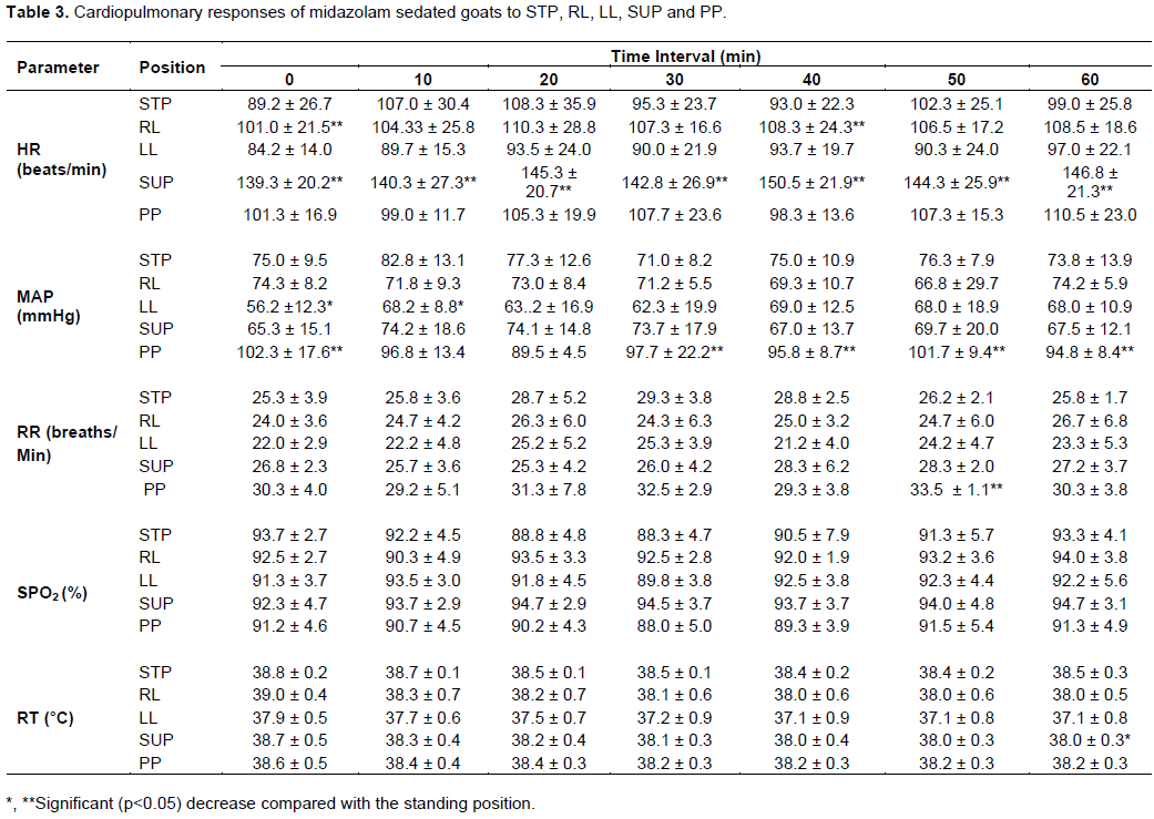
In all the five trials involving midazolam, the sedative agent produced pliable animals that tolerated head and rope restrain. Sedation produced muscle relaxation with no side effect on the goat. No ruminal tympany or salivation was observed. Cardiopulmonary responses of midazolam-sedated goats to STP, RL, LL, SUP and PP are shown in Table 3.
Stress responses to midazolam sedation
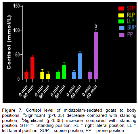
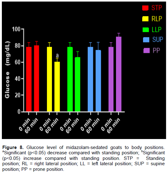
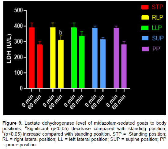
Plasma cortisol responses of midazolam-sedated goats to all the studied body position are as shown in Figure 7. Mean plasma cortisol levels at 60 min rose above corresponding baseline values for STP, LL, SUP and PP while it was lower in RL. After 60 min, mean plasma cortisol levels were significantly (P<0.05) higher in LL and PP than in STP while it was significantly (P<0.05) lower in RL than in STP. Mean glucose responses of midazolam-sedated goats to all studied body positions are as shown in Figure 8. At 60 min, mean blood glucose levels rose above corresponding baseline levels for STP and PP while it was lower in RL, LL and SUP. Mean blood glucose levels in RL was significantly (P<0.05) lower than in STP. There were no significant (P>0.05) differences in mean blood glucose in LL, SUP and PP compared with STP. Figure 9 shows the mean plasma LDH of midazolam-sedated goats to all the studied body positions. Mean plasma LDH levels for STP, RL, LL SUP and PP were lower compared to corresponding baseline values. Only mean plasma LDH in LL was significantly (P<0.05) higher when compared with STP.
DISCUSSION
The experimental goats were sedated in addition to physical restrain in order to make their body positioning somewhat comfortable with minimal movement in line with current clinical practice. The sedative agents employed were chosen on the basis of their local availability and frequent use in caprine practice. Specialized chute was constructed to aid standing position and immobilize the animals’ head during the course of the experiment.
Arterial blood pressure can be assessed directly or indirectly in animals. Direct method of blood pressure measurement involves canulation of a suitable peripheral artery with an attached transducer (Haskin et al., 2005). This invasive method provides accurate blood pressure measurement, facilitates blood gas analysis and blood collected can be used for extended period of time. Indirect method involves the use of inflatable cuffs that are about 40% of the circumference of the appendage which are applied firmly but not tightly over an artery. In this study, the indirect method was employed because it is quicker, non-invasive, less risky and minimal professional skill is required compared to direct method (Clarke et al., 2014).
Responses of xylazine-sedated goats to STP, RL, LL, SUP and PP
Cardiopulmonary responses
In the xylazine study, the HR of standing sedated goats ranged from 66 to 78 beats/min, which is on the lower side of normal range of 70 to 90 bpm reported for awake goat (Muir and Hubbell, 2013). Excessively slow heart rate allows enough diastolic filling time but the rate cannot maintain cardiac output (Haskin et al., 2005). This finding of reduced heart rate might be explained by the action of xylazine, an alpha2 adrenoceptor agonist (Pypendop and Verstegen, 1998). Whereas, each goat received equal dose of xylazine sedation, mean HR in the RL, LL and SUP were significantly (p<0.05) lower than STP (Table 1). This finding might be explained by the baroreflex response to recumbency. In the recumbent position, the circulating blood volume could possibly stretch the aortic arch and carotid sinus walls which then send nervous signal to the CNS, exciting the nuclei of vagus and inhibiting sympathetic outflow. The net result is vasodilation, decreased heart contraction and heart rate (Guyton and Hall, 2006).
Also, the MAP of standing sedated goat ranged from 61 to 69 mmHg, which is below the normal range of 80 to 110 mmHg reported for awake goats (Haskin et al., 2005). This lower blood pressure correlated with the slow heart rate discussed earlier. Furthermore, MAP in the RL, LL and SUP were significantly (p<0.05) lower than in the STP. This could be explained by baroreceptor response to recumbency causing vasodilation, reduced heart contraction and heart rate (Merin, 1986). Vasodilation causes pulling of blood in large veins leading to reduced venous return, preload, stroke volume and reduced contractility, resulting from centrally-induced sympathetic outflow (Merin, 1986; Guyton and Hall, 2006). In addition, xylazine also causes bradycardia and myocardia depression leading to 50% reduction in cardiac output (Paddleford and Harvey, 1999; Valverde and Doherty, 2008). Minimum MAP required for adequate tissue perfusion is put at 60 mmHg (Haskin, 2015), MAP in RL, LL and SUP were below this value indicating severe hypotension requiring blood volume support. It is however surprising that the goats used for this experiment recovered without any therapeutic intervention. This survival might be explained by the fact that healthy goats have large physiological reserves from which they can draw as the need arises.
The ranges of respiratory rate in STP, RL, LL, SUP and PP (Table 1) were generally above normal range of 16 to 24 breath/min quoted for the awake goats (Thomas and Lerche, 2016). This finding indicates that experimental goats exhibited tachypnoea probably due to xylazine administration (Mohammed et al., 1996). Tachypnoea has been recorded in sheep given xylazine (Raptopoulos et al., 1995). The net result is reduced tidal volume with compensatory increased RR to maintain minute volume.
Animals with tachypnoea may have low effective alveolar minutes volumes because they are rebreathing dead space gas and therefore may be ventilating very inefficiently (Thomas and Lerche, 2016). The impact of the increased RR on pulmonary gas exchange was not determined in this study because neither a capnograph nor arterial blood gas analysis was performed. Capnography requires endotracheal intubation under general anaesthesia while blood gas analysis requires canulation of a peripheral artery for blood sampling, procedures which were not included in this study protocol.
Generally, SpO2 in all the body positions ranged from 79 to 96% which is below the normal range of 95 to 98% indicating mild to moderate hypoxaemia (Clarke et al., 2014). The causes of hypoxaemia include hypoventilation, decrease fractional inspired oxygen, shunting of blood flow and decreased ability of oxygen to diffuse from the lung into the blood stream in animal breathing room air (Thomas and Lerche, 2016). It has been reported that sheep placed in lateral recumbency under xylazine sedation may show reduced arterial oxygen tension despite no injection of sedative or chemical restraint (Mitchell and Williams, 2008). It is also interesting to note that healthy experimental goats recovered without oxygen supplementation presumably because of their physiological reserves.
Mean rectal temperature of xylazine sedated goats in STP ranged from 37 to 38°C (Table 1); this is within the normal value of 35 to 39°C adequate for metabolic activities (Thomas and Lerche, 2016). This means that the animals were able to maintain normothermia under xylazine sedation in the absence of such risk factors such as clipping of the hair, use of cold scrub solutions, surgical exposure of large body cavity, application of cold lavage solution and administration of cold intravenous fluid.
Stress responses to xylazine sedation
Cortisol is generally considered a stress hormone because its levels rise during episode of acute stress (Brook and Marshal, 2001). Physical or mental stress increase ACTH secretion, which in turn stimulate the adrenal cortex to secrete cortisol (Nwe et al., 1996; Allen et al., 2014). A spike in cortisol level mobilizes necessary resources, such as by tapping into the body’s reserve to produce energy and ensure individuals return to a stable state (Guyton and Hall, 2006). In this study, mean plasma cortisol levels for RL, SUP and PP at 60 min was significantly more elevated (p<0.05) than in the standing position (Figure 1). This finding suggests that these body positions were more stressful. It is noteworthy that even xylazine sedation could not prevent the stress response; this report does not corroborate the study by Sanhouri et al. (1992), who attributed decrease in cortisol level because of inhibitory action of xylazine during goat transportation. In practice, prolonged stress could lead to increased morbidity of the patient if not mitigated. The experimental goats were able to cope with the degree of stress because of short duration procedure and large physiological reserves. Mean blood glucose levels in the RL was significantly (p<0.05) lower than the standing position value while it was elevated in prone position than standing position (Figure 2). This finding suggests that in stressful periods, insulin resistance causes hyperglycaemia and muscle protein breakdown, which are consequences of severe stress response (Carli, 2014). However in this study, the increased blood glucose recorded might have been caused by temporary hyperglycaemic effect of xylazine but also blood glucose would appear to be non-specific for stress. Plasma LDH values for RL, LL, SUP and PP were significantly (p<0.05) lower than the standing value. This finding does not correlate with that of cortisol and a study conducted in deer where no correlation between cortisol concentration and LDH activity was recorded (Jones and Price, 1992).
Responses of acepromazine-sedated goats to STP, RL, LL, SUP and PP
Cardiopulmonary responses
In this study, the HR in STP ranged from 123 to 136 bpm, which is far above the normal range of 70 to 90 bpm quoted for awake goats (Muir and Hubbell, 2013). This finding of increased HR is probably due to the effect of acepromazine, which is an alpha blocker causing vasodilation with subsequent hypotension and reflex bradycardia (Taylor, 1991; Alvaides et al., 2008). Mean HR in LL only was significantly (p<0.05) lower than the standing value in the first 30 min. Mean HR in all body positions was generally not significantly different (p>0.05) from STP. This finding implies that recumbency drug-induced baroreflex response was overridden by reflex increases in the heart rate (Pequito et al., 2012). Excessively fast heart rate can reduce cardiac output by reducing diastolic filling time and stroke volume (Haskin et al., 2005).
In this study, MAP in the STP ranged from 67 to 87 mmHg which is lower than the normal range of 80 to 110 mmHg accepted for awake goat (Haskin et al., 2005). This finding implies acepromazine-induced hypotension (Alvaides et al., 2008). Except in PP in which MAP was not significantly (p>0.05) different from STP, mean arterial pressure in RL, LL and SUP were significantly (p<0.05) lower than in the STP. This finding could be the result of combined effect of recumbency-induced baroreflex responses, vasodilation and hypotensive effect of acepromazine and aortocaval compression in the supine position, resulting in reduced venous return and preload. As the end diastolic volume decreases, cardiac muscle fibre length decreases, and stroke volume decreases, resulting in decreased cardiac output and blood pressure in accordance to Frank Sterling law of the heart (Guyton and Hall, 2006). Since the minimum MAP of 60mmHg is required for adequate tissue perfusion (Haskin et al., 2005), MAP obtained particularly in SUP was generally life threatening. These experimental goats were able to cope with the recorded hypotension possibly because of their large physiological reserves from which they could draw. Certainly, acepromazine-sedated sick goats would require blood volume support to maintain adequate tissue perfusion.
The mean respiratory rate in all body positions were generally higher than the minimum of 16-24 breath/min recorded for the awake goats (Thomas and Lerche, 2016). This finding of tachypnoea is generally considered inefficient ventilation. The impact of tachypnoea on pulmonary gas exchange was also not known, although the goats recovered without supportive intervention.
Mean SPO2 values ranged from 87 to 95, indicating mild to moderate hypoxia. In clinical practice, oxygen supplementation would be required to ensure optimum oxygen delivery to tissue.
The RT in all the studied body positions was within the normal range of 37 to 38°C accepted for awake goat (Haskin et al., 2015). This indicates that acepromazine did not interfere with thermoregulation in the CNS.
Stress responses
Mean plasma cortisol values in both right and left lateral body positions were significantly (p<0.05) lower than in the standing position, but in supine and prone body positions cortisol level was significantly (p<0.05) higher compared with the standing position suggesting these latter positions were be more stressful. It has been documented that cortisol secretion increases in response to any stress in the body (Brook and Marshal, 2001). Mean blood glucose level in RL was significantly lower compared with STP while it was significantly (p<0.05) higher in LL and SUP compared with STP. However, this finding does not completely correlate with that of cortisol. Mean plasma LDH value was significantly (p<0.05) lower in RL only compared with the standing position. This finding also agrees with the report documented by Jones and Price (1992) in deer.
Responses of midazolam-sedated goats to STP, RL, LL, SUP and PP
Cardiopulmonary responses
In the midazolam sedation study, the HR in STP ranged from 89 to 108 bpm, which is within and mildly above the normal range of 70 to 90 bpm quoted for awake goats (Muir and Hubbell, 2013). This finding of mild increase in HR in the STP could be due to the cardiovascular effect of midazolam via its central effect on the vasomotor center (Lemke, 2007; Dzikiti et al., 2014). Since all the experimental goats received the same dose of midazolam, this sedative would appear to stimulate the HR to rise above the normal range in the goats. This is interesting considering that midazolam reportedly has minimal cardiovascular effect (Dzikiti et al., 2014). Mean HR in RL and SUP only were significantly (p<0.05) higher (Table 3) than the standing value. This finding could be possibly attributed to the action of midazolam, which has been shown to induce diminished cardiac output and depress baroreflex thus limited ability to compensate for haemodynamic changes associated with hypovolaemia (Marty et al., 1986).
Also, the non-invasive method for the evaluation of systemic blood pressure has been unequivocally established as a reproducible way of obtaining reliable clinical parameters in healthy and sick goats. The MAP in the STP ranged from 71 to 82 mmHg (Table 3), which is slightly below the normal range of 80 to 110 mmHg quoted for awake goat (Haskin et al., 2005). This finding corroborates the central effect of midazolam on the vasomotor centres by reducing arterial blood pressure due to decrease in systemic vascular resistance (Katzung, 2004). Mean arterial pressure obtained in LL was significantly (p<0.05) lower (Table 3) than in standing while it was elevated in prone position. Certainly, recorded MAP fluctuations were not due to midazolam, which has lower cardiovascular effect in the animal breathing room air. However, the reason behind this finding was not clear but the overall MAP was generally above the minimum of 60 mmHg which is the minimum level needed for adequate tissue perfusion (Haskin, 2015).
In the STP, RL, LL, SUP and PP, mean RR was above the normal range of 16 to 24 breath/min accepted for awake goat (Thomas and Lerche, 2010). The cause of the increase respiratory rate was not clear, but might be a response from stress as corroborated below by increased serum cortisol levels. Tachypnoea largely results in dead space ventilation, which does not participate in gas exchange and is therefore considered inefficient ventilation (Thomas and Lerche, 2016).
In all the studied body positions, mean SPO2 value ranged from 88 to 94 (Table 3), indicating mild to moderate hypoxia (Clarke et al., 2014). In clinical practice, oxygen supplementation would be required to ensure optimum oxygen delivery to tissue. The monitor used to assess oxygen-haemoglobin saturation (SPO2) is well acceptable in veterinary clinical practice, it can also help to detect desaturation prior to evidence of cyanosis (White and Taylor, 2000; Hofmeister et al., 2005).
Also, the RT in all the studied body positions was within the normal range of 38 to 39°C accepted for awake goat (Haskin, 2015). This indicates that midazolam did not interfere with thermoregulation in the CNS.
Stress responses to midazolam sedation
Mean plasma cortisol in the prone position only was significantly (p<0.05) elevated compared with standing value while it was significantly lower in RL and LL than in STP (Figure 7). The finding of higher cortisol level in the PP might be due to midazolam-induced relaxation of the limb muscle (Evert et al., 2002). This might have put excess weight on the other musculoskeletal structures such as tendon, ligament and joint. These factors were absent in the laterally recumbent goat. This might relate to the animal’s body weight been suspended on the rumen or due to other reasons. Mean blood glucose level was significantly (p<0.05) lower in RL only compared with STP. This finding does not also completely correlate with that of cortisol. Plasma LDH value in LL only was significantly (p<0.05) higher compared with the standing position and this finding does not corroborate the finding recorded in cortisol.
CONCLUSION
Body positioning under sedation provoked measurable cardiopulmonary changes and stress responses in healthy goats. Xylazine sedation was associated with slowing of heart rate, whereas sedation with acepromazine and midazolam was associated with increases in the heart rate in anybody position. In addition, xylazine-sedated goat placed in RL, LL and SUP positions developed severe hypotension that would require blood volume support for adequate tissue perfusion. Goats sedated with acepromazine or midazolam and placed in similar body positions developed only moderate hypotension that could still maintain adequate tissue perfusion. All other measured parameters in all body positions showed no significant changes. Each body position elicited some stress response detectable by increases in plasma cortisol. In spite of these recorded physiological changes, all the goats withstood the experiment and recovered on their own without support.
CONFLICT OF INTERESTS
The authors have not declared any conflict of interests.
ACKNOWLEDGMENTS
The authors appreciate the support of Dr Abraham Adeyemo and Mr Holifield. They also acknowledge the support of Mrs Ogah, Mrs Famuyiwa and Odedoyin and other staff of Chemical Pathology Laboratory, University Teaching Hospital, Ibadan, Nigeria.
REFERENCES
|
Abu AH, Mhomha LI, Akogwu, EI (2013). Assessment of Udder Characteristics Of West African Dwarf (WAD) Goats Reared Under Different Management Systems In Makurdi, Benue State, Nigeria. African Journal of Agricultural Research 8(25):3255-3258. |
|
|
Adetunji A, Pascoe PJ, McDonnell WN, Horney FD (1984). Retrospective evaluation of xylazine/halothane anaesthesia in 125 cattle. Canadian Veterinary Journals 25(9):342-346. |
|
|
Adetunji A, McDonell WN, Pascoe PJ (1985). Cardiopulmonary effects of xylazine, acetylpromazine and chloral hydrate in supine cows. Presented at the 2nd international Congress of Veterinary Anaesthesia, Sacramento, Oct 7-10. |
|
|
Ajala OO, Iyo GJ, Lasisi OT, Leigh OO, Akusu MO (2008). The Effects Of Mixed Leucaena And Gliricidia Browse As Supplements In The Diet Of West African Dwarf (WAD) Sheep On Some Aspects Of Reproduction. Nigerian Veterinary Journal 29(1):11-18 |
|
|
Allen PA, Paul Kennedy J, Cryan, JF, Dinan TG, Clarke G (2014). Biological And Psychological Markers Of Stress In Humans: Focus On The Trier Social Stress Test. Neuroscience and Biobehavioral Reviews 38:94-124. |
|
|
Armario A, Marti O, Molina T, De Pablo J, Valdes M (1996). Acute stress markers in humans: response of plasma glucose, cortisol and prolactin to two examinations differing in the anxiety they provoke. Psychoneuroendocrinology 21(1):17-24. |
|
|
Alvaides RK, Teixeira, Neto FJ, Aguiar AJA (2008). Sedative and Cardiorespiratory effects of acepromazine or atropine given before dexmedetomidine in dogs. Veterinary Record 162(26):852-856. |
|
|
Breazile JE (1987). Physiologic basis and consequences of distress in animals. Journal of Veterinary Medicine Association 191:1212-1215. |
|
|
Brook CG, Marshal NJ (2001). Essential Endocrinology (4th ed). London: Blackwell; Science P 48. |
|
|
Carli F (2014). Physiologic Considerations Of Enhanced Recovery After Surgery (ERAS) programs: implications of the stress response. Canadian Journal Anesthesia/Journal of Canadian Anesthesia 62(2):110-119. |
|
|
Chiejina SN, Behnke JM, Nnadi PA, Ngongeh LA, Musongong GA (2009). The response of two ecotypes of Nigerian Dwarf goat to experimental infections with Trypanosoma brucei and Haemonchus contortus. Small Ruminant Research 85(2-3):91-98. |
|
|
Chiejina SN, Behnke JM, Fakae BB (2015). Hamoncho-tolerance in West African Dwarf , Contribution to sustainable, anthelmintic-free helminth control intraditionally managed Nigerian dwarf goats. Parasites and Vectors 22:7. |
|
|
Clarke KW, Trim CM, Hall LW (2014). Patient monitoring and clinical measurement. In: Clarke KW, Trim CM, Hall LW, editors. Veterinary Anaesthesia. eleventh ed. St. Louis, Missouri: Elsevier pp. 30-38. |
|
|
Desborough JP (2000). The stress response to trauma and surgery. British Journal of Anaesthesia 85(1):109-117. |
|
|
Dzikiti TB, Zeiler GE, Dzikiti LN, Garcia ER (2014). The effects of midazolam and butorphanol administered alone or combined on the dose and quality of anaesthetic induction with alfaxalone in goats Journal of South Africa Veterinary Association 85(1):1047. |
|
|
Evert U, Frey H, Schulz R (2002). Pharmakologie des zentralen nervensystems (ZNSZ0, In: Lehrbuch der Pharmakologie and Toxikologie fur die Veterinarmedizine, 2nd edn H. Frey and W. Loscher (eds). Enke Verlay, Stuttgart pp. 87-138. |
|
|
Fubini SL, Ducharme NG (2004). In: Farm Animal Surgery 2nd edn. St Louis (NJO), Saunders pp. 551-600. |
|
|
Fasae OA, Amos AO, Owodunni A, Yusuf AO (2015). Performance, Haematological Parameters And Faecal Egg Count Of Semi-Intensively Managed West African Dwarf Sheep To Varying Levels Of Cassava Leaves And Peels Supplementation. Pertanika Journal of Tropical Agricultural Science 38(1):1-12. |
|
|
Finnerty CC, Mabvuure NT, Kozar RA, Herndon DN (2013). The Surgery Induced Stress Response. Journal of Parenteral and Enteral Nutrition 37(S1):21S-29S |
|
|
Guyton AC, Hall JE (2006). In: Textbook of Medical Physiology 11th edn, WB Saunders. |
|
|
Haskins SC, Pascoe PJ, Llkw JE (2005). Reference cardiopulmonary values in normal dogs. Comparative Medicine 55(2):156-161. |
|
|
Haskins SC (2015). Monitoring Anaesthetized Patients. In: Grimm KA, Lamont LA, Tranquilli WJ, Greene SA and Robertson SA (eds) Veterinary Anaesthesia and Analgesia 5thedn. |
|
|
Hemsworth PH, Rice M, Karlen MG, Calleja L, Barnett JL, Nash J, Coleman GJ (2011). Human-animal interactions at abattoirs: Relationships between handling and animal stress in sheep and cattle. Applied Animal Behaviour Science 135(1-2):24-33. |
|
|
Hofmeister EH, Read MR, Brainard BM (2005). Evaluation of veterinarian and veterinary student knowledge and clinical use of pulse oximetry. Veterinary Anaesthesia Analgesia 32(4):2-3. |
|
|
Jones AR, Price SE (1992). Measuring the responses of fallow deer to disturbance. In: The biology of Deer. Ed R.D Brown. New york, Springer Verlag pp. 211-216. |
|
|
Katzung BG (2004). Basic and Clinical Pharmacology, 9TH edn, New York: McGraw Hill. |
|
|
Lebbie SHB (2004). Goats under household conditions. Small Ruminant Research 51(2):131-136. |
|
|
Lemke KA (2007). Anticholinergic and sedatives.In: J.C Tranquilli, Thurmon and K.A Trimm (eds) Lumb and Jones Veterinary anaesthesia and analgesia. Blackwell publishing, Iowa pp. 203-239. |
|
|
Marty J, Gauzit R, Leferre P, Couderc E, Farionotti R, Henzel C, Desmait JM (1986). Effects of diazepam and midazolam on baroreflex control of heart rate and on sympathetic activity in humans. Anaesthesia Analgesia 65(2):113-119. |
|
|
Merin RG (1986). Pharmacology of autonormic nervous system. In: Anaesthesia 2nd edn. Miller, R.D. Newyork Churchill-Livingstrone pp. 945-982. |
|
|
Mitchell B, Williams JT (2008). Respiratory function changes in sheep associated with lying in lateral recumbency and with sedation of xylazine. Veterinary Anaesthesia Analgesia 6(1):30-36. |
|
|
Mohammed FK, Wahed RA, Dabbagh BK (1996). Stimulation of food intake by xylazine in sheep. Jourrnal of Veterinary Medical Association 43(1?10):387-391. |
|
|
Muir WW, Hubbell JAE (2013). Anaesthetic procedures and Techniques in Ruminants. In: William W. Muir, John A.E Hubbell, Richard M. Bednarski and Phillip Lerche (eds) Handbook of Veterinary Anaesthesia. Elsevier Mosby Inc St Louis, Missouri pp. 419-429. |
|
|
Musewe VO, Gillespie JR, Berry, JD (1979). Influence of ruminal insufflation on pulmonary function and diaphragmatic electromyography in cattle. American Journal of Veterinary. Research 40(1):26-31. |
|
|
Nwe TM, Hori M, Manda E, Watanabe S (1996). Significance of catecholamines and cortisol levels during transportation stress in goats. Small Ruminants Research 20(2):129-135. |
|
|
Resource Inventory and Management Limited (RIM) (1992). Nigerian Livestock Research, Vol. III, National Synthesis. Report by Resource Inventory and Management Limited (RIM) to Federal Department of Livestock and Pest Control Services, Abuja, Nigeria. |
|
|
Olatunji-Akioye AO, Adeyemo OK (2009). Live weight and chest girth correlation in Commercial sheep and goat herds in southwestern Nigeria. International Journal of Morphology 27(1):49-52. |
|
|
Okpeku M, Yakubu A, Peters S, Ozoje M, Ikeobi C, Adebambo O, Imumorin I (2011). Application of Multivariate Principal Component Analysis to Morphological Characterization of Indigenous Goats in Southern Nigeria. Actaargiculturae Slovenica 98(2):101-109. |
|
|
Paddleford RR, Harvey RC (1999). Alapha2 agonists and antagonists. Veterinary Practice 29(3):77-745. |
|
|
Pequito M, Amory H, Serteyn D (2012). Comparison of the sedative and haemodynamic effects of acepromazine and promethazine in the standing horse. Journal Equine Veterinary Science 32(12):799-804. |
|
|
Pypendop BH, Verstegen JP (1998). Hemodynamic effects of medetomidine in the dog: a dose titration study. Veterinary Surgery 27(6):612-622. |
|
|
Raptopoulos D., Weaver BMQ, Papanastassopoulou M., Staddon GE., Parkinson, TJ (1995). The effect of xylazine on plasma thromboxane B2 concentration in sheep. Journal of Veterinary Pharmacology and Therapeutics 18(6):438-441. |
|
|
Saidu AM, Bokko PB, Mohammed A, Bukbuk DN, Igwenagu E (2016). Serum cortisol of Sahel goats following rumenotomy with assorted anaesthetics and sutures. International Journal of Veterinary Science and Medicine 4(1):23-26. |
|
|
Sanhouri AA, Jones RS, Dobson H (1992). Effects of xylazine on the stress response to transport in male goats. British Veterinary Journal 148(2):119-128 |
|
|
Seddighi R, Doherty TJ (2016). Field sedation and anaesthesia of ruminants. Veterinary Clinic Food Animal 32(3):553-570. http://dx.doi.org/10.1016/jcvfa.2016.05.002 |
|
|
Siyasinghe NM, Sooriyanrachi MR (2011). Guidelines for calculating sample size in 2×2 crossover trials: a simulation study. Journal of the National Science Foundation of Sri Lanka 39(1):77-89. |
|
|
Tagawa M, Okano S, Soko T, Ouma H, Steffey EP (1994). Effect of change in body position on cardiopulmonary function and plasma cortisol in cattle. Journal Veterinal Medicine Science, 56(1):131-134 |
|
|
Taylor P (1991). Anaesthesia in sheep and goats. In Practice, 13:31-36. |
|
|
Taylor SE (2008). Health Psychol. 6th ed. Washington: McGraw-Hill Humanities pp. 3-9. |
|
|
Thomas J, Lerche P (2016). Anaesthesia and Analgesia for Veterinary technicians. 5th ed., Elsevier Mosby. |
|
|
Udegbunam RI, Adetunji A (2007). In: Comparison of three Ketamine drug combinations for short term anaesthesia in West African Dwarf goats. Journal of Agriculture, Environment and Extension 6(2):67-71. |
|
|
Valverde A, Doherty TJ (2008). Anesthesia and analgesia in ruminants. In: Fish R, Danneman PJ, Brown M, et al, editors. Anesthesia and analgesia in laboratory animals. 2nd edition. London: Academic Press pp. 385-411. |
|
|
Wagner AE, Muir WW, Grospitch EJ (1990). Cardiopulmonary effects of position in conscious cattle. American Journal of Veterinary Research 51(1):7-10. |
|
|
White K, Taylor P (2000). Anaesthesia in sheep. In Practice 22(3):126-135. |
|
Copyright © 2024 Author(s) retain the copyright of this article.
This article is published under the terms of the Creative Commons Attribution License 4.0


