Full Length Research Paper

ABSTRACT
Testicular ultrasound analysis is a non-invasive diagnostic method and an important step in andrological examination, which enables the early assessment of fertility disorders. Given the importance that asinines have for low-income families, and the need for experiments to establish these patterns of andrological evaluation for donkeys, this study aims to quantitatively determine the testicular echogenicity without a defined racial pattern, as well as to evaluate the testicular biometry and ultrasound changes observed in the testicles of these animals. 30 asinines without a defined racial pattern were used, divided into two groups according to age group (AG1 - 15 animals from 16 to 50 months of age and AG2 - 15 animals over 50 months of age and maximum 130 months). Ultrasound scanning was performed in longitudinal and transverse sections of the testicles. The images obtained were analyzed using GIMP 2.8 software (2012) and a standard gray scale graph (histogram) was generated. The AG2 group showed a significant difference in testicular length, width, and height (P < 0.05), when compared to the AG1 group. When comparing the volume and testicular index between the groups, it was observed that the animals of the AG2 group presented values ??higher than that of AG1 (P < 0.05). There was no significant difference (P > 0.05) between echogenicity (gray scale histogram) of the parenchyma and left testicular mediastinum, between the studied age groups. There was a significant difference (P < 0.05) between the echogenicity of the mediastinum of the right testicle between the age groups.
Key words: Diagnostic imaging, reproduction, ultrasound, asinines.
INTRODUCTION
Ultrasonography is an imaging technique that allows the diagnosis of changes in several organs. The obstacle to its use is the disagreement in the interpretation of the findings under subjective and individual analysis (Nyland and Matton, 2004). To avoid such divergences, methods have been developed to quantify echogenicity and echotexture, such as the gray-scale histogram (Feeney et al., 2008). This allows quantification with the measurement of gray levels, with the determination of echogenicity by quantifying the echoes that return from the transducer and ecotexture by the luminous points that appear most frequently (Santos et al., 2009). This is efficient in the evaluation of testicular echogenicity during physiological changes in the reproductive phases (Silva et al., 2015).
Ultrasound evaluation can provide data that can aid in the diagnosis of disorders in the male reproductive tract, which can course in testicular dysfunction and subfetility (Ortiz-Rodriguez et al., 2017; Segabinazzi et al., 2018).
In animal andrological evaluation, testicular ultrasound is the technique of choice to evaluate the testicular parenchyma and mediastinum (Clark et al., 2003) at different stages of maturation (Ahmad and Noakes, 1995). In this context, testicular biometrics is one of the components of andrological evaluation, constituting an important parameter in the choice of a breeder (Pimentel and Silva, 2010). In Brazil, the main interest in donkeys is summarized in the production of mules and the use as work animals. Although some studies have demonstrated reproductive evaluation techniques for donkeys (Costa et al., 1991), they do not yet have well-established andrological standards and do not present a standardized quantitative analysis of echotexture and testicular biometrics (Canisso, 2008).
In recent years, there have been many studies evaluating the reproductive tract of cattle through ultrasound, however, studies related to donkeys are scarce (Gacem et al., 2020). The difficulty in performing studies on donkeys is also due to the limited number of individuals that meet the selection criteria and the reduced breeding and reproduction of these individuals (Navas et al., 2017). This study aims to quantitatively determine the testicular echogenicity of asinines without a defined racial pattern, as well as to evaluate testicular biometrics and ultrasound changes observed in the testicles of these animals.
MATERIALS AND METHODS
Animals
The experiment was carried out in the city of São Luís, Maranhão state, Brazil (02° 31’ 48” South Latitude and 44° 18’ 10” West Longitude). Thirty male asinines without a defined racial standard breed, weighing between 90 and 140 kg, were used. These were divided into two groups according to age: Age group 1 (AG1) - 15 animals ranging from 16 to 50 months old and Age group 2 (AG2) - 15 animals older than 50 months and maximum 130 months. After clinical evaluation, animals with low body score, testicular morphological alterations, and with clinical alterations were excluded from the experiment.
Ultrasound examination
The ultrasound evaluation of the asinines testicles was performed using a portable ultrasound device1 (Kaixin, model KX5500®), coupled to a linear transducer of 7.5 MHz frequency. The animals were evaluated under physical restraint (restraint trunk), without the need for sedation. To assess the echogenicity of the testicular parenchyma and mediastinum, the transducer was positioned longitudinally to the longest axis of each testis. Scans were performed in sagittal and frontal planes on the right and left testicles of each animal. To minimize the variables that may interfere with the results, the evaluations were carried out by a single professional and always with the same gray scale adjustment of the ultrasound equipment (brightness intensity, positioning of the main focus and gain curve as time).
Grayscale histogram
The images were analyzed, and the gray-scale histogram graphs generated by the GIMP 2.8 software (2012). For analysis of the gray scale, three areas of the parenchyma and three of the mediastinum were selected in each scan plane of each testicle, ending with the calculation of the average of the three measurements. The delimitation of the areas was carried out using a 6.3 mm2 square for the parenchyma and 2 mm2 for the testicular mediastinum, without selecting areas of other testicular structures, as previously described (Cardilli et al., 2009). The pixel intensity scale of the image ranged from 0 to 255, indicating, respectively, dark image (testicular parenchyma more echogenic) and white image (testicular parenchyma more echogenic).
Testicular biometrics
The biometry of the testicles of each animal was performed on the day of the ultrasound exams, with the aid of a pachymeter. Testicular length (LEN), height (HEI) and width (WID) were evaluated. With these data, the individual and combined testicular volumes were calculated from the sum of the volumes of the two testicles (El Wishy, 1974).
Statistical analysis
Analysis of variance (ANOVA) was performed comparing the means of the variables (length, width, height, index, volume, and testicular echogenicity) between the right and left testicles, by comparing the means using the paired t test. For the comparison between the age groups (AG1 and AG2), ANOVA was used with multiple comparison of the means by the t test for the variables tested (length, width, height, echogenicity of the parenchyma, testicular mediastinum, volume, and testicular index). The analyzes were performed using the GraphPad Instat version 3.05 statistical package. All statistical analyzes were performed considering a minimum significance level of 5% (P < 0.05).
RESULTS
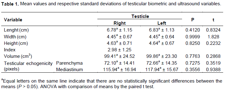
There was no difference between the biometric variables and echogenicity of the testicular parenchyma and mediastinum of the right and left testicles (P > 0.05), with a testicular index (TI) of 2.98. The testicular volume results did not have a statistically significant difference between the antimers, maintaining bilateral symmetry (Table 1).
When analyzing testicular biometric variables according to age groups, a statistically significant difference was found for length (P < 0.05), width (P < 0.05) and testicular height (P < 0.05) between AG1 and AG2 for both the right testicle as for the left, as observed in Table 2. The testicular index and volume also showed statistically significant differences (P < 0.05) between the first and the second group of animals.
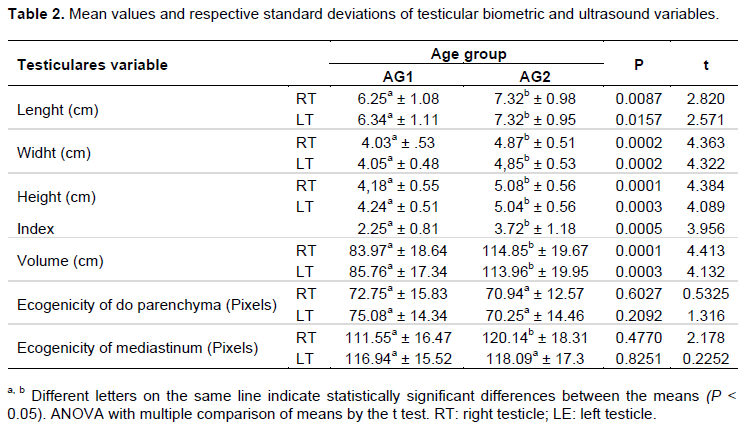
However, the echogenicity of the testicular parenchyma measured by the gray-scale histogram ranged from 70.25 to 75.08 pixels with homogeneous echotexture between animals, there was no correlation between echogenicity and age group. Regarding the testicular mediastinum, identified in all animals, an event similar to the testicular parenchyma was noted, with no statistical difference between the age groups in the left testicle. However, there was a statistical difference between the age groups when comparing the echogenicity of the right testicular mediastinum with increased echogenicity in the right testicular mediastinum correlated with age. After ultrasound scans of the testicles, the testicular parenchyma of homogeneous echotexture was evident, varying from low to moderate intensity, the hyperechogenic testicular mediastinum in relation to the testicular parenchyma and the hyperechogenic tunica surrounding the testicular parenchyma.
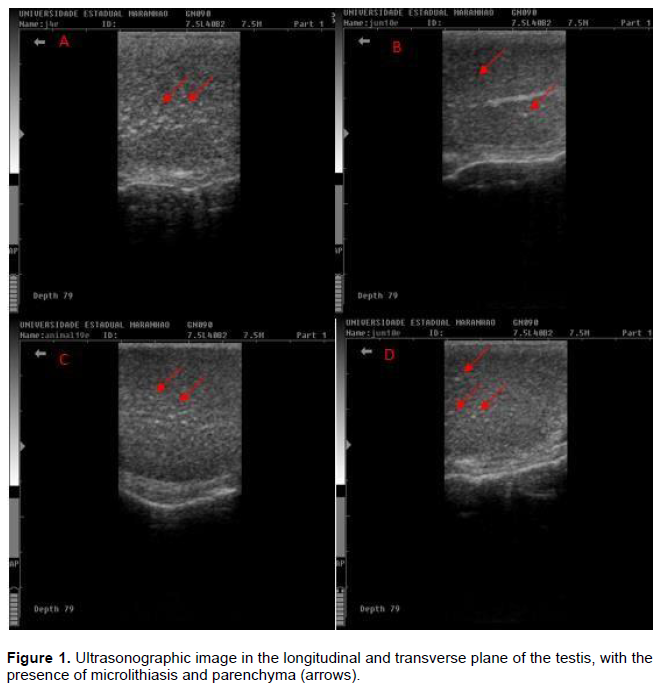
In the evaluation of the spermatic cord, the hypoechoic pampiniform plexus was observed in relation to the testicular parenchyma and the presence of anechoic circular structures with anechoic content, visualizing the lumen of the pampiniform plexus veins. Ultrasonographic changes were observed in four animals in AG1 and in five in AG2, with heterogeneous testicular parenchyma, with the presence of microlithiasis (hyperechoic foci not formers of acoustic shadow), suggesting points of fibrosis (Figure 1).
DISCUSSION
Symmetry between the gonads was also observed in studies carried out with Nordestina asinines (Gastal, 1991). In Campolina stallions, it was observed that the biometric values ??of the testicles showed no difference between the variables (Borges et al., 2010). These similar results demonstrate that even with different management and without an adequate nutritional condition, testicular symmetry was maintained, having no relationship with the development of testicular antimers.
However, this symmetry was not observed when studying the testicular biometry of asinines, observing in these animals’ different biometric values ??for right and left testicles, with significant difference in testicular length, as well as the height and width of the left testicle, smaller than that of the right one (El Wishy, 1974; Morais, 1990). In horses, this difference can be attributed to the early growth of the right testicle, in relation to the left one, during the initial reproductive development phase of the asinine (Gastal, 1991).
The biometric values ??of the animals evaluated in the present study were lower than the descriptions made for
asinines by El Wishy (1974) (LEN: 8.39 cm; HEI: 5.8 cm; WID: 4.6 cm), Kreuchauf (1984) (LEN: 8.58 cm; WID: 5.78 cm; HEI: 6.30 cm), Morais (1990) (LEN: 10.2 cm; WID: 7.0 cm; HEI: 7.4 cm), Gebbers (1995) (LEN: 8.7 cm, HEI: 7.4 cm; WID: 6.7 cm), Gastal (1991) (LEN: 7.6 cm; HEI: 5.5 cm; WID: 5.1 cm) Canisso et al. (2009) (LEN: 8.8 cm; HEI: 6.2 cm; WID: 68.35 cm). This difference in values ??is due to the difference in body weight between the animals evaluated since body weight directly influences testicular volume and size. In this study the average weight was 100 kg, while in the other studies the average weight varied between 150 to 275 kg.
However, in a study conducted by Aissanou and Ayad (2020), with testicular biometry of donkeys from Algeria, with an average weight of 193 kg and 10 years of age, they observed that the length (7.1 cm), width (4.87 cm), height (3.65 cm) and volume (73.6 cm3). This result is close to the values of the present study, even with the variation in body weight among the animals studied, this value in testicular biometry may be influenced by the arid location, with food scarcity and seasonal changes that these animals are subjected to.
The testicular index (TI) in the present study was lower than that recommended for stallions (Morais, 1990) and Pêga donkeys (Morais, 1990; Costa, 1991; Gebbers, 1995; Canisso et al., 2009). This fact can be explained by the body difference between the animals evaluated. In addition, management conditions can influence testicular and TI biometric characteristics. According to several studies, animals with TI less than or greater than eight proved to be reproductively efficient (Canisso et al., 2009; Costa, 1991; Gebbers, 1995). Asinines, even with an TI below that recommended for horses, have among domestic animals the highest spermatogenic efficiency per gram of testicular parenchyma (Neves, 2001).
The present testicular volume results are lower than those found by El Wishy in 1974. This author studied the testicles of nine donkeys (averages: age 5.1 years and weight 257 kg) and developed a formula for evaluating testicular volume and he obtained the volumes of 103.44 and 103.89 ml for the right and left testicles, respectively. The difference in testicular volume can be attributed by age and management conditions (Gacem et al., 2020).
The relationship between biometric values ??and age observed in the present study is similar to that obtained for donkeys of the Pêga breed (Canisso et al., 2009), sheep of the Santa Inês breed (Jucá et al., 2009), bubalus (Ayala, 2011), and Creole stallions (Mendes, 2012). In these studies, the authors observed that the biometric values ??in young animals are lower, with a proportional increase in the biometric characteristics with age. A progressive increase in testicular width was observed after 10 months in donkeys, more pronounced after 16 months of age, even in patients before puberty (Rota et al., 2018). This relationship is influenced by testicular and body development in young animals, since in adult animals’ testicular conformation, as well as sperm and hormonal production are already established (Canisso et al., 2009).
The measurement of testicular volume in different age groups can be a useful tool in andrological evaluation and in the prediction quality of sperm production, since it provides global dimensions of the organ and would facilitate the comparison between animals under different conditions of creation (Canisso et al., 2009; Gacem et al., 2020). When comparing the results of the testicular biometric characteristics of Pêga donkeys with the other breeds, it was observed that these, because they are medium-large animals within the species, present testicular biometric characteristics superior to those reported for Nordestina donkeys (Gastal, 1991) and for crossbred animals of African origin (Kreuchauf ,1984).
The echogenicity of the testicular parenchyma with homogeneous echotexture without alteration of echogenicity among the age groups, for this study, differed from the results found for bubalus (Ayala, 2011), in which a progressive growth of testicular echogenicity was observed between the age groups. It is justified by the fact that the testicular parenchyma of young animals is homogeneous and with low echogenicity, increasing in direct proportion to the age of the animals (Cardilli et al., 2009a, 2009b, 2010; Pastore, 2008).
Although it has been described that the male's reproductive organ is more resistant to nutritional changes in immature animals (Leathem, 1975), this fact was not evidenced in the present study, since the evaluated asinines did not have any nutritional control and the echogenicity remained without statistical difference, even among animals in the pubertal phase.
The increase in echogenicity may be associated with the increase in daily sperm production (Brito et al., 2002).
The similar echogenicity between the age groups in the present study can be influenced by the full function of the gonads in both groups, because in the pubertal phase the animals already produce sperm and hormones.
It was believed that the changes in the echogenicity of the testicular parenchyma of animals in sexual development would be due to the differentiation of the Sertoli cells (CS) and the formation of the hematotesticular barrier (Brito et al., 2012). However, in a study carried out with pantaneiro horses (Varoni, 2014), no correlation of pixel intensity with CS was observed, suggesting that the change in the echogenicity of the testicular parenchyma of animals in sexual development would be caused by the formation of the tubular lumen.
The echogenicity of the testicular parenchyma was not influenced by the production of fluids inside the seminiferous tubules during maturation. According to Evans et al. (1996), the beginning of fluid production promotes a decrease in the pixel average, which was also observed in bunalos in the age group from 36 to 60 months of age (Ayala, 2011). Young asinines in the present study had testicular echogenicity similar to mature animals, differing from previous studies (Aravindakshan et al., 2000; Brito et al., 2004; Chandolia et al., 1997), which described that the initial reduction in numerical pixel values ??in prepubertal animals probably reflects the production of liquid in testicles, commonly seen before the onset of spermatogenesis or explained by the significant increase in tubule volume.
The evaluation of the spermatic cord was similar to that observed in sheep, which visualized the vas deferens with hyperechoic walls and hypoechoic lumen. As the age group increases, the testicles become more pendulous, providing better ultrasound access to the pampiniform plexus and the vas deferens, allowing for better assessments (Andrade et al., 2012). As in the present study, in cattle and sheep, a higher frequency of fibrosis lesions was observed in the testicular parenchyma (Pinho et al., 2013a, b, Silva et al., 2015). In cattle, this fact was attributed to the intense proliferation of fibroblasts in the testicular parenchyma (Varoni, 2014).
CONCLUSION
Through testicular ultrasonography, it was possible to quantitatively determine the testicular echogenicity of asinines without a defined racial pattern, used in animal-drawn vehicles in the city of São Luís, Maranhão state, as well as the evaluation of testicular biometry and observed ultrasound changes. The gray scale histogram technique proved to be efficient in determining testicular echogenicity for the studied groups, allowing the observation of testicular parenchyma and mediastinal echogenicity among the studied age groups.
ETHICAL APPROVAL
The study was approved by the Animal Ethics and Experimentation Committee of the Veterinary Medicine Course, State University of Maranhão (SUMA), with protocol number 13/2015, approved on 11/24/2015.
CONFLICT OF INTERESTS
The authors have not declared any conflict of interests.
ACKNOWLEDGMENTS
The author express gratitude to FAPEMA, the institution that finances the research; the State University of Maranhão, for the availability of the necessary equipment and infrastructure, and to the asinine breeders, for the availability in giving the animals for the experiment.
REFERENCES
|
Ahmad N, Noakes DE (1995). A clinical and ultrasonographic study of induced testicular and epididymal lesions in goats and a ram. Animal Reproduction Science 39(1):35-48. |
|
|
Aissanou S, Ayad A (2020). Influence of age, bory weight and season no testicular and epididymis biometrics in donkeys (equus asinus). International Journal of Morphology 38(5):1434 1443. |
|
|
Andrade AKG, Soares AT, Cartaxo FQ, Peña-Alfaro CE, Guerra MMP (2012). Achados ultrassonográficos nos testículos e epidídimos de carneiros deslanados jovens e clinicamente sadios. Arquivo Brasileiro de Medicina Veterinária e Zootecnia 64(2):371-379. |
|
|
Aravindakshan JP, Honaramooz A, Bartlewski PM (2000). Pattern of gonadotropin secretion and ultrasonographic evaluation of developmental changes in the testis of early and late maturing bull calves. Theriogenology 54(3):339-354. |
|
|
Ayala HDM (2011). Ultrassonografia testicular, em machos bubalinos criados em regime extensivo no Estado do Pará. 64f. Belém-PA. (Mestrado) - Universidade Federal do Pará. |
|
|
Borges GS, Melo MIV, Mamvrini JVM, Snoek PPN.(2010). Biometria testicular de garanhões da raça campolina. Boletin de Indústria Animal 67(2):157-162. |
|
|
Brito LF, Silva AE, Unanian MM, Dode MAN, Barbosa RT, Kastelic JP (2004). Sexual development in early an late maturing Bos indicus and Bos indicus x Bos taurus crossbred bulls in Brasil. Theriogenology 62(7):1198-1217. |
|
|
Brito LFC, Barth AD, Wilde RE, Kastelic JP (2012). Testicular ultrasonogram pixels intensity during sexual development and its relationship with quality, sperm production, an quantitative testicular histology in beef bulls. Theriogenology 78(1):69-76. |
|
|
Brito LFC Silva AEDF, Rodrigues LH, Vieira FV, Deragon LAG, Kastelic JP (2002). Effect of age and genetic group on characteristics of the scrotum, tests and vascular cones, and on sperm production and semen quality in AI bulls in Brazil. Theriogenology 58(6):1175-1186. |
|
|
Canisso IC, Carvalho GR, Silva EC, Rodrigues AL, Ker PG, Guimarães JD (2009). Alguns aspectos biométricos do aparelho genital externo de jumentos doadores de sêmen da raça Pêga. Ciência Rural 39(9):2556-2562. |
|
|
Canisso IF (2008). Alguns aspectos fundamentais do exame clínico andrológico de jumentos (Equus asinus). Revista Brasileira de Reprodução Animal 32(4):233-239. |
|
|
Cardilli DJ, Toniollo GH, Pastore AA, Canola JC, Mercadante MEZ, Oliveira JA (2010). Padrão ultrassonográfico do parênquima, mediastino e túnicas testiculares em bovinos jovens da raça Nelore. Ciência Animal Brasileira 11(4):899-905. |
|
|
Cardilli DJ, Toniollo GH, Pastore AA, Canola JCC (2009a). Ultrasonographic study of testicular development in young Nelore bulls raised in extensive management system. Animal Reproduction 64(1):75-82. |
|
|
Cardilli DJ, Toniollo GH, Pastore AA, Canola JCC, Mercadante MEZ (2009b). Alterações do padrão ultrassonográfico do parênquima testicular em bovinos jovens da raça Nelore. Acta Scientia e Veterinariae 37(4):367-370. |
|
|
Chandolia RK, Bartlewski PM, Omeke BC. Beard AP, Rawlings NC, Pierson RA (1997). Ultrasonography of the developing reproductive tract in ram lambs effects of a GnRH agonist. Theriogenology 48(1):99-117. |
|
|
Clark SG, Schaeffer DJ, Althouse GC (2003). B-mode ultrasonographic evaluation of paired testicular diameter of mature boars in relation to average total of sperm numbers. Theriogenology 60(6):1011-1023. |
|
|
Costa AJSA (1991). Avaliação clínico andrológica do jumento da raça Pega. 1991, 66f. Belo Horizonte- MG. Dissertação (Mestrado em Medicina Veterinária) - Universidade Federal de Minas Gerais, Escola de Veterinária. |
|
|
El Wishy AB (1974). Testicular and epididymal sperm reserves in the ass (Equus asinus) and stallion (Equus caballus). Z Tierzuecht Zuechtungsbiol 91(4):334-344. |
|
|
Evans ACO, Pierson RA, Garcia A, Mcdougall LM, Hrudka F, Rawlings NC (1996). Changes in cirlating hormone concentrations, testes histology and testes ultrassonography during sexual maturation in beef bulls. Theriogenology 46(2):345-357. |
|
|
Feeney DA, Anderson KL, Ziegler LE, Jessen CR, Daubs BM, Hardy RM (2008). Statistical relevance of ultrasonographic criteria in the assessment of diffuse liver disease in dogs and cats. American Journal of Veterinary Research 69(2):212-221. |
|
|
Gacem S, Catalán J, Valverde A, Soler C, Miró, J (2020). Optimization of CASA-Mot Analysis of Donkey Sperm: Optimum Frame Rate and Values of Kinematic Variables for Di_erent Counting Chamber and Fields. Animals 10, 1993; |
|
|
Gastal MMFO (1991). Estudo das características seminais e do comportamento sexual de jumentos. 105f. Belo Horizonte- MG. Dissertação (Mestrado em Medicina Veterinária) - Escola de Veterinária, Universidade Federal de Minas Gerais. |
|
|
Gebbers AM (1995). Emissão diária de espermatozóides e algumas características reprodutivas de jumentos da raça Pega. 90f. Viçosa-MG. Dissertação (Mestrado em Zootecnia) - Universidade Federal de Viçosa. |
|
|
Jucá AF, Moura JCA, Gusmão AL (2009). Avaliação ultra-sonográfica dos testículos e das glândulas sexuais anexas de carneiros Santa Inês. Ciência Animal Brasileira 10(2):650-659. |
|
|
Kreuchauf A (1984). Reproductive physiology in the jackass. Animal Research and Development 20(1):51-78. doi.org/10.1590/1678-4162-7939. |
|
|
Leathem JH (1975). Nutricional influences on testicular composition and function in mammals. Handbook of Physiology 5:225-32. |
|
|
Mendes LQ (2012). Aspectos biométricos e histológicos de testículos de garanhões da raça crioula. 31f. Porto Alegre-RS. Dissertação (Mestrado em Ciências Veterinárias) - Universidade Federal do Rio Grande do Sul. |
|
|
Morais RN (1990). Contribuição ao estudo da biologia reprodutiva de jumentos (Equus asinus). 105f. São Paulo- SP. Dissertação (Mestrado em Medicina Veterinária) -Universidade de São Paulo. |
|
|
Navas FJ, Jordana J, León JM, Barba C, Delgado JV (2017). A model to infer the demographic structure evolution of endangered donkey populations. Animal 11(12):2129-2138. |
|
|
Neves ES (2001). Estudo comparativo da estrutura do testículo e do processo espermatogênico em jumentos (Equus asinus) e burros (Equus mulus mulus). 135f. Belo Horizonte, MG. Tese (Doutorado em Ciências Biológicas) - Instituto de Ciências Biológicas, Universidade Federal de Minas Gerais. |
|
|
Nyland TG, Matton JS (2004). Próstata e testículo. In: Nyland T.G. & Matton J.S. (Eds). Ultrasson: diagnóstico em pequenos animais. 1.ed. São Paulo: Editor Roca pp. 255-271. |
|
|
Ortiz-Rodriguez JM, Anel-Lopez L, Martín-Muñoz P, Álvarez M, Gaitskell-Phillips G, Anel L, Ortega Ferrusola C (2017). Pulse Doppler ultrasound as a tool for the diagnosis of chronic testicular dysfunction in stallions. PLoS One 12(5):e0175878. |
|
|
Pastore AA (2008). Contribuição da ultra-sonografia na avaliação andrológica de bovinos Nelore. 63f. Jaboticabal-SP. Tese (Doutorado em Medicina Veterinária) - Universidade Estadual Paulista. |
|
|
Pimentel SM, Silva EA (2010). Correlação entre perímetro escrotal e características reprodutivas da progênie. FAZU em Revista 7:177-185. |
|
|
Pinho PO, Costa DS, Siqueira JB (2013a). Lack of relationship between testicular echotexture and breeding soundness evaluation in adult Nelore bulls. Livestock Science 154(1-3):246-249. |
|
|
Pinho PO, Costa DS, Siqueira JB (2013b). Testicular fibrotic lesions and semen quality in adult montana tropical compound bulls. Revista Brasileira de Medicina Veterinária 35(2):105-110. |
|
|
Rota A, Puddu B, Sabatini C, Panzani D, Lainé AL, Camillo F (2018). Reproductive parameters of donkey jacks undergoing puberty. Animal Reproduction Science, 192(2010):119-125. |
|
|
Santos WG, Monteiro JNM, Oliveira D, Borlini DC, Martins Filho S, Machado FM, Nunes LC, Costa FS (2009). Ultrassonografia quantitativa do fígado de gatos com tirotoxicose induzida. Brazilian Journal of Veterinary Research and Animal Science 46(6):438-447. |
|
|
Segabinazzi LG, Silva LF, Okada C, Medrado F, Papa F, Alvarenga MA (2018). Plugged Ampullae in a Donkey Stallion (Equus asinus). Journal of Equine Veterinary Science 63:24-26. |
|
|
Silva EG, Gonçalves MTC, Pinto SCC, Soares DM, Oliveira RA, Alves FR, Araújo ACV, Guerra PC (2015). Análise quantitativa da ecogenicidade testicular pela técnica do histograma de ovinos da baixada ocidental maranhense. Pesquisa Veterinária Brasileira 35(3):297-303. |
|
|
Varoni MS (2014). Avaliação da espermatogênese e da ecotextura testicular do cavalo pantaneiro. 57f. Campo Grande- MS. Dissertação (Mestrado Ciência Animal) - Universidade Federal de Mato Grosso do Sul. |
|
Copyright © 2024 Author(s) retain the copyright of this article.
This article is published under the terms of the Creative Commons Attribution License 4.0


