Full Length Research Paper

ABSTRACT
Both cross-sectional and retrospective studies were conducted from October, 2011 to June, 2012 in and around Harar to determine prevalence, public health and economic significance of bovine hydatidosis. Out of 384 cattle examined with thorough carcass inspection at Harar city municipal abattoir, 36 (9.4%) were found to be infected with hydatid cyst. Significant differences were found among adult and young, poor, medium and good body condition cattle. However, no significant differences were found between sexes. Infected cattle harbored one or more hydatid cysts that were unequally distributed to lung, liver, kidney and spleen. In this study, 27 (52.94%) fertile cysts were observed and the rate of cyst calcification was higher in kidney (50%) than in other visceral organs. The direct and indirect annual financial loss from organ condemnations and carcass weight loss at the abattoir was estimated to be about 841,419.3 ETB. The retrospective case-book survey (2008 to 2011) of hospitals, health centers and clinic were indicated to be 0.195% (n=98,349) prevalence of human hydatidosis. A questionnaire survey and interview were also supplemented to a society in and around Harar to assess public awareness on hydatidosis. For this, 600 individuals (500 for questionnaires and 100 for interviews) were included using stratified random sampling in which educational level and profession were considered for stratification. This questionnaire survey and interview were made giving emphasis to way of acquiring hydatidosis and the individuals’ source of information about the disease as a zoonosis; the result showed schools and health extension workers were the leading information sources in the society. In conclusion, results of the present study showed bovine hydatidosis as an importantly producing great economic loss and public health hazardous at the study area. Therefore, public awareness creation, and appropriate control and prevention mechanisms in animal population should be done.
Key words: Abattoir, bovine, financial loss, hydatidosis, prevalence, public health, Harar, Ethiopia.
INTRODUCTION
In spite of large livestock population in Ethiopia, the productivity remains marginal due to many factors like malnutrition, management problems, prevalent diseases, etc. Among these hydatidosis is one of the important parasitic disease of livestock that has both economic and public health significance (FAO, 1995). Hydatidosis (Cystic Echinococosis) is a zoonotic disease caused by the larval stages of Echinococosis granulosus for which domestic intermediate hosts (cattle, sheep, goats and camels) are major reservoirs for the occurrence of human hydatidosis (Torgerson and Deplazes, 2009). Dogs are the obligate final host and infected by ingesting infected offals (lung, liver, kidney, spleen, etc). Human and wide varieties of other animals also serve as an accidental intermediate hosts for the parasite (Thompson and Mcmans, 2002).
Hydatidosis constitute public health problem worldwide, particularly it causes heavy burden in developing countries (Eckert and Deplazes, 2004; Chhabra and Singla, 2009). Its transmission is most intensive in livestock raising regions where veterinary service is unsatisfactory and offal from slaughtered animals is accessible to dogs. Cattle and other intermediate hosts contract hydatidosis by grazing on pastures contaminated with dog faeces that contain eggs of the cestode (Acha and Szyfres, 2003). Man is an intermediate host and Plays no role in the transmission of the hydatidosis, unless the individual is eaten by carnivores. Nevertheless, people’s sanitary habits make human being the main agent responsible for perpetuating the infection by feeding dogs viscera that contain hydatid cysts (Jobre et al., 1996; Chai, 1995). The life cycle of this parasite is completed when organs containing hydatid cysts are consumed by dogs (Torgerson and Deplazes, 2009). Therefore, theoretically the infection would die out if man ceased re-infecting dogs by feeding them raw viscera (Chai, 1995). Close contact with dogs and deficient personal hygiene practices such as not washing hands and vegetables before eating, and water contaminated with infected dog faeces are important factors in the transmission of human infection. Coprophagic flies may also serve as mechanical vector for eggs of the cestode (Acha and Szyfres, 2003).
The larval stage of hydatid cyst is fluid filled bladder containing cellular laminated layer with internally nucleated germinal layer. If the scolices separate from the inner lining of the capsule they caused hydatid sand (OIE, 2001).
The pathogenesis of hydatidosis heavily depends on the extents and severity of infections and the organ on which it is situated. The occasional rupture of hydatid cysts often leads to sudden death due anaphylaxis, hemorrhage and metastasis (Jobre et al., 1996); hence, the disease is associated with severe morbidity and disability. The status of hydatidosis in animals has been studied in some regions of Ethiopia and indicated hydatidosis is widely spread with great economic and public health significance (Kebede et al., 2009a). Apart from animal population, the status of human hydatidosis is among the most neglected parasitic diseases. In general, hydatidosis results in enormous economic loss in animal population with risk of public health hazards. Therefore, the main objectives of this study were to determine prevalence and financial losses of bovine hydatidosis at Harar city municipal abattoir and to elucidate public health significance of hydatidosis and assess public awareness on the disease in the study area.
MATERIALS AND METHODS
This study was conducted in and around Harar, the capital city of Harari regional state, Ethiopia. This area constitutes 76% mid-subtropical weather of “weynadega” and 24% desert type climate of “kola” (NMSA, 2011). During this study, in average 40 cattle were slaughtered per day at the Harar city abattoir. The abattoir was not provided with proper waste disposal system as even condemned abattoir materials were accessible to dogs.
Study design
Both cross sectional and retrospective studies supported with questionnaires and interviews were conducted.
Study population
Local breed of apparently healthy cattle of different body condition, sex and age groups coming from neighboring provinces of Harar were included in the study. Most of the study animals were males though females with reproductive problem, poor performance and end productive life were also encountered. Concerning public health aspect, questionnaires were distributed to individuals living in and around Harar.
Sample size determination and sampling method
Sample size was calculated by considering 50% expected prevalence at 95% confidence level and 5% precision (Thrusfield, 2005). Accordingly,
N = (1.96)2 Pexp(1-pexp)/d2 = 384.
Then these animals were included in the study using systematic random sampling method. 500 individuals filled the questionnaires and 100 were interviewed applying stratified random sampling, using educational level and profession for stratification. Case book survey was also conducted to determine retrospective prevalence of human hydatidosis in patients that had come to hospitals, health centers and public clinic in and around Harar.
Experimental
Ante-mortem inspection
During ante-mortem inspection, each of the study animals was given an identification number. Age, origin, sex and body condition scoring of the study animals were made. The body condition score was classified into poor, medium and good categories (Heinonen, 1989). Young and adult age groups were encountered following their dentations.
Post mortem inspection
During post mortem examination organs especially liver, lung, spleens, kidney, heart, muscle and head part as a whole were systematically inspected for the presence of hydatid cyst by applying the routine meat inspection procedure of primary examination followed by secondary examination (Alula, 2010). The primary examination involves visualizations of the organs whereas secondary examination involves further incision of each organ into pieces and whenever evidence of the cyst was found, it was classified as live or calcified and the cyst distribution to organs was also recorded.
Fertility and viability tests
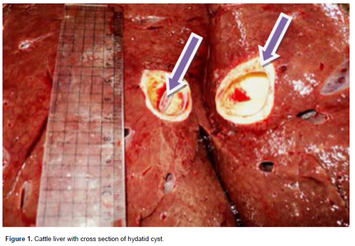
Positive or suspected samples were taken to the laboratory for the cyst identification, fertility and viability tests were performed. Of the collected hydatid cysts, individual cysts where carefully incised and examined for protoscoleces, which are similar to the appearance of white dots on the germinal epithelium (Figure 1); such cysts were characterized as fertile cysts; fertile cysts was subjected to viability test. A drop of the sediment containing the protoscoleces was placed on the microscopic glass slide and covered with a cover slip and observed for amoeboid like peristaltic movements. For clear vision, a drop of 0.1% aqueous eosin solution was added to equal volume of protoscoleces in hydatid fluid on the microscopic slide with the principle that viable protoscoleces should completely or partially exclude the dye while the dead ones took it up. Furthermore, infertile cysts were further classified as sterile or calcified (Soulsby, 1982) (Table 2).
Estimated financial loss
Losses due to organ condemnation was calculated by considering information on the retail market price of condemned organs (Ogunirale and Ogunrinade, 1980), obtained from butcher shops of Harar city during the study period. The average annual slaughter rate of cattle at the abattoir was estimated from the retrospective data of the last two years. In line with this:
LOS=MAK×PH[(P1C1)+(P2C2)+(P3C3)+(P4C4)]
where LOS = loss due to organ condemnation, MAK = annual average number of cattle slaughtered at Harar city municipal abattoir, PH = prevalence of hydatidosis, P1-4 = prevalence of each organ condemned, C1-4 = main retail cost of single organs condemned.
The financial loss encountered from carcass weight losses was also calculated by considering retail market cost of 1 kg beef at Harar city, and 5% carcass weight loss due to hydatidosis was considered as described by Polydrous (1981). Accordingly,
LCWL = MAK × PH × CPB × 5% × 126 kg,
where LCWL = loss from carcass weight loss, NAS = average number of cattle slaughtered annually, CPB = current average price of 1 kg beef, 126 kg = average carcass weight dressing percentage of an adult zebu cattle. The total annual financial loss was the sum of losses from organ condemned and carcass weight lost.
Questionnaire and retrospective studies
Evaluation on public awareness about hydatidosis was done through well designed interview and questionnaires. Stratified random sampling method was considered based on educational level and profession. Accordingly, designed questionnaires were supplemented to 500 selected people of whom 200 were elementary and high school students, 100 were university and college students, 50 were health professionals and 150 non health professionals of different departments. In addition, 20 health and 20 non health professionals, 10 veterinarians and 50 illiterate local people were interviewed about their custom of consuming backyard slaughtered beef, drinking surface water, washing vegetables and their hands before meal, attitude of offering dogs raw viscera and their close contact with stray dogs in their social activity. Mass media, schools they attended and have been attending, health extension workers and other informal sources (including family, religious leader advises, etc.) were also filled in questionnaires and interviewed as a source of information about hydatidosis for individuals.
Retrospective cystic echinococcus cases from the total ultrasound admitted patients during January, 2008 to December, 2011 in hospitals and cases registered as hydatidosis at health centers and clinic in and around Harar were collected from the institutions case book reports.
Data management and analysis
The information or data about bovine hydatidosis was collected and later entered in to Microsoft excel 2010 spreadsheet and analyzed using SPSS statistical software version 20. Prevalence data were analyzed by chi-square, and descriptive statistics were also used. P≤0.05 was considered as statistically significant at 95% CI.
RESULTS
Prevalences
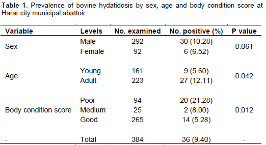
The present study showed 9.4% (n=384) bovine hydatidosis at Harar city municipal abattoir. There was a significant difference in the harboring of hydatid cyst between age groups and body condition scores. The prevalence was significantly higher in adult and poor body condition than in young, and medium, good body condition cattle (p<0.05). However, there was no significant difference between sexes (p > 0.05) (Table 1).
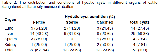
Among the infected organs, the highest prevalence was found in liver and followed by lung, which accounted for 56.86 and 27.45%, respectively. The rate of cyst calcification was by far higher in kidney (50%) than in other visceral organs, the least calcification rate (20.69%) was encountered in liver.
Financial loss assessment
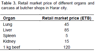
Cystic echinococcus financial loss assessment was made based on the direct and indirect losses. The direct loss was losses due to condemnation of the offals like lung, spleen, liver and kidney, whereas carcass weight loss due to hydatid cyst is indirect loss. The retail market price of different edible offals’ and 1 kg beef in Harar city were considered as a parameter for this calculation (Table 3).
Therefore, by applying the formula of Ogunirale and Ogunrinade (1980) and Polydrous (1981), the annual financial loss due to bovine hydatidosis at the study area could be as followed.
Direct loss (DL)
LOS = MAK × PH [(P1C1) + (P2C2) + (P3C3) + (P4C4)] = (10950 × 9.4%) [(7.84% × 5) + (27.45% × 45) + (56.86% × 85) + (7.84% × 15)] = 63268.50ETB
Indirect loss (IL)
LCWL = MAK × PH × CPB × 5% × 126 kg =126 × 5% × 10950 × 9.4% × 120 =778150.8 ETB
Total financial loss (TFL)
TEL = DL + IL =63268.50 + 778150.8 =841,419.3 ETB; about ~ $46,745.5
Questionnaire and retrospective studies
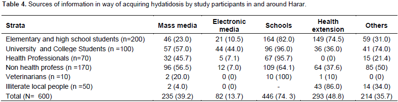
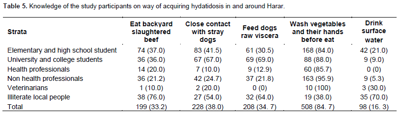
Results of questionnaire and interview showed that 74.3 and 48.8% of the participants got information about way of acquiring hydatidosis from schools and health extension workers, respectively (Table 4). On the other hand, about 38.0% of the participants had direct contact with stray dogs in their daily activities. The results also indicated only 84.7% of our study population washed their hands and vegetables before they eat it (Table 5).
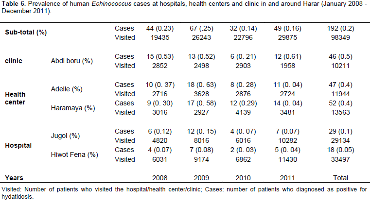
Case book analysis showed that out of 98,349 total patients admitted for ultrasound and clinical examinations at hospitals, health centers and clinic in and around Harar, 192 (0.195%) individuals were registered as positive cases for hydatidosis. There were prevalence variations among the institutions and the highest prevalence (0.5%) was found at Abdi Boru clinic (Table 6).
DISCUSSION
The present 9.4% prevalence of bovine hydatidosis at Harar city municipal abattoir was in agreement with the report of 9.38% (Wubet, 1987), from Hararge region; however, this prevalence was less than the findings of 37.7% (Roman, 1987), 34.05 and 15% (Kebede et al., 2009b; Belina et al., 2012), and 32.1% (Berhe, 2009), from Gonder, Bahir Dar and Mekele, respectively. Relatively lower prevalence in current study may be due to adverse conditions of high temperature and low humidity encountered at the origin of animals on survival of E. granulosus egg (Thompson and Allsopp, 1988), as majority of the study animals came from low land areas. The prevalence variations with geographical regions may also have some connection with cultural and religious taboos such as backyard slaughtering of animals, attitudes in offering uncooked infected offal to pet animals, close contact with stray dogs in social activities and in general poor public awareness about the hydatidosis. Njoroge et al. (2002) explained environmental conditions, livestock stocking intensity and movement in different regions contribute to the prevalence differences. Attributing to the work of Belina et al. (2012), our current study indicated there was no significant difference (p>0.05) between sex groups, though higher prevalence was found in male animals. This may be due to small number of female animals slaughtered at the abattoir during our study. Females were kept for breeding, hence only females with reproductive problem, poor performance and end productive life were slaughtered at the abattoir. However, significant (P=0.01) higher prevalence was found in animals with poor body condition score which probably reflected the effect of relatively high cyst burden. According to Polydrous (1981) and Battelli (1997), moderate to severe infection of the parasite leads to live weight loss, retarded performance and growth with reduced quality of meat and milk. Our finding also showed there was a significant difference between age groups (p< 0.05) in harboring hydatid cyst. Majority of the studied animals were adult and hence, they were exposed to the disease (parasitic ova) over a long period of time with an increased possibility of acquiring the infections than younger ones. It has been stated that the easier development and the fertility rate of hydatid cysts may show the tendency to increase with advancing age of the hosts (Himonas et al., 1987). In the present study, the highest cyst frequency was observed in liver and followed by lung which is in agreement with report of Belina et al. (2012). This is explained by the fact that livers and lung possess the first greater capillary sites encountered by the migrating E. granulosus oncosphere (hexacanth embryo) which adopt the portal vein route and primarily negotiate hepatic and pulmonary filtering system sequentially before any other peripheral organ is involved (Alula, 2010).
The annual financial loss of 841,419.3 ETB, in our study due to bovine hydatidosis from offal condemnation and carcass weight loss was greater than the findings of Yilma (1984), 813,526.46, Wubet (1987), 64,920.00 and Kebede et al. (2009a), 25,608.00 ETB from Debrezeit abattoir, Hararge zone, and Tigray region, respectively; however, by far lower than the report of Regassa et al. (2010), 1791625.89 ETB from Hawassa municipal abattoir. According to Alula (2010), the financial loss varied from region to region and even from abattoir to abattoir based on the prevalence of hydatidosis, mean annual number of cattle slaughtered at different abattoirs and the retail market price of organs.
The results of the questionnaire and interview showed that 74.3% of the participants got information about way of acquiring hydatidosis from schools. Information from school was the leading one in this study. However, Sisay et al. (2012) reported that majority of the elementary and high school students get information about zoonotic diseases from their families in the form of advice, though most health professionals get their information from medical schools they attended. On the other hand, 48.8% of our participants got information about way of acquiring hydatidosis from health extension workers. Health extension workers have high chance of getting all social classes from all corners and they have been working on zoonosis and communicable disease (working on prevention) is their main objective, as Ethiopian govern-ment has been working with the principle of at least one health extension worker to one kebele/PA. Furthermore, the most important social classes (86% of illiterate local people) got information about way of acquiring hydatidosis from health extension workers. The study also showed that none of the illiterate local people got information about the disease from media and schools.
Contact with stray dog, not washing vegetables and their hands before eating are important way of acquiring hydatidosis (Ketema, 2010). However, in the present study, 38% of the participants had close contact with stray dogs in their social activities, and only 84.7% of the study population washed their hands and vegetables before they ate. Supporting this study, Tamiru et al. (2008) and Avery (2004) reported in their previous study, that eating uninspected backyard slaughtered raw meat had been considered as risk factor for hydatidosis. This could be due to the low level of awareness of the people on the importance of using inspected meat, because of cultural beliefs that raw meat is better than cooked one and the deeply established traditional habit of eating raw meat in the country (Sisay et al., 2012). About 69% of the university and college students, 64% of illiterate local people and even 12.9% health professionals support feeding dogs raw viscera unlike veterinarians. None of the veterinarians were in favor of cultural beliefs and traditional habit of offering uncooked infected offal to dogs. Veterinarians possess better knowledge on animal diseases and food productions, as well as training in ecological, economic and human cultural issues, make them the leaders in developing and implementing new methods of promoting sustainable public health (King and Khabbaz, 2003). Sisay et al. (2012) also stated, besides educating health professionals and directors of public health, it is important to increase the involvement of veterinarians in public health improvement.
Retrospective case book analysis of patients admitted for ultrasound and clinical examinations at hospitals, health centers and clinic in and around Harar showed 0.195% (192/98,349) human hydatidosis. This pre-valence was higher than the report of 0.044% (Belina et al., 2012) from Bahir Dar. The higher current prevalence might be because of the low public awareness, backyard slaughtering practices, poor control measures and presence of a large number of stray dogs that contributed to human infection. In addition to this, our study area lacks modern diagnostic facilities, and there was inability to offer treatment by the most vulnerable sections of the society. However, 1.6 and 0.5% human hydatidosis were also screened with ultrasound from southern part of Ethiopia in 1987 and 1996, respectively (Eckert et al., 2002). Battelli (2003) also reported 0.22% human cases from Portugal.
In conclusion, the present study showed 9.4% preva-lence of bovine hydatidosis with significant estimated financial loss in and around Harar. Body condition score and age groups cattle were statistically risky factors unlike sexes in the study area. The result also showed liver was the most frequently affected organ and cyst calcification was by far higher in kidney among other examined organs. In average, about 841,419.3 ETB financial losses were encountered in the current study. 0.195% human hydatidosis was recorded from retrospective hospitals, health centers and clinic case book analysis that hydatidosis is an important zoonotic disease. The results of the questionnaire and interview data showed that majority of the participants got information about way of acquiring hydatidosis from schools and health extension workers. In addition, people in and around Harar do not have enough knowledge about way of acquiring hydatidosis where majority of the people were accustomed to consuming backyard slaughtered beef and even offering infected raw offals to dogs. Therefore, based on this study, we recommended that effective control and prevention mechanisms in animal population should be done and stray dogs have to be restricted; there must be legislation that will strictly prevent backyard slaughtering practice, and public heath veterinaries should work with medical professionals and reach the rural areas; schools and other public institutions have to teach society at large, and lastly creation of public awareness about hydatidosis as zoonosis and the present works of health extension workers in Ethiopia should be encouraged and further expanded.
CONFLICT OF INTEREST
The authors declare that there is no conflict of interest.
REFERENCES
| Acha PN, Szyfres B (2003). Zoonoses and communicable diseases common to man and animals, 3rd ed. Pan American Health Organization, Washington DC. pp. 192-193. | ||||
| Alula A (2010). Major Metacestodes in cattle slaughtered at Kombolcha Elfora abattoir, North Eastern Ethiopia: Prevalence, Cyst viability, Organ distribution and Socio-economic implications. DVM Thesis, Hawassa University, Department of Veterinary Medicine, Hawassa, Ethiopia. | ||||
| Avery A (2004). Red meat and poultry production and consumption in Ethiopia and distribution in Addis Ababa. Borlaug Ruan World Food Prize. International Livestock Research Institute Addis Ababa, Ethiopia. pp. 4-15. | ||||
| Battelli G (1997). Evaluation of the economic costs of echinococosis. Int. Arch. Hydat. 32:33-37. | ||||
| Battelli G (2003). Socio-Economic impact of CE. WHO Mediterr. Zoonoses Control Center Inform Circular 57:1020-1378. | ||||
| Belina T, Alemayehu A, Moje, Yechale A, Girma S (2012). Prevalence and public health significance of ovine hydatidosis in Bahir Dar town, Ethiopia. J. Vet. Med. Anim. Health 4(8):110-115. | ||||
| Berhe G (2009). Abattoir survey on cattle hydatidosis in Tigray region of Ethiopia. Trop. Anim. Health Prod. 32:56-68. | ||||
| Chai JJ (1995). Epidemiological studies on cystic echinococcosis in China. National Hydatid Center of China, Xinjiang Institute. Endemic Dis. Cont. Res. 8(2):122-136. | ||||
| Chhabra MB, Singla LD (2009). Food-borne parasitic zoonoses in India: Review of recent reports of human infections. J. Vet. Parasitol. 23(2):103-110. | ||||
| Eckert J, Schantz PM, Gasser RB (2002). WHO/OIE manual in echinococcosis in humans' and animals. Geographic distribution and prevalence. World Health Organization and World Organization for Animal Health, Paris. 101-143. | ||||
|
Eckert J, Deplazes P (2004). Biological, epidemiological and clinical aspects of echinococcosis, a zoonosis of increasing concern. Clin. Microbiol. Rev. 17(1):107-135. Crossref |
||||
| FAO/Food and Agricultural Organization (1995). Livestock development strategies for low income countries. Proceedings of the Joint, FAO. pp. 6-8. | ||||
| Heinonen M (1989). Body condition scoring as of cattle in Ethiopia. MOA. pp. 3-12. | ||||
|
Himonas C, Frydas S, Antoniadou S (1987). The fertility of hydatid cyst in food animals in Greece In: Geerts S, Kumar V, Brandt J (eds.), Helminth Zoonoses. Martinus Nijjhoff Publishers, Netherlands. pp. 1-21. Crossref |
||||
| Jobre Y, Lobago F, Tiruneh R, Abebe G, Dorchies P (1996). Hydatidosis in three selected region in Ethiopia an assessment trial. On its prevalence, economic and public health importance. Rev. Med. Vet. 143:797-804. | ||||
|
Kebede N, Mitiku A, Tilahun G (2009a). Hydatidosis of slaughtered animals in Bahir dar abattoirs, North Western Ethiopia. Trop. Anim. Health Prod. 41:43-50. Crossref |
||||
|
Kebede N, Hagos A, Girma Z, Lobago F (2009b). Echinococcousis/hydatidosis of slaughtered animals in Mekele Abattoir, North Western Ethiopia. Trop. Anim. Health Prod. 41:41-45. Crossref |
||||
| Ketema T (2010). Assessments of the awareness the community about zoonotic diseases, habit of consuming raw animal products and the importance of veterinary public health in the health institutes. Research work submitted to University of Gondar research and publication office, Gonder, Ethiopia. pp. 5-11. | ||||
|
King L, Khabbaz R (2003). Converging Issues in Veterinary and Public health. Emerg. Inf. Dis. 9:4. Crossref |
||||
|
Njoroge EN, Mmbithi PM, Jathuma JM, Wachira TM, Gashura TM, Magambo JK, Zeyhle E (2002). The study of cystic echinococcousis in slaughter animals in three selected areas of Northern Turkana, Kenya. Vet. Parasitol. 194:85-91. Crossref |
||||
| NMSA (2011). National Meteorology Service Agency. Addis Ababa, Ethiopia. | ||||
| OIE/Office International des Epizootics (2001). WHO/OIE Manual on Echinococcosis in humans and animals. Public health problem of global concern. Paris, France. pp. 53-64. | ||||
| Ogunirale AF, Bola L, Ogunrinade BI (1980). Bovine fasciolosis in Nigeria. Inter current parasitic and bacterial infections. Trop. Anim. Health Prod. 14:121-125. | ||||
| Polydrous K (1981). Animal health and economic case study: Echinococcousis with reference to Cyprus. OIE Technical Series 93:195-203. | ||||
|
Regassa F, Molla A, Bekele J (2010). Study on the prevalence of cystic hydatidosis in cattle slaughtered at Hawassa Municipal abattoir, Ethiopia. Trop. Anim. Health Prod. 42:977-984. Crossref |
||||
| Roman T (1987). Study on economic significance of bovine fasciolosis and hydatidosis at Gonder. DVM thesis, FVM, AAU, Debrezeit, Ethiopia. pp. 5-18. | ||||
| Sisay G, Girma Z, Ketema T, Tariku J (2012). Assessment of awareness on food borne zoonosis and its relation with Veterinary Public Health Services in and around Addis Ababa, Ethiopia. Ethiop. Vet. J. 16 (1):15-22. | ||||
| Soulsby EJ (1982). Helminth, Arthropod and Protozoa of domestic animals, 7th ed. Bailliere Tindall, London. P 809. | ||||
| Tamiru N, Getachew T, Medhin G (2008). Seroprevalence of Toxoplasma gondii in Nazareth Town, Ethiopia. E. Afr. J. Pub. Health 5:3. | ||||
| Thompson RC, Allsop CE (1988). Hydatidosis: Veterinary perspectives and annotated Bibliography. CAB International. pp. 45-57. | ||||
| Thompson RC, McManus DB (2002). Aetiology: parasites and life cycles. WHO/OIE Manual in echinococcosis in humans and animals. WHO/OIE, Paris. pp. 1-19. | ||||
|
Torgerson PR, Deplazes P (2009). Echinococosis, diagnosis and dignistic interpretation in population studies. Trends Parasitol. 23:164-170. Crossref |
||||
| Thrusfield M (2005). Veterinary Epidemiology 2nd ed., University of Edinburgh, Black well science. pp. 180-188. | ||||
| Wubet M (1987). A preliminary study of echinococcous hydatidosis in Hararge region. DVM thesis, Addis Ababa University, Faculty of Veterinary Medicine, Debrezeit, Ethiopia. pp. 3-32. | ||||
| Yilma J (1984). Preliminary study of the economic and public health significance of echinococcous in Debrezeit abattoir. DVM thesis, FVM, AAU, Debrezeit, Ethiopia. pp. 12-27. | ||||
Copyright © 2024 Author(s) retain the copyright of this article.
This article is published under the terms of the Creative Commons Attribution License 4.0


