Case Report

ABSTRACT
A case of atresia ani with recto-vaginal fistula was observed in a two-month old Sudanese crossbred lamb. On physical examination, the abdomen was distended, and there was bulging around the perineal region and severe straining. Upon palpation, the anal ring was felt at the anal area, voiding of feaces and urine from the vaginal opening was seen, while the fistula was felt upon vaginal palpation. Reconstructive surgery by incising the bulged, palpable region was done. The rectum was located and sutured to the perineal wall. The patency of the newly constructed anal opening was however maintained with the use of fabricated syringe barrel. Full recovery was attained within 21 days. The animal was relived of pressure strain, pain through the surgical intervention with resultant increase in body condition score.
Key words: Atresia ani, fistula, anal, palpation.
INTRODUCTION
Congenital anomalies of gastro intestinal tract occurs among different species of animals with an incidence of about 4.3% (Leipold et al., 1971).
Atresia ani is a developmental anomaly of the new born which occurs as a result of an autosomal recessive gene (Bademkiran et al., 2009). This condition is characterized by absence of anal opening and may be associated with recto-vaginal fistula, recto-cystic fistula, vagino urethral agenesis, taillessness, hypospadias (Singh et al., 1993) and diphallus (Loynachan et al., 2006). Recto-vaginal fistula or anus vaginalis is an inherited lethal abnormality in which there is an abnormal passage between rectum and vagina; also, feaces are passed through the vagina as a result of the imperforate anus (Oehme and Prier, 1974). Atresia ani associated with recto-vaginal fistula has been reported in many species. These include calves (Shakoor et al., 2012; Mahesh et al., 2014 ), lambs (Kamalakar et al., 2014, 2015), dogs (Rahal et al., 2007) and pigs (Monsang et al., 2014).
These anomalies are usually noticed at birth whereas in some cases, usually diagnosed at a later age. Early diagnosis of non-lethal anomalies aids in efficient management of the condition.
There are four major types of atresia ani which consists of type I-IV. In type I, a mucosal barrier obstructs the lumen of the intestine. Animals with type II have an intestine with two segments without any communication usually with a fibrous cord joining them together. In type III, two segments of intestine separated completely which may be coiled at the distant end in some animals. Type IV atresia involves multiple site of atresia (Bademkiran et al., 2009 ; Rahal et al., 2007). Congenital rectovaginal fistula usually associated with type II atresia ani in which the rectum ends as a blind pouch immediately cranial to the imperforated anus (Rahal et al., 2007).
CASE HISTORY AND CLINICAL EXAMINATION
A 2-month old female Sudanesse crossbred lamb was presented to the Veterinary Teaching Hospital, University of Maiduguri, Nigeria with the complaint of inability to void feaces normally. An examination of perineal region, revealed absence of anal opening, tenesmus, bulging at the anal region and communication between rectal floor and vaginal roof, through which the feaces was voiding out. Based on the prevailing clinical signs, the condition was however diagnosed as congenital atresia ani associated with recto-vaginal fistula.
Treatment plan
Complete blood count was conducted so as to ascertain whether the animal is anemic or not; ascultation of the heart and lungs was done to know the status of the cardiopulmonary system, whether or not the animal can withstand both aesthesia and surgery. Unfortunately, ultrasound scan was not done to know the type of atresia ani we were dealing with and lastly, reconstructive surgery was performed to correct the condition.
Vital parameters
Respiratory rate: 65 breaths/min (60-90)
Heart rate 120 beats/min: (70 - 100)
Temperature: 38.5 (38 - 40) (Table 1).
Surgical treatment
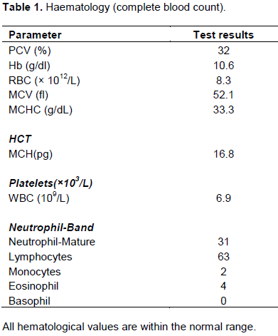
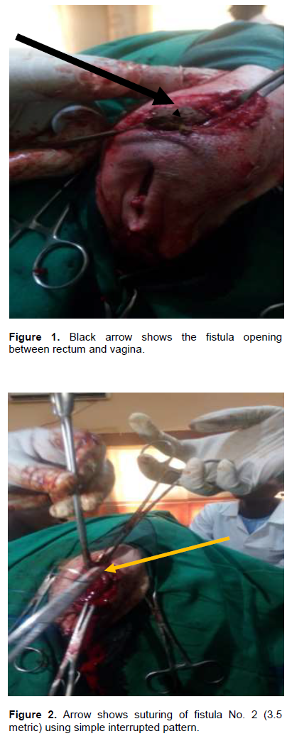
The rectum and vagina were evacuated of feaces, the perineum was shaved and prepared for aseptic surgery (Figure 1). Fluid therapy using 5% normal saline was instituted. Anesthesia was effected with xylazine at 0.084 mg i.m. and local infiltration of the perineum using 20% lignocaine hydrochloride. The animal was placed on lateral recumbency and a cruciate skin incision was made on the skin at the bulging area. The rectum was opened and the contents were evacuated. The fistulous orifice, which was about 3 cm in diameter and 2” away from anus, was reached through anal route and was closed using simple interrupted suture pattern with chromic catgut size 0/2 (Figure 2). The area was irrigated with normal saline and rectal mucosa was sutured to the skin in simple interrupted pattern using black braided silk. A sterile 20-mL syringe barrel (Figure 3) was cut at non winged end and two holes were made at the centre of each wing. The non-winged end was lubricated with liquid paraffin and inserted into rectum in other to hold the rectum tightly to the skin, to avoid closure through healing. The wings of barrel were secured to the perineal skin by passing nylon suture material from skin through hole in that side wing and tied to outside using simple interrupted sutures.

Outcome
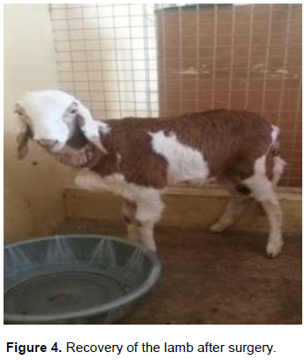
The animal fully recovered 90 min after the surgery and stood on its entire limbs (Figure 4). After another 90 min, a rising appetite was noticed, hence small quantity of feed (wheat bran) was provided. After 24 h, normal defecation even though softer than normal was noticed, which may be due to the wheat bran.
Post-operative care
Post operatively, 5% dextrose for rehydration, procine penicillin with streptomycin was given at 9 mg/kg × 3/7. Diclofenac sodium was given at 3 mg/kg × 3/7 as an analgesic. The animal was fed wheat bran continuously for 7 days at the Veterinary Hospital so that it could pass out soft feaces and for monitoring, should there be any complication.
DISCUSSION
Atresia usually arises during the embryonic period which results from autosomal recessive gene (Loynachan et al., 2006). Though environmental teratogens, plant toxins and some viruses (Loynachan et al., 2006) are recognised complicating factors in calves. In the present case, the reason could not be ascertained and unspecific as reported by Johnson et al. (1980). The increased faecal pressure may have caused an abnormal opening between rectal wall and vagina forming recto-vaginal fistula and thus causing defecation via vulva (Norrish and Rennie, 1968). Atresia ani is frequently associated with recto-vaginal fistula between dorsal wall of vagina and ventral wall of terminal rectum. A clinical sign is mainly the absence of anal opening; however, while Amith et al. (2017) reported tenesmus, abdominal discomfort in a 5- day old lamb, Prasad et al. (2016) did not report such clinical signs in a 3-day old lamb. Tenesmus, abdominal distension, abdominal discomfort was equally not observed in this present study, which means there are variations in clinical signs regardless of age. Furthermore, radiographs are considered important to determine the position of the fistula and to differentiate the four types of congenital atresia ani (Rahal et al., 2007). However, in our present case, the defects were rectified individually as reported by Rahal et al. (2007).
CONCLUSION
Surgical intervention (anal reconstruction) is the only possible solution to cope with these congenital anomalies in animals and to make affected animals economically profitable for the keepers. However, it is worthy of note that female animals, especially the Sudaneese breed with these conditions may live up to 2 months or more and still have a good body condition score until corrected.
CONFLICT OF INTERESTS
The authors have not declared any conflict of interests.
REFERENCES
|
Amith NG, Sandesh KV, Nagaraja BN (2017). Surgical repair of congenital rectovaginal fistula and atresia ani in a lamb. International Journal of Science and Research pp. 12-14. |
|
|
Bademkiran S, Icen H, Kurt D (2009). Congenital recto vaginal fistula with atresia ani in a heifer: a case report. YYU Veteriner Fakultesi Dergisi 20(1):61-64. |
|
|
Johnson EH, Nyack B, Marsh A (1980). Surgical repair of atresia ani and rectovaginal fistula in a goat. Veterinary medicine, Small Animal Clinician 75(12):1833. |
|
|
Kamalakar G, Devaratnam J, Brahmaji R, Jyothsna B (2015). Congenital atresia ani associated with recto-vaginal fistula in Ongole calf. Journal of Livestock Science 6:80-84. |
|
|
Kamalakar G, Sumiran N, Mahesh R, Prasad VD (2014). Repair of acquired recto- vaginal fistula associated with atresia ani in a lamb. |
|
|
Leipold HW, Dennis SM, Huston K (1971). Congenital defects of cattle: Nature, cause, and effect. Advances in Veterinary Science and Comparative Medicine 16:103-150. |
|
|
Loynachan AT, Jackson CB, Harrison LR (2006). Complete diphallia, imperforate ani (type 2 atresia ani), and an accessory scrotum in a 5-day-old calf. Journal of veterinary diagnostic investigation 18(4):408-412. |
|
|
Mahesh R, kamalakar G, Devi Prasad V (2014). Surgical management of atresia ani in a calf: A case report. International Journal of Veterinary Science and Medicine 2(2):51-53. |
|
|
Monsang SW, Madhu DN, Sarode IP, Kumar R, Amarpal, Pawde AM, Kinjavdekar P, Aithal HP (2014). Surgical repair of a rare case of congenital recto-vaginal fistula and atresia ani in a crossbred piglet. |
|
|
Norrish JG, Rennie JC (1968). Obervations on the Inheritance of atresia ani in swine. Journal of Heredity 59(3):186-187. |
|
|
Oehme FW, Prier JE (1974). Text book of large animal surgery: Williams and Wilkins, Baltimore London pp. 425-509. |
|
|
Prasad VD, Mahesh R, Kamalakar G, Devarathnam J (2016). Congenital recto-vaginal fistula with atresia ani in a lamb: A case report. |
|
|
Rahal SC, Vicente CS, Mortari AC, Mamprim MJ, Caporalli EH (2007). Rectovaginal fistula with anal atresia in 5 dogs. The Canadian Veterinary Journal 48(8):827. |
|
|
Shakoor A, Muhammad SA, Younus M, Kashif M (2012). Surgical repair of congenital recto-vaginal fistula with atresia ani in a cow calf. Pakistan Veterinary Journal 32(2):298-300. |
|
|
Singh J, Singh AP, Patil DB (1993). From Digestive system: In Ruminant Surgery 1st edition, Edited by Tyagi RPS and Singh J, CBS Publishers, New Delhi P 222. |
|
Copyright © 2024 Author(s) retain the copyright of this article.
This article is published under the terms of the Creative Commons Attribution License 4.0


