Journal of
Veterinary Medicine and Animal Health

  • Abbreviation: J. Vet. Med. Anim. Health
  • Language: English
  • ISSN: 2141-2529
  • DOI: 10.5897/JVMAH
  • Start Year: 2009
  • Published Articles: 416

Full Length Research Paper

Prevalence of bovine brucellosis, tuberculosis and dermatophilosis among cattle from Benin’s main dairy basins

Nestor Denakpo Noudeke*
  • Nestor Denakpo Noudeke*
  • Département de Production et Santé Animales, Ecole Polytechnique d’Abomey-Calavi, Université d’Abomey-Calavi, 01 BP 2009, Cotonou, République du Bénin.
  • Google Scholar
Gerard Dossou-Gbete
  • Gerard Dossou-Gbete
  • Laboratoire des Recherches Zootechnique, Vétérinaire et Halieutique (LRZVH), Centre de Recherches Agricoles d'Agonkanmey (CRA-Agonkanmey), Institut National des Recherches Agricoles du Bénin(INRAB), 01 BP 884 Recette Principale, Cotonou 01, Bénin.
  • Google Scholar
Charles Pomalegni
  • Charles Pomalegni
  • Laboratoire des Recherches Zootechnique, Vétérinaire et Halieutique (LRZVH), Centre de Recherches Agricoles d'Agonkanmey (CRA-Agonkanmey), Institut National des Recherches Agricoles du Bénin(INRAB), 01 BP 884 Recette Principale, Cotonou 01, Bénin.
  • Google Scholar
Serge Mensah
  • Serge Mensah
  • Laboratoire des Recherches Zootechnique, Vétérinaire et Halieutique (LRZVH), Centre de Recherches Agricoles d'Agonkanmey (CRA-Agonkanmey), Institut National des Recherches Agricoles du Bénin(INRAB), 01 BP 884 Recette Principale, Cotonou 01, Bénin.
  • Google Scholar
Luc Gilbert Aplogan
  • Luc Gilbert Aplogan
  • Laboratoire de Diagnostic Vétérinaire et Sérosurveillance des maladies animales de Parakou, Ministère de l’Agriculture, de l’Elevage et de la Pêche, Bénin.
  • Google Scholar
Germain Atchade
  • Germain Atchade
  • Laboratoire Vétérinaire de Bohicon, Ministère de l’Agriculture, de l’Elevage et de la Pêche, Bénin.
  • Google Scholar
Jacques Dougnon
  • Jacques Dougnon
  • Département de Production et Santé Animales, Ecole Polytechnique d’Abomey-Calavi, Université d’Abomey-Calavi, 01 BP 2009, Cotonou, République du Bénin.
  • Google Scholar
Issaka Youssao
  • Issaka Youssao
  • Département de Production et Santé Animales, Ecole Polytechnique d’Abomey-Calavi, Université d’Abomey-Calavi, 01 BP 2009, Cotonou, République du Bénin.
  • Google Scholar
Guy Apollinaire Mensah
  • Guy Apollinaire Mensah
  • Laboratoire des Recherches Zootechnique, Vétérinaire et Halieutique (LRZVH), Centre de Recherches Agricoles d'Agonkanmey (CRA-Agonkanmey), Institut National des Recherches Agricoles du Bénin(INRAB), 01 BP 884 Recette Principale, Cotonou 01, Bénin.
  • Google Scholar
Souaïbou Farougou
  • Souaïbou Farougou
  • Département de Production et Santé Animales, Ecole Polytechnique d’Abomey-Calavi, Université d’Abomey-Calavi, 01 BP 2009, Cotonou, République du Bénin.
  • Google Scholar


  •  Received: 21 February 2017
  •  Accepted: 18 April 2017
  •  Published: 31 May 2017

 ABSTRACT

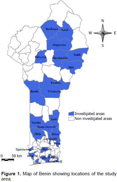
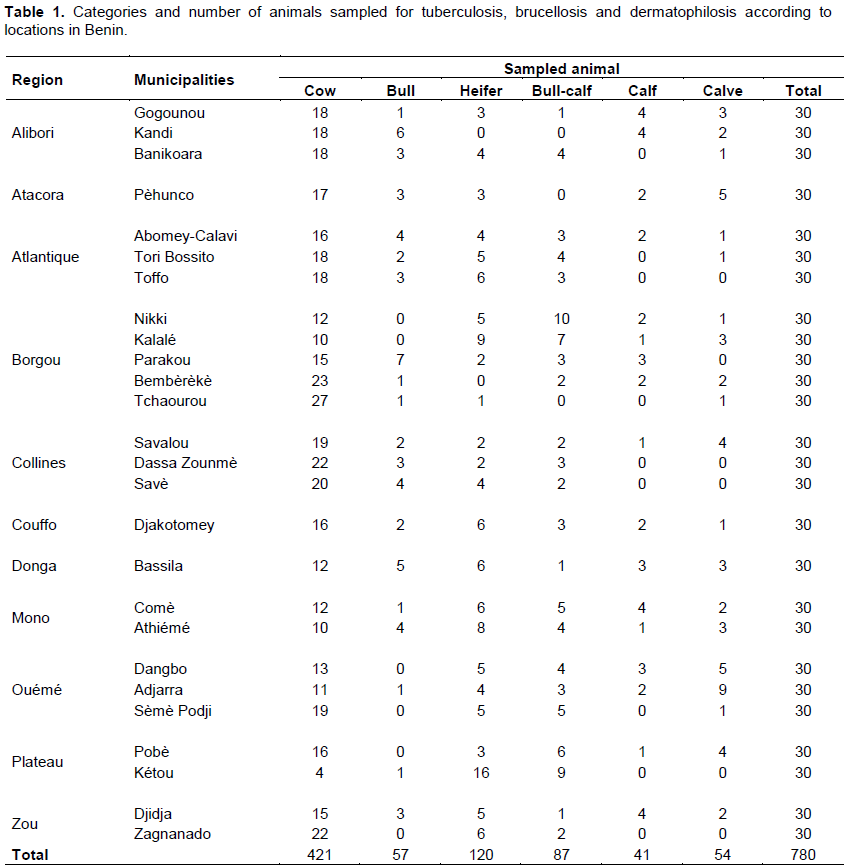
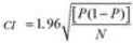
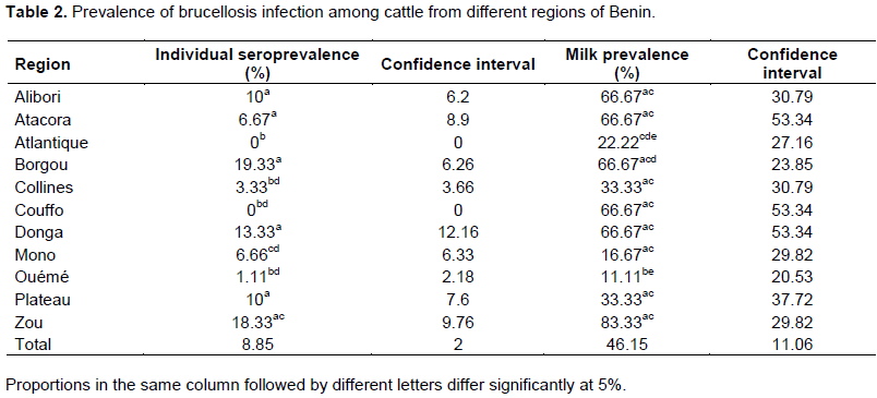
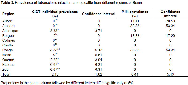
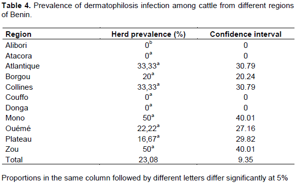
In order to determine the prevalence of bovine brucellosis, tuberculosis and dermatophilosis, a study was carried out in main dairy areas of Benin from April to September 2015. For brucellosis, 780 sera and 78 milk samples were analyzed by indirect enzyme-linked immunosorbent assay (iELISA). For tuberculosis, 780 cattle underwent a comparative intradermal tuberculin test and 78 milk samples were used for Ziehl-Neelsen’s staining. About dermatophilosis, 78 samples of scabs were collected for Giemsa’s staining. For brucellosis, the overall individual animal seroprevalence was 8.85%. The regions of Borgou with 19.33% and Atlantique with 0% prevalence showed significant differences (p <0.05) with the other regions. For tuberculosis, the overall individual animal prevalence was 2.18%. The regions of Borgou and Alibori, with 0% prevalence each, showed significant differences (p <0.05) with most other regions. Taking into account the individual animal prevalence, Zou (brucellosis 18.33%, tuberculosis 6.67%) and Plateau (brucellosis 10%, tuberculosis 6.67%) were the areas at risk for these two diseases. For dermatophilosis the overall herd prevalence was 23.08%. There was significant difference (p<0.05) between Alibori and Mono but also between Alibori and Zou. It is urgent, therefore, to put in place an adapted control strategy taking into account these geographical realities.

Key words: Brucellosis, tuberculosis, dermatophilosis, prevalence, cattle, Benin


 INTRODUCTION

Brucellosis and tuberculosis are considered as the most important and prevalent zoonotic diseases (WHO, 2004). Both diseases are under control in developed countries, but remain prevalent in sub-Saharan Africa, affecting both livestock and human populations (Abbas and Agab, 2002; Schelling et al., 2003; Mostowy et al., 2005; Zinsstag et al., 2007). In addition to being a threat to public health, both diseases can have serious economic implications. Bovine tuberculosis has a negative impact on livestock production in developing countries by reducing production efficiency, seizure of carcasses or organs and restricting international trade. It has implications not only for the economies of livestock communities, but also for human health through the consumption of raw dairy products and / or close contact with infected animals or animal tissues (OIE, 2009). Brucellosis also causes significant reproductive losses in animals (Cutler et al., 2005). Bovine tuberculosis and brucellosis remain a major public and animal health problem in many developing countries, where cattle are a major source of food and income (Omer et al., 2000). Understanding the epidemiology of bovine tuberculosis and brucellosis is therefore essential to develop evidence-based disease control strategies. However, this information is insufficient in Africa’s sub-Saharan. Therefore, appropriate preventive measures have not been taken (McDermott and Arimi, 2002). Bovine dermatophilosis is distributed worldwide, but mainly recorded in African countries (Kassaye et al., 2003; Kusina et al., 2004; Hamid and Musa, 2009). The disease leads to great economic losses in African countries due to inferior wool and leather quality, death and culling, decrease meat and milk production (Yeruham et al., 2000). Among the skin diseases, bovine dermatophilosis is one of the common economically important diseases of cattle with high economic significance in decreasing the productivity (Awad et al., 2008). As bovine tuberculosis and brucellosis, it is also a zoonotic disease.
Bovine tuberculosis, brucellosis and dermatophilosis are endemic in Benin. This is from the reports of the Direction of Animal Production (DAP) and authors mentioning cases from slaughter houses for tuberculosis and suspicions of clinical signs for brucellosis and dermatophilosis (Ali-Emmanuel et al., 2002; DAP, 2012, 2013, 2014, 2015, 2016). But there is no control program. However, in order to implement Milk and Meat Support Project (PAFILAV), it was imperative to investigate the current situation of the major pathologies affecting milk and meat production in the Project Intervention Zone (ZIP). The main objective of this project is to improve production systems and competitiveness of milk and meat sectors. Then, bovine brucellosis, bovine tuberculosis and bovine dermatophilosis have been retained to determine their prevalence throughout the national territory. These are diseases for which data on their prevalence in Benin are rare. Indeed, for bovine brucellosis, Akapko et al. (1984) found a seroprevalence of 10.4% in extensive herds. Koutinhouin et al. (2003) studies on herds supervised by Livestock Development Project gave a seroprevalence of 6.20 at 15.21%, while those of Adéhan et al. (2005)  gave  a  seroprevalence  of 2.06 to 3.4% on state farms. It should be noted that all these studies have focused on serum analysis only.
Concerning the prevalence of bovine tuberculosis, Farougou et al. (2006) conducted a study at the state farms of Samiondji and Bétécoucocu with single intradermal skin test. Prevalences obtained were 8.25 and 2.64% respectively for Samiondji and Bétécoucou. In addition, Dossou et al. (2016) conducted a study on milk through detection of Brucella abortus and Mycobacterium tuberculosis in the state farms of Kpinnou, Bétécoucou, Okpara and a private farm in Adjohoun, with no case of infection found. It is clear that all these previous studies, in geographical terms, took far more account state farms. No studies have considered both serum and milk for bovine brucellosis. Similarly, no studies have considered a comparative intradermal skin test and milk for bovine tuberculosis. Concerning bovine dermatophilosis there is no study about it prevalence in Benin. Thus, the aim of our study is to provide information on bovine dermatophilosis herd prevalence and to determine the bovine brucellosis and bovine tuberculosis in the Projet d'Appui aux Filières Lait et Viande (PAFILAV)’s ZIP with the identification of areas at risk through the analysis of serum and milk associated with comparative intradermal skin test.


 MATERIALS AND METHODS

Study area
 
The PAFILAV’s ZIP has 27 municipalities out of 77 of the country, and extends throughout the national territory. Benin is part of the intertropical zone. Depending on the latitude in which they occur, rainfall periods combine in different ways to define rainfall regimes. In the south of the 7° 45 'parallel is a bimodal regime with four (4) seasons, two dry and two rainy seasons. North of parallel 8° 30 ', there is a unimodal regime with two seasons, one dry season and one rainy season. Thus the South experiences a climate with four seasons: a great rainy season from April to July; a small dry season from August to September; a small rainy season from October to November and a great dry season from December to March. The North has two seasons: a dry season from November to early May and a rainy season from May to October. The administrative division of Benin comprises 4 hierarchical levels, which are in decreasing order: Regions, municipalities, districts, villages or wards. So we have 12 regions; 77 municipalities; 546 districts and 3557 villages/wards.
The intervention zones are targeted by region and municipality according to the potential in livestock and milk production. The study was conducted in 26 municipalities from eleven of the twelve regions of the country. These selected municipalities are 10 in the northern area and 16 in the southern and central areas of the country (Figure 1). 
 
 
These include:
 
1. Nikki, Kalalé, Parakou, Bembèrèkè, Gogounou, Tchaourou, Kandi,  Banikoara,  Bassila  and  Pehunco  in  the   northern   zone;
 
2. Djidja, Zagnanado,Djakotomey, Comé, Athiémé, Pobè, Kétou, Savalou, Dassa-Zoumè, Savè, Dangbo, Adjarra, Sèmè Podji, Abomey-Calavi, Tori Bossito and Toffo in the southern and central zones.
 
Sample collection
 
The sampling was carried out from April to September 2015. Respondents were selected based on their accessibility and availability to cooperate. Recruitment of animals for testing was based entirely on the owner’s willingness. From the twenty-six (26) municipalities of the ZIP, three (3) herds were retained in each of them. Ten (10) identified animals were selected in each of those herds. In total, seventy-eight (78) herds and seven hundred and eighty (780) animals were included in the study (Table 1). On each of the 10 animals, blood is taken from the jugular vein and the two tuberculins are injected. In each herd, all animals with exudative dermatitis were examined. From these animals, three samples of scabs were collected in clean, sterile tubes for isolation of Dermatophilus congolensis.
 
 
For tuberculins’ injection, animal is maintained in position of lateral decubitus. Two sites located to the right of the collar (the flat of the neck), at 20 cm intervals, were shaved and the thickness of skin is measured with caliper measurement. A first site is injected with 0.1 ml containing 2500 IU/ml bovine PPD. Similarly, 0.1 ml of avian PPD of 2500 IU/ml was injected into the second site. The injection is done in the dermis using an insulin syringe. After 72 h, the skin thicknesses were measured at injection sites which are:
 
1. For bovine tuberculin (B): at the limit of the posterior and middle thirds of the neck and approximately equidistant from the upper and lower edges of the latter;
2. For avian tuberculin (A): in front of the preceding one, at the limit of the anterior and middle thirds of the neck, and approximately equidistant from the upper and lower edges of the latter.
In addition, once all herd cows are milked, 50 ml of the milk mixture is collected. During this study, 78 herds including 780 animals (595 females and 185 males) were investigated throughout the national territory (Table 1). For tuberculosis, 780 tuberculinations were performed with 78 samples of milk for Ziehl-Neelsen’s staining. For brucellosis, 780 sera and 78 samples of milk were analyzed by indirect ELISA.
 
Analysis of samples
 
To analyze serum, milk and scab, the two veterinary laboratories of the country (Laboratory of Parakou and that of Bohicon) were involved.
 
For bovine tuberculosis
 
Reading was done 72 h later. We went back into the herd and we measured the skin thickness at the injection sites. The thickness differences between D0 and D3 were calculated:
 
DB = B3 - B0 for bovine tuberculin
DA = A3 - A0 for avian tuberculin
 
The interpretation of the measures is as follows:
 
If DB - DA is greater than 4 mm: Positive result
If DB - DA is less than 1 mm: Negative result
If DB - DA is between 1 mm and 4 mm inclusive: Doubtful result
 
The milk samples were subjected to Ziehl-Neelsen’s staining.
 
For bovine brucellosis
 
Sera and milk were subjected to the indirect Enzyme Linked ImmunoSorbent Assay (iELISA) using Brucella smooth lipopolysaccharide (S-LPS) as an antigen. Indirect ELISA results were classified as positive or negative using the manufacturer's recommended values.
 
For dermatophilosis
 
Small pieces were taken from the underside of the scab and softened in a few drops of distilled water on a clean microscope slide; a smear was made and stained with Giemsa’s staining as described by Scott (1988).
 
Statistical analysis
 
Data had been integrated into the Excel spreadsheet and then into the software R version 3.1.2. For brucellosis, individual and herd prevalences were calculated by dividing the number of positive iELISA cases by the number of animals or milk taken.
 
For dermatophilosis, herd prevalence was calculated by dividing the number of positive cases by the number of scabs taken.
For tuberculosis, differences in skin thickness were calculated. Results from the Ziehl-Neelsen’s staining were recorded. Thus individual and herd prevalences were estimated by dividing number of positive cases by the number of tuberculinized animals or the number of milk taken.
In both cases, the individual prevalences obtained by region were compared two by two with Fisher’s exact test. For each relative frequency, a 95% confidence interval (CI) was calculated using the formula:
 
 
Where P is the relative frequency and N is the sample size.


 RESULTS

Concerning brucellosis, the overall individual seroprevalence was 8.85%. There was significant difference by sex and also between cow and calve (p<0.05). Moreover, according to the regions, and overall, Borgou with 19.33% and Atlantique with 0% (Table 2) showed significant differences (p <0.05) with the other regions. Ouémé with 1.11% and Zou with 18.33% also showed some significant differences with the  rest  of  the other regions. Furthermore there was no significant difference (p> 0.05) between the four northern regions (Alibori, Atacora, Borgou and Donga); whereas in the South, with Atlantique, Mono, Ouémé and Plateau, there were significant differences between them. The herd seroprevalence was 37.18% (95% CI 26.45 to 47.91%). The milk prevalence was 46.15% (95% CI 35.1 to 57.2%). It relates only to the cows of the herds investigated. Overall, the region of Ouémé had a significant difference with most other regions. No significant difference was observed between milk prevalence and herd seroprevalence. 
 
 
Concerning tuberculosis, the overall individual prevalence was 2.18%. There is no significant difference between sex and categories (p> 0.05). But depending on regions, Borgou and Alibori, with 0% each (Table 3), showed significant differences (p <0.05) with most of the other regions. Plateau and Zou, with 6.67% each, also had some significant differences with the rest of the other regions. There was no significant difference (p> 0.05) between the four northern regions (Alibori, Atacora, Borgou and Donga). Moreover in South, with Atlantique, Mono, Ouémé and Plateau, there were no significant differences between them. The herd prevalence was 15.38% (95% CI 7.38 to 23.38%). The milk prevalence was 6.41% (95% CI 0.98 to 11.84%). It is also related to the cows of the herds investigated. There were no significant differences between regions. No significant difference was observed between milk prevalence and herd prevalence. 
 
 
Concerning dermatophilosis, Table 4 presents the results of herd prevalence by regions. The overall herd prevalence was 23.08% (95% CI 13.73 to 32.43%). There was significant difference (p<0.05) between Alibori and Mono but also between Alibori and Zou. The four northern regions (Alibori, Atacora, Borgou and Donga) had the lowest rates. In addition, taking into account the individual prevalence, Zou (Brucellosis 18.33%, Tuberculosis 6.67%) and Plateau (Brucellosis 10%, Tuberculosis 6.67%) constituted the zones at risk for these two diseases. In the same way, but to a lesser extent, there were also the regions of Mono and Ouémé. Two cows at Pobè and one at Comè were both positive for brucellosis and tuberculosis. Thus mixed prevalence rate was 0.38%. No herds were positive for these three diseases at the same time.


 DISCUSSION

For bovine brucellosis, the overall seroprevalence was 8.85%. This result is similar to that obtained by Akakpo et al. (1984). Moreover, it is much lower than that obtained by Koutinhouin et al. (2003) and much higher than that obtained by Adéhan et al. (2005). The Borgou region had the highest rate (19.33%) and there were no significant differences between the four northern regions which showed significant differences with those of the South. It should be noted that the North is characterized by large herds, unlike the South. This would promote close contact between the animals. Furthermore, in this region of North, animals move a lot, especially transhumance in search of water and grazing, whereas in the South the herds are more sedentary. These two situations could favor transmission and maintenance of the disease at this level. This has been noted by some authors (Berhe et al., 2007; Ragassa et al., 2009; Matope et al., 2010; Makita et al., 2011; Megersa et al., 2011). Nevertheless, study of Cadmus et al. (2013) in Nigeria has shown a higher seroprevalence in sedentary herds compared to transhumants. Significant difference was found by sex. This is in agreement with studies of some authors (Traoré et al., 2004; Dinka and Chala, 2009; Adugna et al., 2013). between cow and calf/calve. Indeed, younger animals are more resistant to primary infection and eliminate Brucella sp. although sometimes latent infection occurs (Walker, 1999).  According  to  Acha  and  Szyfres  (1989),  heifers and cows are classified as the most sensitive. In Africa, some authors have recorded rates ranging between 3 and 13% (Traoré et al., 2004; Boussini et al., 2012, Cadmus et al., 2013). These rates are relatively close to ours. In Ethiopia, Tschopp et al. (2013) found 1.7%. But in Zambia, Muma et al. (2013) found 20.7%. It should be noted that in this case, serum samples were taken only from cows. Our herd prevalence was 37.18%. This is close to the 45.9% found in Ethiopia by Asgedom et al. (2016). The prevalence after analysis of the milk was 46.15%. It is above the 16% found in Egypt by Wareth et al. (2014).
For bovine tuberculosis, although comparative intradermal tuberculin test gives more specific results than single intradermal tuberculin test (Monaghan et al., 1994), our study gave an overall prevalence of 2.18%. This rate is similar to that obtained by Farougou et al. (2006) in Bétécoucou farm which was 2.64%, but very far from the rate they obtained in Samiondji’s farm which was 8.25%. The highest rates were observed in the regions of the South. It was noted that in this region, there are sedentary herds. Indeed, prolonged contact could favor transmission by aerosols. Factors such as water sharing, grazing, or high promiscuity are potential risk factors for bovine tuberculosis transmission (Thoen and Bloom, 1995). This rate is close to that obtained by Asante-Poku et al. (2014) in Ghana which was 2.48%. However, in Burkina Faso, Traoré et al. (2004) found 27.7%. There is no significant difference about sex. This is in agreement with the study of Traoré et al. (2004) in Burkina Faso. The herd prevalence was 15.38% and the milk prevalence was 6.41%. This difference, although not significant, may be due to the fact that M. bovis is rarely isolated from milk, although it is known to be secreted in milk. However, it is not found in milk that has been stored for a few days probably because of competition with lactobacilli (Mariam, 2009). Moreover, the numerous doubtful cases can have several causes. Indeed, considering that the tuberculin test is not a perfect test, some animals would not have been detected, which can lead to an underestimation of the prevalence. In endemic areas, delayed hypersensitivity may not develop for 3 to 6 weeks after infection, and in chronically infected animals with severe disease, tuberculin testing may not respond (OIE, 2010). Thus, it is evident that the initial thickness of the skin fold could confuse the interpretation of reactivity to tuberculin. In Africa, some authors have found relatively low rates ranging from 2 to 6% (Boukary et al., 2011; Boussini et al., 2012; Katale et al., 2013; Muma et al., 2013). In contrast, in Nigeria, Okeke et al. (2014) found 16.17% with PCR on cattle lungs taken from slaughterhouses. In Ethiopia, Tschopp et al. (2013) found 0.3%. The mixed prevalence rate for brucellosis-tuberculosis was 0.38%. It is close to that observed in Burkina Faso by Boussini et al. (2012) which was 0.49%.
For bovine dermatophilosis, about herd prevalence, the four northern regions (Alibori, Atacora, Borgou and Donga) had the lowest rates. This could be in correlation with season. Indeed, it is warmer in the North (only one rainy season) than in the South (two rainy seasons). Dejene et al. (2012) have shown that there was a significant variation between seasons of the year and bovine dermatophilosis which is highly prevalent during the wet season than the dry season. The higher prevalence of the disease during the mentioned season is due to activation of the motile zoospores by rain and increased arthropods population (ticks) so that they may contribute to the occurrence of the disease. Ticks were present in most sampled herds. Furthermore tick Amblyomma variegatum had been associated with transmission of the disease (Morrow et al., 1993; Chatikobo et al., 2004) and there was also an association with tick Boophilus annulatus (Awad et al., 2008) for which macroclimatic factors play a great role in seasonal dynamics (Singh et al., 2000). In the same way the dry season in the north is usually a period of extensive bush burning. Wilson (1988) observed that the disappearance of vegetation in the dry season had a direct effect on the local abundance of questing adult ticks. He reported that tick abundance was reduced by as much as 88% following removal of vegetation by burning.


 CONCLUSION

Knowledge of diseases is a crucial step in the development of prevention and control measures. This study suggests that the overall prevalence of bovine brucellosis, tuberculosis and dermatophilosis in Benin in general and in the PAFILAV’s intervention area in particular is very high and requires urgent intervention. These three diseases are likely to pose a significant risk for the achievement of PAFILAV's objectives. Several recommendations can be made  to  minimize  the  risk  of spread of these diseases between regions. The first and most important is to disseminate knowledge about brucellosis, tuberculosis and dermatophilosis. Then, educate herders and people involved in the cattle trade on risk factors. Finally train herders on how to deal with any signs of suspicion of disease in their flock. In addition, further studies are needed to determine the actual burden of these zoonoses on public health.


 CONFLICT OF INTERESTS

The authors have not declared any conflict of interests.


 REFERENCES

Abbas B, Agab H (2002). A review of camel brucellosis. Prev. Vet. Med. 55(1):47-56.
Crossref


Acha PN, Szyfres B (1989). Zoonoses and communicable diseases common to humans and animals. Paris: O.I.E. P 1063.


Adehan R, Koutinhouin B, Baba-Moussa LS, Aigbe L, Agbadje PM, Youssao AKI (2005). Prevalence of bovine brucellosis in Benin's State farms from 2000 to 2003. R.A.S.P.A. 3:3-4.


Adugna KE, Agga GE, Zewde G (2013).Sero epidemiological survey of bovine brucellosis in cattle under a traditional production system in western Ethiopia. Rev. Sci. Technol. 32(3):765-773.
Crossref


Akakpo JA, Bonarel P, d'Almeida JF (1984). Epidemiology of bovine brucellosis in tropical Africa.Serological survey in Benin's Republic. Rev. Méd. Vét. 37 :133-137.


Ali-Emmanuel N, Moudachirou M, Akakpo AJ, Quetin-Leclercq J (2002). In vitro antibacterial activities of Cassia alata, Lantana camara, Mitracarpus scaber, Dermatophilus congolensis isolated in Benin. Rev. Elev. Méd. Vét. Pays Trop. 55(3):183-187.


Asante-Poku A, Aning KG, Boi-Kikimoto B, YeboahManu D (2014). Prevalence of bovine tuberculosis in a dairy cattle farm and a research farm in Ghana. Onderstepoort J. Vet. Res. 81(2):1-6.
Crossref


Asgedom H, Damena D, Duguma R (2016). Seroprevalence of bovine brucellosis and associated risk factors in and around Alage district, Ethiopia. Springerplus. 5:851.
Crossref


Awad WS, Nadra-Elwgoud, Abdou MI, El-Sayed AA (2008).Diagnosis and Treatment of Bovine, Ovine and Equine Dermatophilosis. J. Appl. Sci. Res. 4(4):367-374.


Berhe G, Belihu K, Asfaw Y (2007). Seroepidemiological investigation of bovine brucellosis in the extensive cattle production system of Tigray region of Ethiopia. Int. J. Appl. Res. Vet. Med. 5:65-71.


Boukary AR, Thys E, Abatih E, Gamatie D, Ango I (2011). Bovine Tuberculosis Prevalence Survey on Cattle in the Rural Livestock System of Torodi (Niger). PLoS One 6(9):e24629.
Crossref


Boussini H, Traoré A, Tamboura HH, Bessin R, Boly H, Ouédraogo A (2012). Prevalence of tuberculosis and brucellosis in intra-urban and peri-urban dairy cattle in Ouagadougou's city in Burkina Faso. Rev. Sci. Tech. Off. int. Epizoot. 31(3):943-951.
Crossref


Cadmus SIB, Alabi PI, Adesokan HK, Dale EJ, Stack JA, (2013). Serological investigation of bovine brucellosis in three cattle production systems in Yewa Division, south-western Nigeria. J. South African Vet. Assoc. 84(1):00-00.


Chatikobo P, Kusina NT, Hamudikuwanda H, Nyoni O (2004).A monitoring study on the prevalence of dermatophilosis and parafilariosis in cattle in a smallholder semi-arid farming area in Zimbabwe. Trop. Anim. Health. Prod. 36(3):207-215.
Crossref


Cutler SJ, Whatmore AM, Commander NJ (2005). Brucellosis: new aspects of an old disease. J. Appl. Microbiol. 98:1270-1281.
Crossref


Dejene B, Ayalew B, Tewodros F, Mersha C (2012). Occurance of Bovine Dermatophilosis in Ambo town West Shoa Administrative Zone, Ethiopia. American-Eurasian J. Sci. Res. 7(4):172-175.


Dinka H, Chala R (2009). Seroprevalence study of bovine brucellosis in pastoral and agro-pastoral areas of East Showa Zone, Oromia Regional State, Ethiopia. American-Eurasian J. Agric. Environ. Sci. 6(5):508-512


Direction of Animal Production (DAP) (2012). Statistical Yearbook of 2011. Benin. P 31.


Direction of Animal Production (DAP) (2013).Statistical Yearbook of 2012. Benin. P 31.


Direction of Animal Production (DAP) (2014).Statistical Yearbook of 2013. Benin. P 28.


Direction of Animal Production (DAP) (2015).Statistical Yearbook of 2014. Benin. P 25.


Direction of Animal Production (DAP) (2016).Statistical Yearbook of 2015. Benin. P 12.


Dossou J, Atchouké GD, Dabadé DS, Azokpota P, Montcho JK (2016). Nutritional and health quality comparative evaluation of milk from different breeds of cows in some Benin's breeding areas. Eur. Sci. J. 12(3).


Farougou S, Agbadjè P, Kpodékon M, Adoligbé C, Akakpo JA (2006). Prevalence of bovine tuberculosis in the state farms of Samiondji and Betecoucou of Benin. R.A.S.P.A. 4(1-2).


Hamid M, Musa MS (2009).The treatment of bovine dermatophilosis and its effects on some haematological and blood chemical parameters. Rev. Sci. Tech. Off. int. Epizoot. 28:1111-1118.
Crossref


Kassaye E, Moser I, Woldemeskel M (2003). Epidemiological study on clinical bovine dermatophilosis in northern Ethiopia.Dtsch. Tierarztl.Wochenschr.110(10):422-425.


Katale BZ, Mbugi EV, Karimuribo ED, Keyyu JD, Kendall S, Kibiki GS, Faussett PG, Michel AL, Kazwala RR, Helden PV, Matee MI (2013). Prevalence and risk factors for infection of bovine tuberculosis in indigenous cattle in the Serengeti ecosystem, Tanzania. BMC Vet. Res. 9(1):267.
Crossref


Koutinhouin B, Youssao AKI, Houéhou AE, Agbadjè PM (2003). Prevalence of bovine brucellosis in the traditional breeding farms supervised by the Project for Livestock Development (PDE) in Benin. Rev. Méd. Vét. 154(4):271-276.


Kusina NT, Chatikobo P, Hamudikuwanda H, Nyoni O (2004). A monitoring study on the prevalence of dermatophilosis and parafilariosis in cattle in a smallholder semi-arid farming area in Zimbabwe. Trop. Anim. Health. Prod. 36:207-215.
Crossref


Makita K, Fèvre EM, Waiswa C, Eisler MC, Thrusfield M, Welburn SC (2011). Herd prevalence of bovine brucellosis and analysis of risk factors in cattle in urban and peri-urban areas of the Kampala economic zone, Uganda. BMC Vet. Res. 7:60.
Crossref


Mariam SH (2009). Interaction between lactic acid bacteria and M. bovis in Ethiopian fermented milk. Appl. Environ. Microbiol. 75:1790-1792.
Crossref


Matope G, Bhebhe E, Muma JB, Lund A, Skjerve E (2010). Herd-level factors for Brucella seropositivity in cattle reared in smallholder dairy farms of Zimbabwe. Prev. Vet. Med. 94(3-4):213-221.
Crossref


McDermott JJ, Arimi SM (2002). Brucellosis in Sub-Saharan Africa: epidemiology, control and impact. Vet Microbiol. 20:111-134
Crossref


Megersa B, Biff D, Niguse F, Rufael T, Asmare K, Skjerve E (2011b). Cattle brucellosis in traditional livestock husbandry practice in Southern and Eastern Ethiopia, and its zoonotic implication.Acta. Vet. Scand. 53:24.
Crossref


Monaghan ML, Doherty ML, Collins JD, Kazda JF, Quinn PJ (1994). The tuberculin test. Vet. Microbiol. 40(1-2):111-124.
Crossref


Morrow AN, Arnott JL, Heron ID, Koney EBM, Walker AR (1993).The effect of tick control on the prevalence of dermatophilosis on indigenous cattle in Ghana. Rev. Elev. Med. Vet. Pays Trop. 46:317-322.


Mostowy S, Inwald J, Gordon S, Martin C, Warren R, Kremer K, Cousins D, Behr MA (2005). Revisiting the evolution of Mycobacterium bovis. J. Bacteriol. 187:6386-6395.
Crossref


Muma JB, Syakalima M, Munyeme M, Zulu VC, Simuunza M, Kurata M (2013). Bovine Tuberculosis and Brucellosis in Traditionally Managed Livestock in Selected Districts of Southern Province of Zambia. Vet. Med. Int. 2013:730367.
Crossref


Okeke LA, Cadmus S, Okeke IO, Muhammad M, Awoloh O, Dairo D, Waziri EN, Olayinka A, Nguku PM, Fawole O (2014). Prevalence and risk factors of Mycobacterium tuberculosis complex infection in slaughtered cattle at Jos South Abattoir, Plateau State, Nigeria. Pan Afr. Med. J. 18(1):7.
Crossref


Omer K, Holstand G, Skjerve E, Woldehiwet Z, MacMillan AP (2000).Prevalence of antiodies to Brucella spp in catte, sheep, goats, horses and camels in the State of Eritrea; influence of husbandry system. Epidemiol. Infect. 125:447-453.
Crossref


Ragassa G, Mekonnen D, Yamuah L, Tilahun H, Guta T, Gebreyohannes A, Aseff A, Abdoel TH, Smits HL (2009). Human brucellosis in Traditional pastoral communities in Ethiopia. Int. J. Trop. Med. 4:59-64


Schelling E, Diguimbaye C, Daoud S, Nicolet J, Boerlin P, Tanner M, Zinsstag J (2003). Brucellosis and Q-fever sero prevalence of nomadic pastoralists and their livestock in Chad. Prev. Vet. Med. 61:279-293
Crossref


Scott DW (1988). Large animal dermatology.Edition Sanders.Philapdelhia. pp.136-146.


Singh AP, Singla LD and Singh A (2000) A study on the effects of macroclimatic factors on the seasonal population dynamics of Boophilus micropus (Canes, 1888) infesting the cross-bred cattle of Ludhiana district. Int. J. Anim. Sci. 15(1):29-31.


Swai ES, Schoonman L (2010).The Use of Rose Bengal Plate Test to Asses Cattle Exposure to Brucella Infection in Traditional and Smallholder Dairy Production Systems of Tanga Region of Tanzania. Vet. Med. Int. 2010:837950
Crossref


Thoen CO, Bloom BR (1995). Pathogenesis of Mycobacterium bovis In: Thoen CO, Steele JH, editors. Mycobacterium bovis Infection in Animals and Humans. First ed. Ames, Iowa: Iowa State University Press. pp: 3-14.


Traoré A, Tamboura HH, Bayala B, Rouamba DW, Yaméogo N, Sanou M (2004). Overall prevalence of major pathologies affecting cow milk's production in intra urban breeding system in Hamdallaye (Ouagadougou).Biotechnol. Agron. Soc. Environ. 8 (1):3-8.


Tschopp R, Abera B, Sourou SY, Guerne-Bleich E, Aseffa A, Wubete A, Zinsstag J, Young D (2013). Bovine tuberculosis and brucellosis prevalence in cattle from selected milk cooperatives in Arsi zone, Oromia region, Ethiopia. BMC Vet. Res. 9:163.
Crossref


Walker RL (1999). "Brucella," in Veterinary Microbiology, C. H. Dwight and C. Z. Yuang, Eds. Blackwell Science, Cambridge, Mass, USA. pp. 196-203.


Wareth G, Melzer F, Elschner MC, Neubauer H, Roesler U (2014).Detection of Brucella melitensis in bovine milk and milk products from apparently healthy animals in Egypt by real-time PCR. J. Infect. Dev. Ctries. 8(10):1339-1343.
Crossref


Wilson ML (1986). Reduced Abundance of adult Ixodesdammini (Acari: Ixodidae) following destruction of Vegetation. J. Econ. Entomol. 79(3):693-696.
Crossref


Wold Health Organization (WHO) (2004).Report of the WHO/FAO/OIE Joint Consultation on Emerging Zoonotic Diseases, Geneva, Switzerland. P 72.


World Organization for Animal Health (OIE) (2009).Bovine brucellosis and bovine tuberculosis.In OIE Terrestrial Manual. Chapter 2.4.3.Paris: France.


World Organization for Animal Health (OIE) (2010).Manual of the Diagnostic Tests and Vaccines for Terrestrial Animals.vol. 1.Office International Des Epizooties.5th edition. Paris: France.


Yeruham I, Elad D, Perl S (2000). Economic aspects of outbreaks of dermatophilosis in first calving cows in nine herds of dairy cattle in Israel. Vet. Rec. 146(24):695-698.
Crossref


Zinsstag J, Schelling E, Roth F, Bonfoh B, Savigny D, Tanner M (2007). Human benefits of animal interventions for zoonosis control. Emerg. Infect. Dis. 13:527-532.
Crossref


          */?>