Full Length Research Paper

ABSTRACT
A total of 332 dairy cow’s sera samples were collected from Kombolcha and Dessie districts of North Ethiopia to determine the sero-prevalence of infectious bovine rhinotracheitis (IBR), and Brucellosis. Sera samples were split into equal portions and competitive ELISA was used for testing IBR in the first portion while Rose Bengal test (RBT) and Complement Fixation Test (CFT) were used for detection of Brucellosis in the second portion. Sero-positivity was 25.6% (85/332) for IBR and 5.4% (18/332) for brucellosis with RBT which was found to be 0 (0%) by CFT. The prevalence of IBR was significantly influenced (P<0.001; OR=2.880) by parity with higher occurrence in multiparous compared to primiparous cows. IBR sero-positivity was highly associated (P<0.001) with repeat breeding, abortion and retained fetal membrane at odds ratio (OR) 8.833; 13.913 and 12.770, respectively. Similarly, IBR sero-positive animals had significantly higher (P=0.001) average days open, number of service per conception and calving interval than sero-negative animals. Conception rate at first service was higher (P<0.05, 74.4%) in sero-negative cows than in sero-positive dairy cows (25.6%). As latent infections and venereal transmissions are very common in IBR, its impact on the newly emerging urban dairy system would be significant unless an urgent control mechanism is designed. The associations of these reproductive infections with repeat breeding implies that they can seriously undermine the effort toward breed improvement in Ethiopia. Further studies with more detailed evaluation into various reproductive performance parameters should be the next step.
Key words: Brucellosis, dairy cattle, infectious bovine rhinotracheitis, reproductive disorders reproductive performance, sero-prevalence.
INTRODUCTION
The family of Herpesviridae subfamily Alphaherpesvirinae mainly bovine herpes virus Type 1 (BoHV-1) is a virus common to cause many diseases in cattle. This includes infectious bovine rhinotracheitis (IBR), Infectious pustular vulvovaginitis, infectious pustular balanopostitis (IPB), abortion and conjunctivitis as well as enteritis. Of these, IBR is an extremely infectious respiratory disease which brings about substantial financial losses into the dairy industry worldwide (Bosco et al., 2011; Gould et al., 2013). The virus can be transmissible from infected cattle usually through contact, inhalation and by natural mating or artificial insemination from virus-contaminated semen. IBR in cattle causes reproductive disorders mainly abortion, retention of fetal membranes and metritis including oophoritis, and subfertility characterized by increased service per conception and prolonged days open (Sibhat et al., 2018).
The virus is accountable for signi?cant economic losses in the cattle industry worldwide, and some countries are working toward controlling or eradicating the infection (Raaperi et al., 2014; OIE, 2010). BoHV-1 is distributed worldwide although only a few countries are known to be free according to OIE (2010). It has been also eradicated from Austria, Denmark, Finland, Sweden, Switzerland and Norway. Control programmes are in place in Australia, Belgium, Canada, India, Poland, Turkey and USA (Noordegraaf et al., 2000; Turin and Russo, 2003; Boelaert, 2005). Abortions due to BHV-1 which occur principally during the last half of gestation are the greatest economic losses in dairy industry. Several countries have recognized the losses caused by the disease and have initiated eradication programmes through vaccination schemes. However, in Ethiopia there were only three previous reports of the sero-epidemiological studies of BHV-1 virus which was limited in central, south and south western parts of the country (Lefevre, 1975; Bekele et al., 1989; Sibhat et al., 2018).
Brucellosis is another reproductive disease that affects fertility in dairy cattle. It is a persuasive zoonotic disease with worldwide occurrence (Mantur and Amarnath, 2008). Cattle brucellosis is mostly due to B. abortus, which is characterized by abortion, still birth, retained placenta, infertility and economic loss (Degefa et al., 2011). The prevalence on the presence of brucellosis in animals and human beings is being reported in the literature frequently (Gul and Khan, 2007; Scacchia et al., 2013). There are serological survey reports indicating the presence of brucellosis in different parts of Ethiopia. However, the previously reported prevalence of brucellosis is very variable in the country ranging from 0% prevalence in Ambo (Bashitu et al., 2013), 0.05% in Arsi zone (Degefa et al., 2011), 0.2% in Debrebrihan (Bashitu et al., 2013), 4.9% in Northern Ethiopia (Hileselassie et al., 2010) to 11% in Central Ethiopia (Kebede et al., 2008). These results vary a great deal indicating site specific study of bovine brucellosis is very important to set appropriate control measures in that specific area.
In Ethiopia, information on the sero-status of IBR in dairy cattle is scant despite three previous studies conducted in some areas of the country. During the last 3-4 decades, a prevalence of 41.8 and 67%, respectively has been reported in Harar and Sidamo provinces (Lefevre, 1975), and in Gobe and Ghibe in Central Ethiopia (Bekele et al., 1989). More recently, sero-prevalence of 41.0% was reported in the main milk sheds in Addis Ababa, central, southern and southwestern parts of Ethiopia (Sibhat et al., 2018). Despite frequent reports of abortion and subfertility in dairy cattle in Dessie and Kombolcha including the surrounding dairy farms, little is known on the potential infectious causes of these problems. Therefore, the objective of this study was to determine the sero-prevalence of IBR and brucellosis in dairy cattle in Dessie and Kombolcha in the north-central Ethiopia. In addition, the significance of the virus as the cause of subfertility and reproductive disorders in cattle was investigated in this study.
MATERIALS AND METHODS
Study area
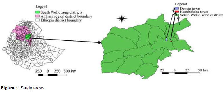
Sero-prevalence investigation of IBR was conducted in randomly selected dairy cattle in Dessie and Kombolcha towns in South Wollo zone of Amhara region, north-central Ethiopia. Dessie municipality is the biggest city in this zone and is located at 401 km Northeast of Addis Ababa. The city is geographically located at latitude of 11°8′N 39°38′E and longitude of 11.133°N 39.633°E and an average altitude of 2,494 m above sea level. The area receives rain fall that ranged from 1100–1200 mm and temperature in the range of 15–27ºC annually. Kombolcha is also located at 380 km Northeast of Addis Ababa at latitude of 11°5′N 39°44′E, longitude of 11.083°N 39.733°E, and elevation between 1842 and 1915 m above sea level (NMSA, 2010). This town has an annual mean temperature in the range of 11.7–24.9°C and annual rain fall of the area ranges from 750 to 900 mm. Sheep are the dominant animal species kept by farmers and these areas are characterized by mixed crop-livestock farming system (Figure 1).
Study animals
A total of 332 animals, 172 cross breed managed under intensive system and 160 local breeds managed under extensive system, were included in this study. Breeding system was mainly by artificial insemination (AI) with some farms also using natural mating (bull) or a combination of both. There was no history of vaccination against IBR or brucellosis. There are many small-scale and large-scale dairy farms that supply milk and milk products to the city and its surroundings. These dairy farms contain either local or exotic breeds depending on the scale of production.
Study design and sample size determination
The study design was cross-sectional and blood samples were collected from all randomly selected animals for serological examination. Using structured questionnaire data on reproductive health such as presence of repeat breeding, abortion and retain fetal membrane (RFM) as well as anestrus was collected from the selected cattle. Besides, information about reproductive performance indicators such as calving interval (CI), days open (DO), number of service per conception (NSPC), as well as first service conception rate (FSCR) were collected during day to day observation.
The minimum number of animals required for this study was determined based on the formula of Thrus?eld, (2007):

Where, n = sample size, Z = values of the standard Normal distribution (= 1.96) at 95% levels of confidence, P = prevalence of 67% (Bekele et al., 1989), d = 5% desired absolute precision. Therefore, the minimum sample size was calculated to be 340, but blood samples were collected from 332 animals for this study.
Sample collection
From the jugular vein around 10 mL of blood sample was collected using a plain vacutainer tube (Becton Dickson, UK). Each sample was labeled using codes identifying each animal and herd from which the blood was taken. At room temperature the tube was set tilted on a table overnight to let clotting. On the subsequent day, the clotted blood in the tubes was centrifuged (at 3000 g for 20 min) to obtain clear serum that was later on stored at –20ºC until testing. Samples were transported either to the National Animal Health Diagnostic and Investigation Center at Sebeta for the IBR test using competitive ELISA or to Kombolcha Animal Disease Investigation and Diagnostic Laboratory for brucellosis test using RBT and CFT.
Serological test
Each of the 332 serum samples was portioned into two equal aliquots for serological examination of IBR and brucellosis. The first portions were subjected to competitive ELISA test (ID.vet IBR gB Competition, Grabels, France) to discover anti-gB glycoprotein antibodies for BoHV-1 virus in serum. Following the manufacturer’s test procedure, briefly, 50 μl of the dilution buffer was deposited to individual well, and then 50 μl of the test sera was added to the test well plates. Likewise, equal amount (50 μl) of control sera (positive and negative bovine serum) was deposited into the BoHV-1 purified lysat coated antigen micro plate wells. The microplate was incubated for 2 h at 37°C, and then each well was emptied and washed three times using wash solution to remove the sera. 100 μl of an anti-gB horseradish peroxidase (HRP) conjugate was added to the wells to fix the remaining free gB epitopes, incubated for 30 min at 37°C, and washed three times. After removing the extra conjugate 100 μl of the substrate solution tetramethylbenzidine (TMB) was added to each well, and then incubated for 15 min at room temperature in the dark. Finally, 100 μl of the stop solution (0.5 M sulfuric acid) was added to each well to stop the reaction, and optical density was read and recorded by ELISA reader. The competition percentage (S/N%) for each sample was calculated and interpreted as per the guidelines of the manufacturer and samples with S/N% ≤ 45% were regarded as positive.
The remaining second portions were subjected to Rose Bengal Plate Agglutination test (RBPT) (Cenogenic Corporation, Morganville, USA) as a screening test for brucella. Briefly, 30 μL of RBT antigen and 30 μL of the test serum were added altogether on the plate, and then allowed to mix by shaking thoroughly for 4 min. If any agglutination was observed the test sera were classified as positive and negative if no agglutination. Further, all RBPT positive samples were subjected to CFT as a confirmatory test for the RBT sero-positivity.
To avoid cross contamination of test wells special caution was taken by using separate plates for each test, keeping personal hygiene by washing hands, and by cleaning and sanitizing all the work surfaces.
Statistical analysis
The collected data were entered into a Microsoft Excel spread sheet and checked for accuracy. The data were then analyzed using SPSS software package version 20.0 (IBM. Corp, 2011). Individual animal’s test response and the covariates were assessed using logistic regression to assess the associations and risk factors in relation to BoHV-1 seroprevalence. Chi square test was used for the analysis of association between the seropositivity and reproductive disorders. In all tests, the p value was considered to be statistically significant at P<0.05. One way ANOVA was used to compare means of reproductive performance indicators among IBR seropositive animals.
RESULTS
Seroprevalence of IBR
Association of BoHV-1 exposure status and possible risk factors
Table 1 reveals the seroprevalence of IBR based on district, parity, and production system as well as breeding method of dairy cattle. From the 332 sera samples tested 25.6% (85/332) were positive for IBR. No significant difference in seroprevalence of IBR was found between districts although the prevalence was higher in Dessie compared to Kombolcha. Similarly, no significant difference in seroprevalence of IBR among cows managed under intensive and extensive production systems was found despite the seroprevalence was higher in intensive system. Moreover, dairy cows that were bred by AI had high prevalence of IBR than those bred by bull although the difference was not significant. However, significant difference in sero-prevalence of IBR was observed between parity, and multiparous cows showed higher seroprevalence than primiparous cows (Table 1).
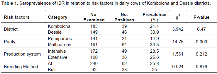
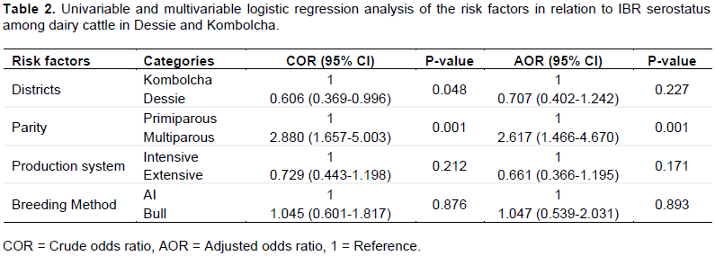
Table 2 shows the results of crude odds ratio and adjusted odds ratio of the risk factors in relation to IBR serostatus among dairy cattle in the study areas. The risk/odds of being positive for BoHV-1 were significantly associated with parity and multiparous cows had higher odds ratio (AOR = 2.6, p < 0.05) compared to primiparous cows. However, no significant association was noted in the risk of BoHV-1 seroprevalence with respect to districts, production system and breeding methods in dairy cows (Table 2).
Association of IBR seroprevalence with reproductive disorders
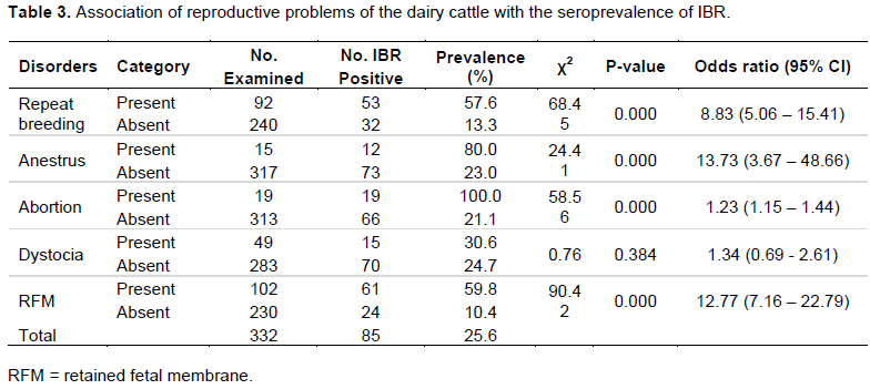
Seroprevalence of IBR among cows with cases of repeat breeding, anestrus, abortion, dystocia and retained fetal membrane was 57.6, 80.0, 100.0, 30.6 and 59.8%, respectively (Table 3). The occurrence of reproductive problems such as repeat breeding, anestrus, abortion and retained fetal membrane was significantly associated with IBR seropositivity. In other words, cows infected with BoHV-1 had higher (p < 0.05) risk of repeat breeding, anestrus, abortion and retained fetal membrane as compared to their counterparts. However, there was no significant relationship between IBR seropositivity and dystocia (Table 3).
Association of IBR sero-prevalence with key reproductive performance indicators
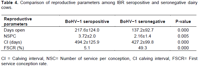
The reproductive parameters including days open, number of service per conception, and CI were significantly higher in seropositive animals compared to seronegative animals (Table 4). This proves the consequences of BoHV−1 infection which prolong the days open, NSC and CI including low first service conception rate of dairy cattle. First service conception rate was significantly higher (P<0.05) in BoHV-1 serologically negative cows (49.3%) than BHV-1 serologically positive cows (5.1%).
Seroprevalence of brucellosis
Seroprevalence of brucellosis based on RBPT was 5.4% (18/332). All the 18 sera were, however, negative for brucellosis on CFT, thus, the final sero-prevalence of brucellosis was reported as 0 (0%).
DISCUSSION
This study was designed to determine the seroprevalence of IBR and brucellosis and to evaluate their association with reproductive disorders and reproductive performance in dairy cows. The seroprevalence of IBR found in this study was higher than the findings of 10.39% in India (Nandi et al., 2009), 19.5% in Turkey (Tan et al., 2006) and 24.19 % in Algeria (Kaddour et al., 2019). On the other hand, very high prevalence was reported in Ethiopia with prevalence of 67% in Gobe and Ghibe (Bekele et al., 1989), 41.8% in Harar and Sidamo provinces (Lefevre, 1975) and 41.0% in Addis Ababa, central and southern including southwestern parts of Ethiopia (Sibhat et al., 2018). Similar studies in other countries indicated that the seroprevalence of IBR was higher than the present finding (64.5%) in southern Veracruz, Mexico (Hussein et al., 2013), 66.12% in India (Trangadia et al., 2010), 60.84% in India (Trangadia et al., 2012), and 93.75% in Egypt from cattle imported from Sudan (Hussein et al., 2019), and 63.54% in southern India (Krishnamoorthy et al., 2015). This indicates that IBR seroprevalence was higher and ranged from 10.39% to as high as 93.75% in various parts of the world. The difference in the seroprevalence across countries may be attributed to variations in geographical locations, variation in management such as housing, feeding, semen quality during breeding, and health care program used by these countries.
All of these findings confirm that IBR is a serious economic problem for livestock production all over the world. Abortion occurring mainly at the late stage of gestation due to IBR infection causes the most economic losses in dairy cattle. This loss is recognized by most of the countries and is using vaccination against IBR to eradicate the disease (Singh et al., 2015). Although there was a disparity in the prevalence of the variation in the risk factors such as district difference, the variation in production system and breeding method, there was no statistically significant difference between the different categories in presence of IBR sero-positivity in the study area. However, there was highly statistical significant difference between the prevalence of multiparous (33.5%) and primiparous (14.9%) dairy cattle being higher in multiparous with above two fold risk for multiparous than primiparous. This might be due to the repeated exposure to breeding where by the infection can occur from the bull or AI.
The prevalence of IBR with respect to reproductive disorders in this study showed strong association between seropositivity and the presence of these disorders. Higher prevalence of IBR in this study was found in animals which had history of reproductive problems such as repeat breeding, anestrus, abortion and retained fetal membrane. The significant association of the IBR seropositivity with the occurrence of reproductive disorders in this study was also approved by previous findings reported as 79.69% abortion cases, 76.32% repeat breeding cases, 76.09% retention of placenta cases were sero-positive for IBR (Sibhat et al., 2018), in Ethiopia, and 65.15% repeat breeding, 100% abortion and 100% RFM cases were seropositive for IBR (Krishnamoorthy et al., 2015) in Southern India. This indicates that IBR has contributed to the occurrence of reproductive disorders. In this study cows with repeat breeders were about 8.8 times at risk than the non-repeat breeders, dairy cows with recorded cases of abortion were about 14 times at risk than the dairy cows that had not been aborted and these cows which had retained fetal membrane were about 13 times at risk than those having no RFM. This finding was similar to previous results; in India where 79.69% abortion cases, 76.32% repeat breeding cases, 76.09% and retention of placenta cases were sero-positive for IBR respectively (Kathiriya et al., 2018). In addition it is discussed that animals with history of reproductive problems showed increased sero-prevalence when compared to apparently healthy animals (Krishnamoorthy et al., 2015).
In the present study, increased length of days open, number of services per conception and inter-calving intervals were noted in IBR sero-positive dairy cattle. This could be due to the influence of reproductive disorders caused by IBR infection implying IBR has indirectly affected reproductive performance indicators. It is also stated that the potential for BoHV-1 infection to have a negative impact on fertility has been recognized for many years and the losses are due to abortions, metritis, retention of placenta, repeat breeding, death of animals, loss of production and trade restrictions etc (Romero-salas et al., 2013). The finding in this study and findings of other researches (Ayhan et al., 2013) indicate that the reproductive disorders such as repeat breeding, abortion and RFM and infertility problems such as longer days open (DO), number of service per conception (NSC) and calving interval (CI) as well as lower first service conception rate (FSCR) were strongly correlated with IBR virus infection in dairy cattle. IBR infection and forced traction of retain fetal membrane as a result of the infection result in lower conception, or estrus expression and inflammations of the endometrium (Nandi et al., 2009; Tikoo et al., 1995). CI in IBR sero-positive animals was significantly higher than sero-negative animals in this study was similar to the finding of Can et al. (2016) who reported 415 days CI in IBR seropositive and 401 days CI in sero-negative dairy cattle with (p< 0.05); similarly, the days open (DO) in the dairy cattle with IBR sero positive was higher (p<.0.05) than the days open of animals with sero-negative animals in this study as well as other findings reported by Ayhan et al. (2012).
The overall prevalence of brucellosis based on RBT was 28.3% in this study, which was much higher than that of previous studies: 3.3% seropositive to RBPT in Tigray (Berhe et al., 2007), 14 (1.2%) tested positive by RBT in Western Ethiopia (Adugna et al., 2013), 28 (3.4%) in Horro Guduru Animal Production and Research Center and its surrounding (Adugna et al., 2013) and 1(0.2%) in Ambo and 3(0.7%) in Deberbrhan (Edao et al., 2018). The higher prevalence by RBT did not indicate the risk of brucellosis in the study area as it also should be confirmed by confirmatory test. The sero-prevalence using CFT is 0 (0%) in the study area which is almost similar to most of the studies who reported 0(0%) prevalence in different part of Ethiopia including in Ambo (Bashitu et al., 2015), in Arsi zone (Degefa et al., 2011), in Addis Ababa (Towns et al., 2015). Although studies indicate that brucellosis is wide spread with a lower percentage in Ethiopia, the prevalence of brucellosis was found to be null (0.0%) in the study area in confirmation test by CFT although it was around 5% in RBT. Since RBT is used as screening test it is clear that the prevalence to be reported is after confirmation by CFT which was 0.0 % in this case indicates that brucellosis is not risk in the study area.
CONCLUSION AND RECOMMENDATIONS
The study was done to the investigate the seroprevalence of IBR and brucellosisin Kombolcha and Dessie districts and to assess if the disease is contributing to the subfertility of dairy cattle. This study reveals the presence of IBR in dairy cattle at high prevalence and shows its impact on reproductive and productive performance in dairy cattle affecting profitability of dairy farms in the study area; when the reproductive performance of dairy cattle is affected it is clear that productivity and profitability of dairy farms is affected. The study on IBR is the third in Ethiopia which also revealed the presence of IBR in dairy cattle at high prevalence. This shows its impact on reproductive and productive performance in dairy cattle affecting profitability of dairy farms. The high prevalence of IBR in dairy cattle warrants immediate consideration to implement preventive measures. It requires implementing complementary measures like regular screening of animals against these diseases, culling of positive reactors, strict vaccination, and quarantine of animals at the time of purchase, use of semen free from infectious agents and strict implementation of zoo sanitary and biosecurity measures to control these diseases. Brucellosis is found to be null prevalence and is not a risk for dairy cattle in the study area.
CONFLICT OF INTERESTS
The authors have not declared any conflict of interests.
ACKNOWLEDGEMENT
Authors would like to thank the sincere collaboration of dairy farm managers/owners for their willingness to cooperate in sample collection and interview. We also appreciate the kind support of Kombolcha Regional Animal Disease Investigation Laboratory and NAHDIC, Sebeta for providing laboratory facilities and technical support for this study.
REFERENCES
|
Adugna KE, Agga GE, Zewde G (2013). Seroepidemiological survey of bovine brucellosis in cattle under a traditional production system in western Ethiopia 32(2805):765-773. |
|
|
Ayhan A Mesih KM, Sibel H, Mehmet K, Mehmet ?G (2012). Investigation of Relationship Between Bovine Herpesvirus-1 (BHV-1) Infection and Fertility in Repeat Breeding. Dairy Cows in Family-Type Small Dairy Farms. 18(4):579-583, |
|
|
Bashitu L, Afera B, Tuli G, Aklilu F (2015). Sero-prevalence study of bovine brucellosis and its associated risk factors in Debrebirhan and Ambo towns. Journal of Advances in Dairy Research 3:131. |
|
|
Bekele T, Cecchini G, Kassali OB, Scholtens RG, Mukassa-Mugurewa E (1989). Infectious bovine rhinotracheitis/infectious pustular vulvovaginitis (IBR/IPV) in cattle in central Ethiopia. Bulletin of Animal Health and Production in Africa 37(1):97-98. |
|
|
Berhe G, Belihu K, Asfaw Y (2007). Seroepidemiological Investigation of Bovine Brucellosis in the Extensive Cattle Production System of Tigray Region of Ethiopia pp. 65-71. |
|
|
Boelaert F, Speybroeck N, de Kruif A, Burzykowski T, Molenberghs G and Berkven DL (2005). Risk factors for bovine herpesvirus-1 seropositivity. Preventive Veterinary Medicine 69:285-295. |
|
|
Bosco Cowley DJ, Clegg TA, Doherty ML, More SJ (2011). Aspects of bovine herpesvirus-1 infection in dairy and beef herds in Republic of Ireland. Acta Veterinaria Scandinavica 53(1):40. |
|
|
Can MF, Ataseven VS, Yalç?n C (2016). Estimation of production and reproductive performance losses in dairy cattle due to bovine herpesvirus 1 (BoHV- 1) infection. Veterinary arhiv 86:499-513. |
|
|
Degefa T, Duressa A, Duguma R (2011). Brucellosis and some reproductive problems of indigenous Arsi cattle in selected Arsi Zones of Oromia Regional State, Ethiopia. Global Veterinary 7(1):45-53. |
|
|
Edao BM, Hailegebreal G, Berg S, Zewude A, Zeleke Y, Sori T, Wood JLN (2018). Brucellosis in the Addis Ababa dairy cattle: the myths and the realities. BMC Veterinary Research 14(1):1-9. |
|
|
Gould S, Cooper V, Reichardt N, O'Connor A (2013). An evaluation of the prevalence of bovine herpesvirus 1 abortions based on diagnostic submissions to five U.S. based veterinary diagnostic laboratories. Journal of Veterinary Diagnostic Investigation 25:243-247. |
|
|
Gul ST, Khan A (2007). Epidemiology and epizootiology of brucellosis: A review. Pakistan Veterinary Journal 27(3):145-151. |
|
|
Hileselassie M, Shewit K, Moses K (2010). Serological Survey of Bovine Brucellosis in Barka and Arado Breeds of Western Tigray, Ethiopia. Preventive Veterinary Medicine 94(1-2):28-35. |
|
|
Hussein S, Hekal A, Al-gaabary MH, El-sayed MM, Sobhy HM, Abdul A, Fayed A (2019). Seroprevalence of some Infectious transboundry diseases in cattle imported from Sudan to Egypt pp. 92-99. |
|
|
IBM Corp. Released (2011). IBM SPSS Statistics for Windows, Version 20.0. Armonk, NY: IBM Corp. |
|
|
Kaddour A, Bouyoucef A, Fernandez G, Prieto A, Geda F, Moula N (2019). Bovine herpesvirus 1 in the northeast of Algiers, Algeria: Seroprevalence and associated risk factors in dairy herd. Journal of Advanced Veterinary and Animal Research 6(1):60-65. |
|
|
Kathiriya J, Sindhi S, Mathapati B, Bhedi K (2018). Seroprevalence of Infectious Bovine Rhinotracheitis (BHV-1) in Dairy Animals with Reproductive Disorders in Saurashtra of Gujarat, India Seroprevalence of Infectious Bovine Rhinotracheitis (BHV-1) in Dairy Animals with Reproductive Disorders in Saurashtra of Gujarat, India. |
|
|
Kebede T, Ejeta G, Ameni G (2008). Sero Prevalence of Bovine Brucellosis in Small holder Dairy farms in Central Ethiopia (Wuchale-Jida district). The Journal of Livestock and Veterinary Medicine in Tropical Countries 159:3-9. |
|
|
Krishnamoorthy P, Patil SS, Shome R, Rahman H (2015). Sero-epidemiology of infectious bovine rhinotracheitis and brucellosis in organised dairy farms in southern India. Indian Journal of Animal Sciences 85(7):695-700. |
|
|
Lefevre PC (1975). Report on infections bovine rhinotracheitis in Ethiopia. Preliminary serological survey. Revue d'Elevage et de Medecine Veterinaire des Pays Tropicaux 28(2):115-124. |
|
|
Mantur BG, Amarnath SK (2008). Brucellosis in India - A review. Journal of Biosciences 33:539-547. |
|
|
Nandi S, Kumar M, Manohar M, Chauhan RS (2009). Bovine herpes virus infection in cattle. Animal Health Research Reviews 10(1):85-98. |
|
|
National metrology agency (NMSA) (2010). National Meteorology Service Agency. Kombolcha Branch, Kombolcha, Ethiopia. Daily record book. P 89, Not Published. |
|
|
Noordegraaf AV, Jalvingh AW, de Jong MCM, Franken P and Dijkhuizen AA (2000). Evaluating control strategies for outbreaks in BHV-1 free areas including stochastic and spatial simulation. Preventive Veterinary Medicine 44:21-42. |
|
|
Raaperi K, Orro T, Viltrop A (2014). Epidemiology and control of bovine herpesvirus1 infection in Europe. Veterinary Journal 201:249-256. |
|
|
Romero-salas D, Ahuja-aguirre C, Montiel-palacios F, García Z, Cruz-romero A, Aguilar-domínguez M (2013). Seroprevalence and risk factors associated with infectious bovine rhinotracheitis in unvaccinated cattle in southern Veracruz, Mexico 7(17):1716-1722. |
|
|
Scacchia M, Di Provvido A, Ippoliti C, Kefle U, Sebhatu TT, D'Angelo A, De Massis F (2013). Prevalence of brucellosis in dairy cattle from the main dairy farming regions of Eritrea. Onderstepoort Journal of Veterinary Research 80(1):4. |
|
|
Sibhat B, Ayele G, Skjerve E, Gebremedhin EZ, Asmare K (2018). Bovine herpesvirus-1 in three major milk sheds of Ethiopia: Serostatus and association with reproductive disorders in dairy cattle. Preventive Veterinary Medicine 150:126-132. |
|
|
Singh R, Yadav S (2010). Seroprevalence of bovine herpes virus-1 in Uttar Pradesh. Haryana Vet. 49(2003):54-55. |
|
|
TAN MT, Yildirim Y, Erol N, Güngör AB (2006). The Seroprevalence of Bovine Herpes Virus type 1 (BHV-1) and Bovine Leukemia Virus (BLV) in selected dairy cattle herds in Ayd?n province, Turkey. Turkish Journal of Veterinary and Animal Sciences 30(4):353-357. |
|
|
Terrestrial Manual (OIE) (2010). Infectious bovine rhinotracheitis/ Infectious pustular vulvovaginitis. Chapter 2.4.13. |
|
|
Thrus?eld M (2007). Veterinary epidemiology. 3rd Edition, Blackwell Science Ltd., Oxford. |
|
|
Tikoo SK, Campos M, Babiuk LA (1995). Bovine herpesvirus 1 (BHV-1): biology, pathogen- esis, and control. Advances in Virus Research 45:191-223. |
|
|
Towns A, Bashitu L, Afera B, Tuli G, Aklilu F (2015). Sero-Prevalence Study of Bovine Brucellosis and its Associated Risk Factors in Advances in Dairy Research 3(1):1-4. |
|
|
Trangadia BJ, Rana SK, Nagmani K, Srinivasan VA (2012). Serological Investigation of Bovine Brucellosis, Johne's disease and Infectious Bovine Rhinotracheitis in Two States of India. Journal of Advanced Veterinary Research 2(1):38-41. |
|
|
Trangadia B, Rana SK (2010). Prevalence of brucellosis and infectious bovine rhinotracheitis in organized dairy farms in India, |
|
|
Turin L, Russo S (2003). BHV-1 infection in cattle: an update. Veterinary Bulletin 73(8):16-21. |
|
Copyright © 2024 Author(s) retain the copyright of this article.
This article is published under the terms of the Creative Commons Attribution License 4.0


