ABSTRACT
A cross sectional study on bovine fasciolosis was carried out from October 2009 to April 2010 at Arba Minch Municipal abattoir with the aim of determining the prevalence and estimating financial loss. Out of the total 600 cattle examined during the study period, 203 were positive for Fasciola spp. infection with the prevalence rate of 33.83%. Fasciola gigantica was found to be the predominant Fasciola species affecting cattle slaughtered in the study area, 179 (88.18%) of the total livers positive for bovine fasciolosis were infected by F. gigantica, while 15 ( 7.39%) livers had F. hepatica and 9 (4.43) were infected by both species (Fasciola hepatica and F. gigantica). From positive livers for the parasite, 44.33, 33.50 and 22.17% of the livers had slight, moderate and severe gross lesions, respectively. There was a significant difference in the prevalence of fasciolosis (P<0.01) among different body conditions and also among different origins. Higher prevalence of the parasite was observed in animals with poor body condition and lowland origin. The total estimated annual financial losses due to fasciolosis in the abattoir during the study period was726,561.5 ETB ($52,649.38 US) of which 49,493.29 ETB ($3,586.47 USD) was due to liver condemnation (direct) and 677,068.21 ETB ($49,062.91 USD) was because of carcass weight loss (indirect). The estimated annual financial loss showed that fasciolosis is an economically important disease in the abattoir. Therefore, there is a need for further detailed studies on the epidemiology of the disease and snail intermediate hosts found in the area and strategic measure should be taken to control the disease.
Key words: Abattoir, Arba Minch, cattle, Ethiopia, Fasciolosis, financial loss, prevalence.
Ethiopia owns huge number of ruminants with high contribution to the economy and livelihood to the people. Despite the significantly large livestock population, its contribution to the national economy is below the potential due to poor management practices, poor nutrition or low response to improved nutritional inputs, high disease incidence, and low genetic potential. Moreover, improper evaluation of public health importance is due to various individual parasitic disease and in adequate knowledge of epidemiology of disease which otherwise is of great relevance where the distribution of disease determine the type and scope of control measures to be applied (Zegeye, 2003; Taylor et al., 2007; ILRI, 2009).
Fasciolosis is an important parasitic disease of domestic animals, especially cattle and the significance of fasciolosis as emerging helminthic zoonoses is highlighted (Chhabra and Singla, 2009). The two species most commonly implicated as the aetiological agents of fasciolosis are Fasciola hepatica and Fasciola gigantic (Pal, 2007). It is serious hazards to efficient production of cattle particularly in its sub clinical form (Radostitis et al, 2007). Occasionally, fasciolosis can infect human beings (CDC, 2013) and has recently been shown to be a re-emerging and widespread zoonosis affecting many people (Esteban et al., 2003).
Fasciola hepatica has a worldwide distribution but predominates in temperate zones, while F. gigantica is found on most continents, primarily in tropical regions (Andrews, 1999). Mixed infection by both species of Fasciola may occur in area where the ecology is conducive for replication of intermediate host. The economic impact of fasciolosis may greatly vary from year to year depending on the weather condition, management, level of infection, host immunity status and age of animals in endemic areas. In this, cumulative substantial loss could be very high in fluke remaining untreated and suffering from sub clinical fasciolosis (Taylor et al., 2007).
The development of infection in definitive host is divided into migratory phase and the biliary phase (Dubinsky, 1993). The parenchyma phase begins when encysted juvenile flukes penetrate the intestinal wall. After the penetration of the intestine, flukes migrate within the abdominal cavity and penetrate the liver or other organs and cause lesion. The young flukes tunnel through the parenchyma then enter the small bile ducts where they migrate to the larger bile ducts and cause lesion (Behm and Sangsten, 1999; Taylor et al., 2007).
Important economic losses associated with fasciolosis include great expenses on anthelmintics for treatment; liver condemnation; production loss due to mortality; lower production of meat, milk and wool; reduced weight gain; metabolic diseases and impaired fertility (Mason, 2004; Hillyer and Apt, 1997).
Economic losses in Africa and other parts of the world is due to the condemnation of liver as unsuitable for human consumption and associated with poor carcass conformation and predispose the animal to other infectious principally clostridium disease, weight loss, loss in productivity and loss due to death (Andrew et al., 2003)
A review of available literature strongly suggests that fasciolosis exists in almost all parts of Ethiopia. The prevalence and economic significance has been reported form different parts of the country by different researchers (Moje et al., 2015; Petros et al., 2013; Regassa et al., 2012; Miheretab et al., 2010; Manyzewal et al., 2014; Terefe et al., 2012). Even though, different researchers in the country studied the parasite prevalence and economic significance, there is limited work in the southern part of Ethiopia, specifically, there was no work done at Arba Minch Municipal abattoir so far. Therefore, the main objectives of this study were to determine the prevalence of fasciolosis, identify the species of liver flukes involved in fasciolosis, compare the intensity of the infection with the liver lesion involved in fasciolosis and assess the financial loss due to fasciolosis in cattle slaughtered at Arba Minch Municipal Abattoir
Study area
The study was conducted in Arba Minch, Southern Nation Nationality and People’s Regional State (SNNPRS), from October, 2009 to April, 2010. The town is located geographically at 37°5' East of longitude and 6° North of latitude with altitude ranging from 1200 to 3125 m above sea level. The average annual rain fall ranges from 750 to 930 mm with mean average temperature of 30°C. The town is situated in the well-known East African Rift valley and surrounded by Lake Chamo and Abaya as well as the Nech Sar National Park. Mixed livestock agriculture farming system was practiced (GZARDO, 2007).
Study population
A total of 600 indigenous cattle are slaughtered at Arba Minch municipal abattoir. The animals were brought from district markets and brought to the abattoir for slaughtering purpose which originated from different origins with in Gamo Gofa Zonea and nearby areas mainly from highlands (Bonke, Chencha and highlands around Arba Minch), midlands (Kamba and Daramalo) and lowlands (in and around Arba Minch, Jinka and Borena).
Study design, sampling and sample size determination
A cross sectional study was carried out from October, 2009 to April, 2010 by collecting data on events associated with fasciolosis on cattle slaughtered at Arba Minch Municipality abattoir. The study was made on the slaughtered cattle at abattoir by the regular visiting. During abattoir survey includes both ante mortem and postmortem examination
The sample size was calculated according to Thrustfield (1995) by considering estimated prevalence of 50% since there was no previous abattoir survey conducted in the study area. The sample size calculated was 384 with 95% confidence interval and 5% expected error. However, in order to increase the precision, a total of 600 cattle were examined at Arba Minch Municipal abattoir by using systemtic sampling method.
The study animals were selected from the slaughter line using systematic random sampling and examined following ante-mortem and post mortem inspection procedure. Body condition was scored following the guidelines set by Nicholson and Butterworth (1986). Each animal was identified based on the enumerate marks on its body tagged before slaughter. Information regarding age, origin and body condition of the study animals was recorded during ante-mortem examination. Assessment of body condition was carried out using a modified method described by Nicholson and Butterworth (1986). All cattle presented to the abattoir for slaughter were local indigenous breed and male cattle. The liver of each study animal was carefully examined through palpation and incision on each liver and bile duct for presence of lesions indicative of Fasciola infection externally and sliced for confirmation. Liver flukes were recovered for differential count by cutting the infected liver into fine, approximately 1 cm slices with a sharp knife.
Then, positive livers with adult parasites were collected by squeezing into universal bottle containing 10% formalin preservative and then examined to identify the involved fluke species by their size and morphological character. The size of F. gigantica is larger (20-75 x 3-12 mm) than F. hepatica (20-30 x 10 mm) and the anterior cone F. gigantica is not prominent as that of F. hepatica and the body is more transparent (Soulsby, 1982).
Characterization of gross liver lesion
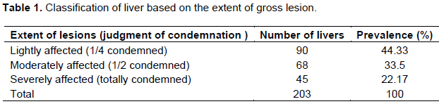
Hepatic lesions in Fasciola positive livers were further grouped in to three different pathological categories depending on the severity of damage inflected by the parasite. The task of categorization was based on the criteria forwarded by Ogurinade and Ogurnrinade (1980). The criteria were:
Lightly affected liver: A quarter of liver was affected or one bile duct was prominently enlarged on the ventral surface of the liver and cutting revealed enlarged or calcified bile ducts and/or thick.
Moderately affected liver: Half of the organ was affected or two or more bile ducts are enlarged and visible before cutting.
Severely affected liver: The entire organ was involved or the liver was cirrhotic or the left lobe atrophy and hyperplasia of the right lobe is seen giving the liver triangular.
Financial loss assessment
The total financial loss due to fasciolosis in cattle slaughtered at Arba Minch municipal abattoir was estimated from the summation of annual liver condemnation (direct loss) and due to carcass weight reduction and poor quality (indirect loss).
Direct financial loss
All livers affected with fasciolosis were totally condemned. The annual direct financial loss was assessed by considering the overall prevalence rate of the disease, the total annual slaughtered animal in the abattoir and retail price during the time of sample collection of an average animal liver. The information obtained was subjected to mathematical compilation using the formula set by Ogurinade and Ogurnrinade (1980).
ALC= CSR*MLC*P
Where, ALC = Annual loss from liver condemnation; CSR = Mean annual cattle slaughtered per year at Arba Minch abattoir; MLC= Mean cost of one liver at Arba Minch town, P = Prevalence rate of the fasciolosis at Arba Minch abattoir.
Indirect financial loss
The indirect (carcass weight reduction) economic loss due to fasciolosis was calculated by considering an estimated 10% carcass weight loss due to fasciolosis in cattle as reported by Robertson (1976) and average carcass weight of an Ethiopian zebu was taken as 126 kg (ILCA, 1992). According to Ogunrinade and Ogunrinade (1980), the annual economic loss because of carcass weight reduction due to bovine fasciolosis was assessed using the formula:
(ACW) = CSR* CL * BC * P*126 kg
Where, ACW is annual loss from carcass weight reduction; CSR, average number of cattle slaughtered at Arba Minch abattoir per year; CL, carcass weight loss in individual cattle due to fasciolosis; BC, an average price of 1 kg beef at the study abattoir; P, prevalence rate of fasciolosis at the study abattoir; 126 kg, average carcass weight of Ethiopia Zebu cattle.
Data management and analysis
The collected data were coded and stored in Microsoft Excel spread sheet. Statistical analysis was done using STATA Version 11.0 (Stata Corp. College Station, TX)) statistical software. Prevalence of fasciolola infection was calculated by descriptive statistics as percentage value, whereas association of Fasciola prevalence with origin, body condition score of the animals and others was analyzed using Chi-square analysis.
Prevalence of bovine fasciolosis
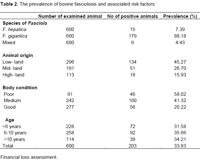
Out of 600 livers examined of cattle slaughtered at the abattoir during the study period, 203 were positive, indicating 33.83% over all prevalence rate. From the total positive livers for fluke 7.39% possess F. hepatica, 88.18% were found infected with F. gigantica and 4.43% have both F. hepatica and F. gigantica mixed infection (Table 2).
Prevalence of bovine fasciolosis from highland, midland and lowland origins were 15.93, 26.7 and 45.27%, respectively. Prevalence of bovine fasciolosis was significant (P< 0.01) based on three origins of animals from different ecological condition. There was significant statistical difference (p<0.001) among different body condition scores (good, medium and poor). More than half of the animals brought to the abattoir (58.02%) were with poor body condition which indicates that fasciolosis is chronic disease of cattle, and the main sign of is emaciation or loss of weight (Taylor et al., 2007). Statistical analysis of the effect of age on prevalence indicated no significance variation (P>0.05) among different age groups of animals.
Hepatic lesion characterization
Based on the severity of gross liver lesion, 44.33, 33.49 and 22.17% were lightly, moderately and severely affected, respectively (Table 1).
Direct financial loss assessment
The average annual cattle slaughtered were estimated to be 4180, while mean retail price of bovine liver, 35 ETB and prevalence of fasciolosis was 33.83%, the estimated annual financial loss from liver condemnation was calculated as
ALC= CSR × LC × P
= 49,493.29 ETB or 3,586.47 $USD
Indirect financial loss assessment
It is the loss due to reduced carcass weight of Fasciola infected animals and was calculated by using average annual cattle slaughtered estimated to be 4180, while the price of carcass weight reduction (indirect) loss of 1 kg beef was 38 ETB. Then, the annual economic loss due to carcass weight reduction was assessed using the formula set by Ogunrinade and Ogunrinade (1980):
ACW = CSR* CL * BC * P*126 kg
= 677,068.21 ETB ($49,062.91 US)
Therefore, the total annual financial loss due to bovine fasciolosis in the Arba Minch Municipality abattoir was the summation of losses from organ condemnation (direct loss) and losses from carcass weight reduction (indirect loss) was about 726,561.5 ETB ($ 52,649.38).
Bovine fasciolosis is economically important and widely distributed disease in almost all region of Ethiopia. The result revealed that the disease is also problem in cattle slaughtered at Ariba Minch municipal abattoir causing high economic loss due to liver condemnation and carcass weight reduction. The overall prevalence of bovine fasciolosis (33%) from the current result was in agreement with the findings of Moje et al. (2015) (30%) in Areka, Mulat et al. (2012) (29.6%) and Miheretab et al. (2010) (32.3%) in Adwa, Ibrahim et al. (2009) (28%) in Kombolcha et al. (2004) (31.7%) from Zimbabwe and Badreldeen and Elfadil (2015) (31.6%) in Sudan.
The present prevalence was much lower than some studies in different parts of the country. In northern Ethiopia, Yilma and Mesfin (2000) reported 90.7% prevalence at Gondar abattoir and also Aregay et al. (2013) found 39.95% in Bahir Dar. In addition, Tolosa and Tigre (2007) recorded a prevalence of 46.2% at Jimma abattoir. Manyazewal et al. (2014) and Abebe et al. (2011) presented 47.1 and 53.7% in Southwest Ethiopia, respectively. The current prevalence findings are also lower than previous studies from other countries in sub-Saharan Africa with prevalence of 53.9% from Zambia, 63.8% from Tanzania and 38.5% from Uganda reported by Phiri et al. (2005), Keyyu et al. (2006) and Ssimbwa et al. (2014), respectively.
On the other hand, the finding is higher than some reports from different parts of Ethiopian and nearby east African country with prevalence of 14.0, 14.4, 24.3, 20.3, 21.9, 25.2, 21.5, 21.6, 24.4 and 26% in Wolaita Soddo (Abunna et al. 2009), Diredawa (Daniel, 1995), Mekele (Gebretsadik et al., 2009), Addis Ababa (Aragaw et al. 2012), Bishoftu (Regassa et al., 2012), Dessie, (Belay et al 2012), Adigrat (Afera, 2012), Nekemte (Petros et al., 2013), Haramaya (Yusuf et al., 2016) and Kenya (Mungube et al., 2006), respectively. Difference in prevalence among geographical locations is attributed mainly to the variation in the climatic and ecological conditions such as altitude, rainfall and temperature. Fasciola spp. prevalence has been reported to vary over the years mainly due to variation in amount and pattern of rainfall.
In relation to risk factors, there was a significant difference in the infection rate (P<0.05) among the animal origins and body condition scores condition groups but, revealed no statistical difference among age groups. The study revealed that there was a statistically significant association (P <0.001) in bovine fasciolosis prevalence among different body condition groups of the animals. Higher prevalence of fasciolosis in cattle with poor body condition as compared to cattle in medium and good body condition (Hagos, 2007; Terefe et al., 2012; Aragaw et al., 2012) as chronic fasciolosis is characterized by progressive loss of condition (Urquhart et al., 1996). However, it must be borne in mind that cattle coming from feedlots, which are expected to be in good body condition, are most likely to be de-wormed than cattle coming directly from grazing (Aragaw et al., 2012).
Out of the total positive livers for fasciolosis species identified, 88.18% of them were infected by F. gigantica where as 7.39% were infected by F. hepatica and 4.43% were mixed infections (both F. hepatica and F. gigantica). The predominant species involved in bovine fasciolosis in the study area was F. gigantica. The high prevalence of F. gigantica as compared to F. hepatica may be associated with the presence of intermediate host L. natalensis and may be explained by the fact that most cattle for slaughter came from low land and mid altitude zones and also as described by Troncy (1989), the favorable condition the snail was border of lakes, flood prone area and low lying marshy and drainage ditches for favorable habitat. Aribaminch and the surroundings were surrounded by lakes Abaya and Chamo and rivers with spring and forest which may be conducive for the development of the intermediate host, L. natalensis.
Analysis of gross liver pathology showing 44.33, 33.5 and 22.17% were affected lightly, moderately and severely, respectively. According to Dwinger et al. (1982) and Yilma and Mesfin (2000), the number of fluke has no direct relationship with the liver gross lesion as it was observed that relatively less flukes in severely affected livers of beef cattle.
However, severe fibrosis impedes the passage of immature flukes and acquired resistance and calcification of bile ducts that impaired the further passage of young flukes and play a role by creating unfavorable microenvironment which results in the expulsion of flukes (Dwinger et al., 1982; Ramato, 1992).
The total annual financial loss estimated at 726,561.5 ETB ($ 52,649.38) which is summation of liver condemnation (direct) and carcass weight reduction (indirect) in this study account for 49,493.29 ETB ($3,586.47) and 677,068.21 ETB ($49,062.91). The direct financial loss due to liver condemnation is comparable to 47,124 ETB by Moje et al. (2015) but, lower than that of Manyzewal et al. (2014) (47,570 ETB) and Petros et al. (2013) (63,072 ETB) at Mettu and Nekemte abattoirs, respectively.
The total financial loss at Arba Minch municipal abattoir in this study was lower than the findings of Belay et al. (2012) at Dessie and Terefe et al. (2012) at Jimma. Moreover, the total financial loss is comparable to the report of Mulugeta et al. (2011) in and around Asella. The existing variation might be correlated with slaughter capacity and number of condemned organs at those specific areas.
The study revealed that bovine fasciolosis is a prevalent disease in the study area causing great financial loss due to condemnation of affected liver and carcass weight reduction. Predominant species involved in bovine fasciolosis in the study area was F. gigantica. This may be due to the fact that most cattle originated from low land and suitable ecological condition for the existence and multiplication of the intermediate host snail. Therefore, proper attention should be paid to control this disease in the study area in particular and in the country in general.
The authors declare that there is no conflict of interest.
REFERENCES
|
Abebe F, Meharenet B, Mekibib B (2011). Major Fasciolosis infections of cattle slaughtered at Jimma municipality abattoir and the occurrence of the intermediate hosts in selected water bodies of the zone. J. Anim. Vet. Adv. 10:1592-1597
Crossref
|
|
|
|
Abunna F, Asfaw L, Megersa B, Regassa, A (2009). Bovine fasciolosis: coprological, abattoir survey and its economic impact due to liver condemnation at Soddo municipal abattoir, Southern Ethiopia. Trop. Anim. Health Prod. 42(2):289-292.
Crossref
|
|
|
|
|
Afera B (2012). Prevalence of bovine fascilosis in municipal Abbatoir of Adigrat, Tigray, Ethiopia. Available at:
View
|
|
|
|
|
Andrews S (1999).The Life Cycle of Fasciola hepatica. In Fasciolosis, Ed. Dalton, J.P. CABI Publishing. pp:1-29.
|
|
|
|
|
Aragaw K, Negus Y, Denbarga Y, Sheferaw De (2012). Fasciolosis in Slaughtered Cattle in Addis Ababa Abattoir, Ethiopia. Global Vet. 8 (2):115-118.
|
|
|
|
|
Aregay F, Bekele J, Ferede Y, Hailemelekot M (2013). Study on the prevalence of bovine fasciolosis in and around Bahir Dar, Ethiopia. Ethiop. Vet. J. 17(1):1-11.
Crossref
|
|
|
|
|
Badreldeen BM, Elfadil AA (2015). A Cross-Sectional Survey of Bovine Fasciolosis at Elkadaro Abattoir, Khartoum State, Sudan. Glob. J. Med. Res. 15(2):1-9.
|
|
|
|
|
Behm C, Sangster N (1999). Pathology, pathophysiology and clinical aspects. In: Dalton, JP ed, Fasciolosis. CABI Publishing. Wallingford. pp: 185-224.
|
|
|
|
|
Belay E, Molla W, Amare A (2012). Prevalence and Economic Losses of Bovine Fasciolosis in Dessie Municipal Abattoir, South Wollo Zone, Ethiopia. Europ. J. Biol. Sci. 4 (2):53-59.
|
|
|
|
|
Centre of Disease Control and Prevention (CDC): Va. USA. (2013). Available at:
View
|
|
|
|
|
Chhabra MB, Singla LD (2009) Food-borne parasitic zoonoses in India: Review of recent reports of human infections. J. Vet. Parasitol. 23(2):103-110.
|
|
|
|
|
Daniel F (1995). Economic Importance of organ condemnation due to Fasciolosis and Hydatidosis in Cattle and Sheep slaughtered at Dire Dawa abattoir, DVM, Thesis, FVM, AAU Debre zeit, Ethiopia. pp: 18-26.
|
|
|
|
|
Dubinsky P (1993). Trematody atrematodozy. In: Jurasek V, Dubinsky P, kolektiv A, Vet. Parasitol. Priroda AS, Bratislava. pp:158-187.
|
|
|
|
|
Dwinger RH, Leriche PD, Kuhne GI (1982). Fascioliasis in beef cattle in North Western Argentina. Trop. Anim. Health Prod. 14(3):167-171.
Crossref
|
|
|
|
|
Esteban JG, Gonzalez C, Curtale F, Mun˜oz-Antoli C, Valero MA, Bargues MD, El-Sayed M, El Wakeel A, Andel-Wahab Y, Montresor A, Engels D, Savioli L, Mas-Coma S (2003). Hyperendemic fascioliasis associated with schistosomiasis in villages in the Nile Delta of Egypt. Am. J. Trop. Med. Hyg. 69:429-437.
|
|
|
|
|
Gamo Goffa Zone Agricultural and Rural Development Office (GZARDO) (2007). Livestock Resource Development and Animal Health Department Annual Report, Arbaminch, Ethiopia.
|
|
|
|
|
Gebretsadik B, Kassahun B, Gebrehiwot T (2009). Prevalence and economic significance of fasciolosis in cattle in Mekelle Area of Ethiopia. Trop. Anim. Health Prod. 41:1503-1504.
Crossref
|
|
|
|
|
Hagos A (2007). Study on prevalence and economic impact of bovine Hydatidosis and Fasciolosis at Mekelle Municipal Abattoir, DVM Thesis, FVM, AAU, Debre zeit, Ethiopia. pp:15-23.
|
|
|
|
|
Hillyer GV, Apt W (1997). Food-borne trematode infections in the Americas. Parasitol. Today 13:87-88.
Crossref
|
|
|
|
|
Ibrahim N, Wasihun P, Tolosa T (2009). Prevalence of Bovines Fasciolosis and Economic Importance due to Liver Condemnation at Kombolcah Industrial Abattoir, Ethioipia. Internet J. Vet. Med. 8(2).
|
|
|
|
|
ILRI (2009). Management of vertisols in Sub-Saharan Africa, Proceedings of a Conference Post-mortem differential parasite counts FAO corporate document Repository.
|
|
|
|
|
International Livestock Center for Africa (ILCA) (1992). Debre Berhan experimental station annual report. P 46.
|
|
|
|
|
Keyyu JD, Kassuku AA, Msalilwa PL, Monrad J, Kyvsgaard CN (2006). Crosssectional prevalence of helminth infections in cattle on traditional, small-scale and large scale dairy farms in Iringa District, Tanzania. Vet. Res. Commun. 30:45-55.
Crossref
|
|
|
|
|
Manyazewal AZ, Gurnesa M, Tesfaye T (2014). Economic Significance of Fasciolosis at Mettu Municipal Abattoir, Southwest, and Ethiopia. J. Adv. Vet. Res. 4(2):53-59.
|
|
|
|
|
Mason C (2004). Fasciolosis associated with metabolic disease in a dairy herd and its effects on health and productivity. Cattle Pract. 12:7-13
|
|
|
|
|
Miheretab B, Tesfay H, Getachew Y (2010). Bovine Fasciolosis: Prevalence and its economic loss due to liver condemnation at Adwa Municipal Abattoir, North Ethiopia. EJAST 1(1):39-47.
|
|
|
|
|
Moje N, Mathewos S, Desissa F, Regassa A (2015). Cross-sectional study on bovine fasciolosis: prevalence, coprological, abattoir survey and financial loss due to liver condemnation at Areka Municipal Abattoir, Southern Ethiopia. J. Vet. Med. Anim. Health 7(1):33-38.
Crossref
|
|
|
|
|
Mulat N, Basaznew B, Mersha C, Achenef M, Tewodros F (2012). Comparison of coprological and postmortem examination techniques for the determination of prevalence and economic significance of bovine fasciolosis. J. Adv. Vet. Res. 2:18-23.
|
|
|
|
|
Mulugeta S, Begna F, Tesgaye E (2011). Prevalence of Bovine Fasciolosis and itsnEconomic Significance in and Around Assela, Ethiopia. Glob. J. Med. Res. 11(3):1-7.
|
|
|
|
|
Mungube EO, Bauni MS, Tenghagen BA, Wamae WL, Nginyi MJ, Mugambi MJ (2006). The Prevalence and Economic Significance of Fasciola gigantica and Stilesia hepatica in Slaughtered Animals in the Semi Arid Coastal Kenya. Trop. Anim. Health Prod. 38:475-483.
Crossref
|
|
|
|
|
Nicholson MJ, Butterworth HM (1986). A guide to condition scoring of zebu cattle. International Livestock Center for Africa (ILCA), Addis Ababa, Ethiopia.
|
|
|
|
|
Ogunrinade A, Ogunrinade B (1980). Economic importance bovine fasciolosis in Nigeria. Trop. Anim. Health Prod.12(3):155-1590.
Crossref
|
|
|
|
|
Pal M (2007). Zoonoses. (2nd edn.), Satyam Publishers, Jaipur, India
|
|
|
|
|
Petros A, Kebede A, Wolde A (2013). Prevalence and economic significance of bovine fasciolosis in Nekemt Municipal Abattoir. J. Vet. Med. Anim. Health 5(8):202-205.
|
|
|
|
|
Phiri AM, Phiri IK, Sikasunge CS, Monrad J (2005). Prevalence of fasciolosis in Zambian cattle observed at selected abattoirs with emphasis on age, sex and origin. J. Vet. Med. 52:414-416.
Crossref
|
|
|
|
|
Radostitis OM, Gray CC, Hinchcliff KW, Constable PD (2007). Hepatic disease associated with thrematods. In text book of cattle, horse, pigs and goats. Veterinary Medicine 10th ed. pp:1576-1580.
|
|
|
|
|
Ramato A (1992). Fasciolosis: clinical occurrence, coprological, abattoir and snail survey in Around Wolliso. DVM Thesis, FVM, AAU, Debre Zeit. P 35.
|
|
|
|
|
Regassa A, Woldemariam T, Demisie S, Moje N, Ayana D, Abunna F (2012). Bovine Fasciolosis: Coprological, Abattoir Survey and Financial loss Due to Liver Condemnation in Bishooftu Municipal Abattoir, Central Ethiopia. Europ. J. Biol. Sci. 4:83-90.
|
|
|
|
|
Robertson A (1976). Hand book on animal disease in tropics. pp: 3:304.
|
|
|
|
|
Soulsby EJL (1982). Helminths, Arthropods and Protozoa of Domesticated Animals,7th edition. Balliere Tindall, London, UK. pp: 40-52.
|
|
|
|
|
Ssimbwa G, Baluka AS, Ocaido M (2014). Prevalence and financial losses associated with bovine fasciolosis at Lyantonde Town abattoir. Livest. Res. Rural Dev. 26(9) Available at:
View
|
|
|
|
|
Taylor AM, Coop LR, Wall LR (2007). Veterinary Parasitology, 3rd Edition, UK, Wiley-Blackwell prublisher. pp:343-345.
|
|
|
|
|
Terefe D, Wondimu A, Dechasa GF (2012). Prevalence, gross pathological lesions and economic losses of bovine fasciolosis at Jimma Municipal Abattoir, Ethiopia. J. Vet. Med. Anim. Health 4:6-11.
|
|
|
|
|
Thrustfield M (1995). Veterinary epidemiology, University of Edinburgh, Black well Science. 2:180-188.
|
|
|
|
|
Tolosa T, Tigre W (2007). The prevalence and economic significance of bovine fasciolosis at Jimma abattoir, Ethiopia. J. Vet. Med.3(2):1-5.
|
|
|
|
|
Troncy PM (1989). Helminthes of livestock and poultry in Tropical Africa. In: Fischer (ed.), Manual of Tropical Veterinary Parasitology. CAB International, UK. pp. 63-73.
|
|
|
|
|
Urquhart GMS, Armour JL, Dunn AM, Jennings FW, Duncan JL (1996). Veterinary Parasitology 2ed. Blackwell Science, London. pp: 87-185.
|
|
|
|
|
Yilma JM, Mesifin A (2000). Dry season bovine fasciolosis in Northwestern part of Ethiopia. Rev. Med. Vet. 151:493-5050
|
|
|
|
|
Yusuf M, Ibrahim N, Tafese W, Deneke Y (2016). Prevalence of Bovine Fasciolosis in Municipal Abattoir of Haramaya, Ethiopia. Food Sci. Quality Manag. 48:38-43.
|
|
|
|
|
Zegeye Y (2003). Challenges and opportunities of livestock marketing in Ethiopia. In: Proc. of the 10th annual conference of Ethiopian Society of Animal Production (ESAP), Addis Ababa, Ethiopia. pp. 47-54
|
|