Full Length Research Paper

ABSTRACT
A potency trial was conducted to evaluate the potency of four commonly used infectious bursal disease (IBD) live vaccines to determine the most appropriate vaccination response to establish the most appropriate vaccination programme against IBD in Nigeria. A total of 700 day old cockerels were randomly divided into six groups 1, 2, 3, 4, 5, and 6. Groups 1 to 4 were the vaccination cockerels while Groups 5 and 6 were the positive and negative controls. The chicks were sub divided into five groups A, B, C, D and E representing the different schedules that is, 1 and 3 weeks, 2 and 4 weeks, 3 and 5, 1, 3 and 5, 2, 4 and 6 weeks, respectively. After each vaccination, the chicks were observed for clinical signs of IBD. Two weeks after each vaccination, chicks were challenged with IBD virus, onset of clinical signs, morbidity and mortality rates were observed. Sample of bursa was collected from 5 birds at 3 and 7 days post challenge from dead as well as necropsied birds. The lesions were scored on the scale of 0 to 5 (mild to severe). Vaccine 3 appeared to be the best with the mean of 1.8 for gross lesions, 1.2 for histo-pathological lesions, 0.6 for clinical signs, 0.6 for mortality, 0.2 for antibody titre and 1.8 for bursal body weight ratio. Schedule A (vaccination at 1 and 3 weeks) also was the best for all the vaccines. Therefore, Nigerian poultry farmers are advised base on the aforementioned result to use vaccine 3 and schedule A in the control the outbreaks of IBD.
Key words: Cockerels chicks, live Infectious bursal disease (IBD) vaccines, potency, vaccination schedule, vaccination strategies.
INTRODUCTION
Infectious bursal disease (IBD) is an acute and highly contagious viral disease of chicks that is immuno-suppressive (Iftihar et al., 2001). The virus has lymphoid tissue as primary target with special predilection for bursa of Fabricius (Iftihar et al., 2001). The disease is characterized by trembling, incoordination, inflammation, followed by necrosis and atrophy of the bursa of Fabricius and immunosuppresion (Giambrone, 1983; Abdu, 2007, 2010). The disease was first described by Cosgrove (1962); it causes economic loss in the poultry industry primarily in the form of mortality and weight loss in affected birds (Abdu, 2010). Chicken 3 to 6 weeks of age are most susceptible to clinical infection (Abdu, 2007). The effective means of controlling infectious bursal disease is vaccination (Abdu, 2007). Infectious bursal disease vaccines had been categorized into mild, intermediate and hot vaccine strain according to the bursa/body weight ratios values following vaccination (Boudaoud and Alloui, 2008). Mild vaccine did not induce bursal lesions and are used in parent chickens to produce primary response prior to vaccination with inactivated vaccine (Babiker and Tawfeeq, 2008); intermediate strains enlarge the bursa twice the normal size. At present there are many imported vaccines and one indigenous vaccine commonly used in Nigeria against IBD. These imported vaccines strains have become popular as they can be used in the presence of maternal antibodies. But experimental results reflected that vaccination of susceptible chickens with such vaccines caused outbreaks of IBD on commercial poultry based on postmortem observations, increased bursal weight to body weight ratio and agar gel precipitation test (Abdu, 1997). IBD has been a persistent problem for the commercial chicken industry worldwide since its discovery in Gomboro District Delaware, USA in 1950, causing huge economic lost mortality and immuno-suppresion (Giambrone, 2008). The present study was designed to evaluate the potency of the four commonly used vaccines (Vaccines 1, 2, 3, and 4) in Nigeria.
MATERIALS AND METHODS
Study area and environmental conditions
The experiment was performed at the livestock farm of the College of Agriculture and Animal Sciences, Ahmadu Bello University, Kaduna located in the Northern Guinea Savanna zone of Nigeria.
Animals and management
The study was approved by the Post graduate Research Committee of the Faculty of Veterinary Medicine, Ahmadu Bello University, Zaria, Nigeria and conducted according to International guidelines on animal ethics (Farsha, 2006). A total of 700 day-old cockerels were purchased from Zartech Nigeria Limited, Ibadan Nigeria.
Vaccination
Vaccination against IBD was carried out in both control and experimental chicks via oral route administration of National Veterinary Research Institute (NVRI) IBD live vaccine (Batch No 21511063 E).
Ration
Vital feed was obtained from a commercial distributor of the product in Kaduna, Kaduna state. The feed giving to the birds had the following composition in g per 100 g of feed: Crude protein 19, fats 5, crude fibre 5.00, available phosphorus 0.45, calcium 1.00, lysine 1.00, methionine 0.40, and salt 0.3, metabolizable energy 2650 kcal. Feed and water were provided to the chicks ad libtum.
Vaccination and blood sampling for antibody titre
The chicks were all bled through the heart at day old to get a base line data of antibody titre. After one week, sub group A and D (Schedule 1 and 3, and 1, 3 and 5, respectively) were bled using insulin syringe to get 3 ml of blood through the heart, the chicks were then vaccinated individually with 0.5 ml of the vaccine. At two weeks, blood was collected from 10 of the vaccinated chicks, while chicks from sub group B and E were bled and vaccinated accordingly. Sera were obtained then stored in the refrigerator until used (2 weeks). At three weeks old, chicks from sub group A, C and D were bled and vaccinated sera were obtained then stored in the refrigerator until used. At four weeks old, chicks from sub group B and E were bled and vaccinated sera were obtained and stored in the refrigerator until used. At five weeks old, chicks were bled and vaccinated. At six weeks old chicks from sub group B and E were bled for antibody. At seven weeks chicks from sub group C and D were also bled and sera were obtained and then stored in the refrigerator until used. Finally, at eight weeks, chicks from sub group E were bled and sera were obtained and stored in the refrigerator until used. The sera at end of 8 weeks were subjected to enzyme linked immune sorbent assay (ELISA).
Challenge virus preparation
The affected bursae of chickens that died from natural IBD were removed, weighed and diluted 1:1(w/v) with PBS (pH 7.6) and ground with a pestle and mortar with the aid of fine sterile sand. The mixture was then frozen, thawed and ground three times and clarified at 2,000 × g for 30 min. Two hundred and fifty I.U. of penicillin and 250 ug of streptomycin were added to the supernatant fluid (2 ml portion) and stored in screw-cap vials at -20°C until used.
Challenge
A 50% suspension w/v of homogenate bursa of fabricius prepared as stated already was used as challenged virus. Five chicks from each sub group were challenged two weeks after each vaccination of the various sub groups (3, 4, 5, 6, 7 and 8 weeks, respectively). Sample of bursa from 5 chickens each at 3 and 7 days post challenged with IBDV, respectively was collected from dead as well as necropsied birds. After challenged, the birds were observed for the development of morbidity, mortality, gross lesions and histopathological studies at post mortem (Babiker and Tawfeeq, 2008) as seen in Table 1.
Blood sample collection
Blood was collected from the chicks before and after vaccination for a period of 8 weeks for antibodies assessment as shown in Table 2. The blood samples collected were kept in a tilted position on a test tube rack for 1 h to get sera.
Enzyme linked immunosorbent assay kit
IDEXX Flock Check Standard ELISA kit, obtained from the IDEX Drive Company (IDEXX LaboratoryWestbrokmaine 04092 U.S.A.) was used to detect antibody titres. The kit consisted of IBD antigen coated plates (5 ml), one bottle of infectious bursal disease positive control-diluted with sodium azide (1.9 ml), 1 bottle of negative control-diluted chicken sera non-reactive for anti-IBD preserved with sodium azide (1.9 ml), one bottle (goat) anti-chicken. Horseradish peroxidase conjugate preserved with gentimacin (50 ml), 1 bottle sample diluent buffer preserved with sodium azide (235 ml), 1 bottle TMB substrate (60 ml), one bottle of stop solution (60 ml). Materials used but not provided in the kits included precision pipette and multiple delivery pipetting device with disposable pipette tips, 96-well plate reader, tubes for diluting samples, distilled water and device for the delivery and aspiration of wash solution.
Enzyme linked immunosorbent assay
The enzyme linked immunosorbent assay (ELISA) technique was carried out according to the methods described by IDEXX Laboratories Incorporation, USA. The reagents in the ELISA kit were brought to room temperature (18 to 25°C) prior to the test. The test sample was diluted to five hundred folds (1:500), with sample diluents prior to the assay. One hundred microlitres (µl) of the diluted sample was then dispensed into each well of the plate. This was followed by 100 µl undiluted negative control into wells A1 and A2, 100 µl of undiluted positive control was dispensed into wells A3 and A4. The plate was incubated for 30 min at room temperature. Each well was washed with approximately 350 µl of distilled water, 3 to 5 times. Goat anti-chicken peroxidase (100 ml) was added as conjugate into each well and was incubated for 30 min at room temperature. After incubation the liquid content was aspirated with a pipette into a waste reservoir and each well was washed 3 to 5 times with about 350 µl of distilled water and then the water was aspirated completely. Tubular basement membrane solution (100 µl) was dispensed with multiple delivery pipette into each well and then incubated for 15 min at room temperature. Finally 100 µl of stop solution was dispensed into each well to stop the reaction. The absorbance values at 650 nm were measured with an ELISA reader and recorded. Infectious bursal disease antibody titre was calculated automatically using a software by Blankfard and Silk (1989)
Clinical evaluation
After challenge, the chickens were monitored for clinical signs and mortalities for one week. At the end of the experiment clinical signs, mortality rates, gross lesions, microscopic lesions antibody titre was calculated as a percentage of the initial number of the birds as describe by Babiker and Tawfeeq (2008). The clinical signs, morality rates, antibody titre, gross lesions, microscopic lesions, bursa body weight ratio were subjectively graded as normal (0), mild (1), mild to moderate (2), moderate (3), and severe (4) by modified scoring of bursa based on Hair-Bejo (2000) method.
Statistical analysis
Data collected were analyzed using SPSS version 17.0. Analysis of variance was used to compare the means antibody titre. Descriptive statistic was used for clinical sign, gross lesson, histopathology, mortality and bursal body weight ratio. Turkey’s post hoc test was used to compare the means across the groups. P < 0.05 was considered significant.
RESULTS
Vaccine 1
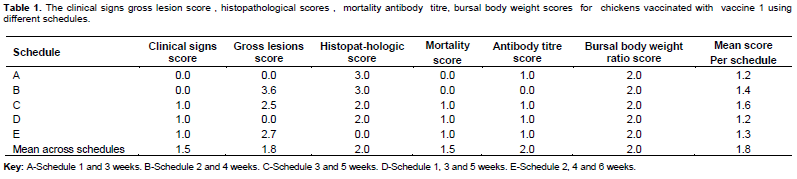
Schedule A (1 and 3 weeks)
Chickens were vaccinated with vaccine 1, at 1 and 3 weeks, the gross lesion score was 0; histopathological score was 3; clinical sign was 0; mortality was 0; antibody titre 1; bursal body weight ratio was 2 and the mean score was 1.2 (Table 1).
Schedule B (1and 2 weeks)
Chickens were vaccinated at 2 and 4 weeks old. The gross lesion score was 3.6; histopathological score was 3.6; clinical sign score was 0; mortality 0; antibody titre was 0 but the bursal body weight ratio was 2 and the mean score was 1.43 (Table 2).
Schedule C (3 and 5 weeks)
Chickens were vaccinated at 3 and 5 weeks old. The gross lesion score was 2.5; histopathological score was 2; clinical sign score was 1; mortality 1; antibody titre was 1; but the bursal body weight ratio was 2 and the mean score was 1.58.
Schedule D (1, 3 and 5 weeks)
Chickens were vaccinated at 1, 3 and 5 weeks old. The gross lesion score was 0; histopathological score was 2; clinical sign was 1; mortality 1 antibody titre was 1; but the bursal body weight ratio was 2 and the mean score was 1.17.
Schedule E (2, 4 and 6 weeks)
Chickens were vaccinated at 2, 4 and 6 weeks old. The gross lesion score was 2.7; histo-pathological score was 0; clinical sign score was 1; mortality 1; antibody titre was 1; but the bursal body weight ratio was 2 and the mean was 1.28 (Table 4).
Vaccine 2
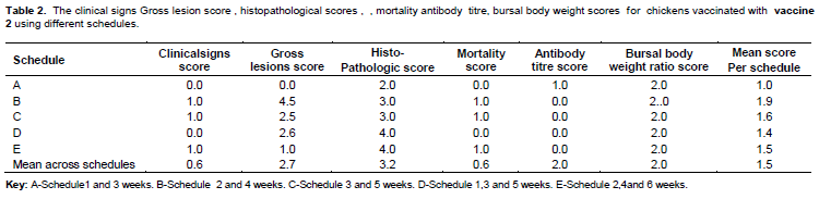
Schedule A (1 and 3 weeks)
Chickens were vaccinated with vaccine 1, at 1 and 3 weeks old. The clinical sign was 0; gross lesion score was 0; histopathological score was 2; mortality was 0; antibody titre 1; bursal body weight ratio was 2 and the mean score for the schedule was 1.0 (Table 2)
Schedule B (1 and 2 weeks)
Chickens were vaccinated at 2 and 4 weeks old. The clinical sign score was 1; gross lesion score was 4.5; histopathological score was 3; mortality 1; antibody titre was 0; but the bursal body weight ratio was 2 and the mean score for this schedule was 1.43 (Table 2).
Schedule C (3 and 5 weeks)
Chickens were vaccinated at 3 and 5 weeks old.
The clinical sign score was 1; gross lesion score was 2.5; histopathological score were 3; mortality 1; antibody titre was 0; but the bursal body weight ratio was 2 and the mean score for this schedule was 1.58 (Table 2).
Schedule D (1, 3 and 5 weeks)
Chickens were vaccinated at 1, 3 and 5 weeks old. The clinical sign was 1; gross lesion score was 0; histopathological scores was 3; mortality 1; antibody titre was 1; but the bursal body weight ratio was 2 and the mean score for this schedule was 1.43 (Table 2).
Schedule E (2, 4 and 6 weeks)
Chickens were vaccinated at 2, 4 and 6 weeks old. The clinical sign score was 1; gross lesion score was 1.0; histopathological score was 4; mortality 1; antibody titre was 0; but the bursal body weight ratio t was 2 and the mean scores for this schedule was 1.50 (Table 2).
Vaccine 3
Schedule A (1 and 3 weeks)
Chickens were vaccinated with vaccine 1, at 1 and 3 weeks old. The clinical sign was 0; gross lesion score was 0; histopathological score was 1; mortality was 0; antibody titre 1; bursal body weight ratio was 2 and the mean score for the schedule was 0.8 (Table 3).
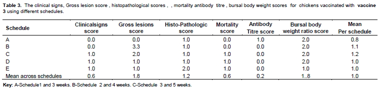
Schedule B (1and 2 weeks)
Chickens were vaccinated at 2 and 4 weeks old. The clinical sign score was 0; gross lesion score was 3.3; histopathological score was 1; mortality 0; antibody titre was 0; but the bursal body weight ratio was 2 and the mean score for this schedule was 1.1 (Table 3).
Schedule C (3 and 5 weeks)
Chickens were vaccinated at 3 and 5 weeks old. The clinical sign score was 1; gross lesion score was 1; histopathological score was 1; mortality 1; antibody titre was 0; but the bursal body weight ratio was 2 and the mean score for this schedule was 1.2 (Table 3).
Schedule D (1, 3 and 5 weeks)
Chickens were vaccinated at 1, 3 and 5 weeks old. The clinical sign was 1; gross lesion score was 1; histopathological score was 1; mortality 1; antibody titre was 0; but the bursal body weight ratio was 2 and the mean score for this schedule was 1.0 (Table 3).
Schedule E (2, 4 and 6 weeks)
Chickens were vaccinated at 2, 4 and 6 weeks the clinical sign score was 1; gross lesion score was 1.0; histopathological score was 2; mortality 1; antibody titre was 0; but the bursal body weight ratio was 2 and the mean scores for this schedule was 1.0 (Table 3).
Vaccine 4
Schedule A (1 and 3 weeks)
Chickens were vaccinated with vaccine 1, at 1 and 3 weeks old. The clinical sign was 0; gross lesion score was 4; histopathological score was 3; mortality was 0; antibody titre1; bursal body weight ratio was 2 and the mean score for the schedule was 1.7 (Table 4).
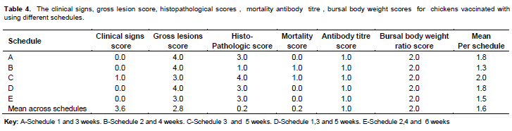
Schedule B (1 and 2 weeks)
Chickens were vaccinated at 2 and 4 weeks old.
The clinical sign score was 0; gross lesion score was 4; histopathological score was 1; mortality 0; antibody titre was 1; but the bursal body weight ratio was 2 and the mean score for this schedule was 1.3 (Table 4).
Schedule C (3 and 5 weeks)
Chickens were vaccinated at 3 and 5 weeks old. The clinical sign score was 1; gross lesion score was 3; histopathological score was 4, mortality 1; antibody titre was 1; but the bursal body weight ratio was 2 and the mean score for this schedule was 2.0 (Table 4).
Schedule D (1, 3 and 5 weeks)
Chickens were vaccinated at 1, 3 and 5 weeks old. The clinical sign was 0; gross lesion score was 4; histopathological score was 3; mortality 0; antibody titre was 1; but the bursal body weight ratio was 2 and the mean score for this schedule was 1.7 (Table 4).
Schedule E (2, 4 and 6 weeks)
Chickens were vaccinated at 2, 4 and 6 weeks old. The clinical sign score was 0; gross lesion score was 3; histopathological score was 3; mortality 0; antibody titre was 1; but the bursal body weight ratio was 2 and the mean score for this schedule was 1.5 (Table 4).
Chickens in positive control group were not vaccinated but challenged showed a clinical sign of 1.0; gross lesion score as 4.9; histopathological score 5.0; mortality 1; antibody titre 0 and a bursal body weight ratio of 6.47; while the negative control group had a clinical sign of 1; gross lesion score of 1.25; histopathological score of 2; motality 1; and antibody titre of 0 and bursal body weight ratio of 2.9 (Table 5).
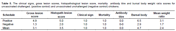
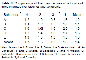
Table 4 shows that for vaccine 1, schedule A (1 and 3 weeks) and scheduled D (1, 3 and 5 weeks) have the lowest mean score of 1.2 each, then followed by schedule E (2, 4 and 6 weeks) with 1.3, then schedule B (2 and 4 weeks) with 1.4, then followed by schedule C (3 and 5 weeks) with mean scores of 1.6. For vaccine 2, schedule A (1 and 3 weeks) had the lowest mean score of 1.2, followed by schedule D (1, 3 and 5 weeks), then schedule E (2, 4 and 6 weeks) with 1.5, and schedule C (3 and 5 weeks) with mean scores of 1.6 and then schedule B with 1.9. For vaccine 3, schedule A (1 and 3 weeks) had the lowest mean score of 0.8 , followed by schedule D (1, 3 and 5 weeks) and E (2,4 and 6 weeks) with 1.0 each ,then schedule B (2 and 4 weeks) with 1.1, and schedule C (3 and 5 weeks) with mean score of 1.2. For vaccine 4, schedule B (2 and 4 weeks) had the lowest mean score of 1.3, followed by schedule E (2, 4 and 6 weeks) with 1.5, schedule A (1 and 3 weeks) with 1.6, scheduled D (1, 3 and 5 weeks) with 1.7, and schedule C (3 and 5 weeks) with mean scores of 2.0.
The mean scores for the vaccines across the schedules was recorded with vaccine 3 having the lowest (1.0), followed by vaccine 1 (1.3) with vaccine 2 (1.5) and then vaccine 4 (1.6). While the mean score across schedule was with schedule A (1 and 3 weeks) with 1.2, followed by scheduled D (1, 3 and 5 weeks) and E (2, 4 and 6 weeks) with 1.3 each, schedule B (2 and 4 weeks) has 1.4, and then schedule C (3 and 5 weeks) with 1.6 (Table 6).
DISCUSSION
The study revealed that vaccinating chickens with vaccine 1 (at 1 and 3 weeks) appeared to be the best schedule. This finding is consistent with what was reported by Segal (2009) who attributed it to the fact that the first vaccination at one week might have neutralized the MDA (Abdu, 1997). It is also important to note that the interval of two weeks before the second vaccination might have given the bursa enough time to recover from the effect of the first vaccination as reported by Abdu (1997). The best schedule for vaccine 2 was also observed at 1 and 3 weeks. The chicks may withstand the infection despite the fact that they were vaccinated at 1 week at an interval of 14 days. This is not surprising because previous researchers have suggested double vaccination against IBD (Segal, 2009). Since the level of MDA usually decline at about 3 week and chicks are highly susceptible, giving the 2nd vaccination at 3 weeks may therefore boost the birds immunity against IBD (Chansiporanchi, 2009).
It was also observed that schedule A was the best schedule for vaccine 3, and was even better than the all the vaccines as the clinical signs, mortality rate, gross lesion scores, histopathological scores, were negligible. This finding agrees with the report of Giambrone (1983). It was also observed that vaccinating chicks at 2 and 4 weeks of age was the best vaccination schedule for vaccine 4 (B). However, despite the fact that this schedule is the most widely adopted by poultry farmers in Zaria, outbreaks of IBD have been reported in vaccinated flocks as reported by Abdul (1997).
The antibody titre observed with vaccine 1 was lower than with vaccines 2, 3 and 4. This implies that the vaccine did not stimulate antibodies to appreciable level; hence its efficacy in vaccination programme may be doubtful (Abdu, 1997; Babiker and Tawfeeq, 2008). In contrasts to the findings of Abdu (1985), the finding in this study was consistent with the report by Naqi et al. (1983) who also observed marked difference in the titre of the antibody produced against IBD by different vaccines.
The clinical signs observed with vaccine 1 was significantly higher (p < 0.05) when compared to vaccine 2, 3 and 4. The least clinical sign scores was also recorded in vaccine 4. This also agrees with the work of Babiker and Tawfeeq (2008). The finding that vaccine 3 showed the least lesions score on the bursa implies that the vaccine is safe and may protect the birds against IBD, while vaccine 1 exhibited the highest lesions and hence the protective ability of this vaccine is questionable, as reported by Abdu (1997). The finding that the gross lesions were highest with vaccine 1 implies that this vaccine may not protect chicks against IBD. It may suffice to also say that this vaccine contained live viruses incapable of multiplying in the bursal cells of the chicks to illicit an immune response. Chickens vaccinated with such vaccine might remain susceptible to IBD outbreaks which may occur following natural exposure to virulent field viruses. These vaccination failures have continued to cause severe psychological stress and huge financial losses to poultry farmers as reported by Abdu (1997). It might have been possible that vaccine 1 had lost its potency through poor handling, transportation, storage or administration, eventhough vaccine 1 was in a freeze-dried state, a condition that improves the thermo stability of live vaccines (Spradbrow, 1992). In addition, the manufacturer of the vaccine 1 stated that the field dose may still be available to birds even after inoculation at 37°C for seven days. Moreover, IBDV is fairly resistant to heat (Benton et al., 1967).
The mortality rate as well as the bursal body weight ratio was worst with vaccine 1. This implies that the vaccine may not be immunogenic. This could be that the long continuous passage of the stock IBDV in the chick’s embryo fibroblast cells may have adversely affected the quality of the batches of vaccine produced. High number of passage might have probably rendered the vaccine virus less able to easily infect and multiply in the bursa and less immunogenic. In contrast vaccine 3 was observed to be immunogenic as the antibody titre was good, the clinical signs, the histo-pathological lesions of the bursa, the gross lesions as well as bursal body weight ratio were low implying that probably the vaccine had maintained its potency. It also implies that the vaccine had been handled, transported, stored and administered appropriately and contained live virus capable of multiplying within bursal cells of the bird to induce an immune response.
CONCLUSION
From the finding of this work, it may be concluded that vaccine 3 was the most potent of all the four live vaccines used and schedule A was the best schedule for vaccines 1, 2 and 3 and Schedule B was the best for vaccine 4. While appropriate handling, transportation, storage and administration is hereby re-emphasized, it is also recommended that the strain (type) of IBD vaccines produced should be defined as either mild, moderate or hot IBD vaccines.
CONFLICT OF INTERESTS
The authors have not declared any conflict of interests.
ACKNOWLEDGEMENTS
The authors are grateful to the Staff of Nutrition Laboratory of Department of Medicine, Faculty of Veterinary Medicine, Ahmadu Bello University Zaria, Nigeria.
REFERENCES
|
Abdu PA (1985). Studies on the profile and relationship of maternal and vaccine antibodies in the prevention of infectious bursal disease. MSc Thesis A.B.U., Zaria. |
|
|
Abdu PA (1997). Studies of the problems associated with vaccination against infectious bursal disease in Nigeria, PhD Dissertation, Ahmadu Bello University, Zaria P 129. |
|
|
Abdu PA (2007). Viral disease. In: Manual of important poultry Diseases in Nigeria 2ndedn Mac Chin Multimedia Designer, Zaria pp. 15-24. |
|
|
Abdu PA (2010). Important viral diseases of chicken in Nigeria and their control. Amo Farm Poultry Workshop on Profitable Poultry Farming 15th September 2010 at Devotion Hotel Jos, Plateau state. |
|
|
Babiker MA, Tawfeeq E (2008). Role of administration of anti IBD VIRUS (Gumboro) vaccine on immunization of chicken. J. Poult. Sci. 7(3):279-282. |
|
|
Benton WJ, Cover MS, Resenberger JK, Lake RS (1967). Physicochemical properties of infectious bursal disease agent. Avian Dis. 11:430-438. |
|
|
Blankfard M, Silk BC (1989). ELISA software R. Gaithersburg, Md. USA. |
|
|
Boudaoud A, Alloui N (2008). Evaluation of the safety of live attenuated vaccine viruses against infectious bursal disease (Gumboro disease) in conventional broiler chicks. Rev. Sci. Technol. 27(3):793-809. |
|
|
Cosgrove AS (1962). An apparently new disease of chicken- avian nephrosis. Avian Dis. 6:385-389. |
|
|
Giambrone JJ (1983). Gumboro vaccines hard hitting advice. Broiler Industry: October issue; pp. 80-87. |
|
|
Giambrone JJ (2008). Variant strain IBDV: Epidemiology and control retrieved June, 25 2008. |
|
|
Farsha (Farm and Ranch Safety Association) (2006). A Health and Safety Guide for Handling Farm Animals and Poultry 2006. |
|
|
Hair-Bejo M (2000). An Infectious bursal disease in broilers. J. Vet. Med. 4:168. |
|
|
Naqi SA, Marquez B, Sahin N (1983). Maternal antibody and its effect on infectious bursa disease immunization. Avian Dis. 27(3):623-631. |
|
|
Segal Y (2009). Gumboro disease. enormix.com, accessed20th /12/20115pm |
|
|
Spradbrow PB (1992). A review of the use of food carriers for the delivery of oral Newcastle disease vaccine. In Newcastle Disease in Village chickens. (Ed. Spradbrow PBS). ACIAR Proc. No. 39. Canberra. |
|
Copyright © 2024 Author(s) retain the copyright of this article.
This article is published under the terms of the Creative Commons Attribution License 4.0


