Full Length Research Paper

ABSTRACT
Although, many studies have been conducted to estimate the prevalence of gastrointestinal tract (GIT) helminths of cattle in Ethiopia, most of them were conducted using simple fecal egg examination rather than larval identification to identify the nematodes. A cross sectional study was conducted from November 2016 to April 2017 with the aim of estimating the prevalence of GIT helminths and identifying nematode genera in cattle in and around Bishoftu, Ethiopia. Of the 300 cattle examined using standard parasitological methods, 42.33% were positive for GIT helminth egg presence. The prevalence rates of GIT helminth based on the age and sex of the cattle were 37.6, 43.9, 60 and 27.9%, in young, adult, male and female animals, respectively. Strongyle type eggs were the most prevalent among the helminthes investigated. The pooled larval culture showed that Haemonchus, Oesophagostomum and Trichostrongylus were the most common nematodes found, with Haemonchus being present in 27.8% of cases, followed by Trichostrongylus (18%) and Oesophagostomum (4.48%). Mixed infection of GIT nematodes (44.4%) is higher than single (mono) infection. Thus, it could be concluded that GIT helminths of cattle cause serious problems in livestock production in the area.
Key words: Control, fecal culture, nematode, mixed infection, season.
INTRODUCTION
Ethiopia possesses about 53.99 million heads of cattle (CSA, 2013). Despite the large cattle population, productivity in Ethiopia is low due to poor nutrition, reproduction inefficiency, management constraints and animal diseases (Alsan, 2012). Helminth parasite infections in cattle’s are a primary factor in the reduction of livestock production and productivity (Wadhawa et al., 2011). Helminthosis, in large part, is caused by nematodes, cestodes and trematodes in domestic animals. Helminthosis lead to a reduction in fertility, work capacity, involuntary culling (Rafiullah et al., 2011), reduction in food intake, weight and milk production, and higher mortality rate (Getachew et al., 2012).
Gastrointestinal nematodes are cosmopolitan parasites that develop within the digestive tract of domestic ruminants. The gastrointestinal tract (GIT) of cattle harbors a variety of parasites, particularly helminthes, which causes clinical and subclinical parasitism. These parasites adversely affect the health status of animals and cause enormous economic losses to the livestock industry (Rafiullah et al., 2011; Singla et al., 2014). The effect of infection by gastrointestinal parasites varies according to the parasite concerned, the degree of infestation and other risk factors such as species, age, season and intensity of worm burden (Perry et al., 2002; Singh et al., 2014).
Analysis of nematodes based on fecal examinations beyond the clinical sign, and the presence of worm eggs or larvae is the most common routine aid to diagnosis. The egg and larvae of nematodes are most often diagnosed via fecal floatation and fecal culture (Hendrix, 1998; Gupta and Singla, 2012). Because of the large differences of the common worms’ genera parasites, it is necessary at least to identify the nematodes genera in order to evaluate the significance of the parasitical infestation or the anthelmintic treatment effectiveness (Lancaster and Hong, 1987). The only practical method, available to the helminthologists to obtain ante-mortem indexes for the parasites genera that are found in ruminants infestation, is larvae identification that are found in fresh faeces or in those developed on the culture faeces. The infective larvae (L3) of the common parasites genera are, generally easier to identify than their eggs (Van Wyk, 2004).
In Ethiopia several studies have been conducted to determine prevalence of GIT helminthes in different agro-ecology of the country. A prevalence of 27.57% GIT nematodes of cattle were reported in Gondar (Tigist et al., 2012), 50.08% gastrointestinal helminths in Tulu district, West Harergae Zone (Tulu and Lelisa, 2016), 41.15% GIT nematodes in Diredawa (Yimer et al., 2015) and 61% GIT helminthosis in the East Showa Zone (Telila et al., 2014). Although, many studies (Tigist et al., 2012; Telila et al., 2014; Yimer et al., 2015; Tulu and Lelisa, 2016) have been conducted to estimate the prevalence of GIT helminths of cattle in Ethiopia, most were concerned with determining the prevalence of the GIT helminths in cattle by using simple fecal egg examination rather than larval identification to identify the nematodes. In identification of parasitic nematode infections of ruminants by egg morphology, faecal worm egg counts is relatively inaccurate method as compared to larva identification, as no indication can be obtained for the identities of most of the common worm genera, except for those genera with morphologically distinct ova, for example, Strongyloides papillosus, Nematodirus spp. and Trichuris spp. Thus, the aim of the present study was to estimate the overall prevalence of GIT helminths, and identify major nematode parasites of cattle at genera level in and around Bishoftu, Ethiopia. This study has paramount importance in evaluating the significance of a specific parasitical infestation and designing appropriate prevention and control strategies against identified nematodes in the area.
MATERIALS AND METHODS
Study area
The study was conducted in and around the town of Bishoftu. The town is located at 9°N latitude and 40°E longitudes at an altitude of 1850 m above sea level, and situated in central highlands of Ethiopia. Bishoftu has an annual rainfall of 866 mm of which 84% falls in the rainy season (June to September). The rainfall is bimodal. The mean annual maximum and minimum temperature ranges are 26 and 14°C, respectively (CSA, 2012).
Study animals
The study animals were 300 cattle of three breeds (local, cross and exotic breeds), both sexes (139 male and 161 female), managed under intensive, semi-intensive and extensive production systems. Body condition score was according to Nicolson and Butterworth (1986) and recorded as poor, medium or good. The age of the animal was estimated by looking at the dentition pattern of the animals according to Frandson (1992) and also by owners’ response. Based on this, study animals were classified as young (1 to 3 years) and Adult (> 3 years).
Sampling method and sample size
Simple random sampling technique was employed to select the study animals. The total number of cattle required for the study was calculated using the formula given by Thrusfield (2005) with 5% acceptable error and 95% confidence level. Accordingly, by taking previous study prevalence of (82.8%) cattle GIT parasites around Holleta by Estehiwot (2004), the total number of cattle calculated for the study was 228. However, to increase precision, a total of 300 cattle were sampled for this study.
N = [1.962 Pexp (1-Pexp)]/d2
Where, N = sample size; Pexp = expected prevalence; D = desired absolute precision.
Study design
A cross sectional study was carried out from November 2016 to April 2017 to collect data on events associated with gastrointestinal helminth of cattle and identify nematode parasites. The study animals in each selected kebele and peasant association are categorized into two age groups, that is, young (1-3 years) and adult (above 3 years). A simple random selection was employed to select cattle reared under different management conditions.
Data collection
Fecal samples were directly collected per rectum. Each fecal sample was put in plastic containers with lids and labeled with animal identification record including age, sex and body condition (thin, moderate and good). Then, transported using ice box, to Addis Ababa University Veterinary Parasitology Laboratory at Bishoftu, and immediately examined or stored in refrigerator at 4°C for later examinations.
Fecal examination
The collected fecal samples were processed and examined by the standard flotation using saturated sodium chloride and sedimentation technique for qualitative investigation of the types of gastro-intestinal helminth eggs (Charles, 2006). Eggs of the different helminths were identified on the basis of morphological appearance and size of eggs according to the literature (Foreit, 1999).
Fecal culture and L3 identification
Fecal samples from nematode eggs positive animals, were pooled and cultured for harvesting of third stage larvae and identification of genera of nematode eggs. Pooled fecal samples were then finely broken using stirring device, kept moist and brittle; the mixtures were transferred to Petri dishes and placed at 27°C in an incubator for 7 to 10 days. The culture was kept moist by adding water every 2 days. During this period, the larvae was hatched from the eggs and developed into L3. Finally, larvae were recovered using the modified Baermann technique (Hansen and Perry, 1994). The presence of larvae was assessed by using a stereomicroscope, when present; two drops of larval suspension were mixed with drop of lugols iodine on glass slide, and examined at low magnification power for identification. From each culture, the third-stage larvae (L3) was morphologically differentiated and identified according to Van Wyk (2004). Conventional characteristics for identification (total length, esophagus length, tail sheath length and the number of intestinal cells) of infective larvae from gastrointestinal nematode genera/species were microscopically examined.
Data management and analysis
The raw data obtained from each animal were coded, entered and filtered into Microsoft Office Excel spread sheet and analyzed using STATA software version 17 (Stata Corp, Texas, USA). The prevalence of each parasite infection was calculated as the number of animals diagnosed positive for a given parasite divided by the total number of animals examined at the particular time (Thrusfield, 2005). Chi-square statistics (χ2) was used to determine the association of the variables (sex, age, breed, body condition and management) with the prevalence of GIT parasitism. The P-value less than 0.05 were taken as statistically significant and greater than 0.05 was taken as statistically insignificant at 95% confidence interval.
RESULTS
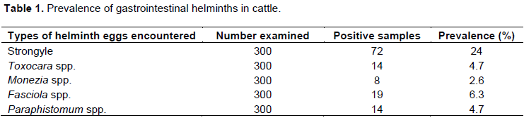
Of the 300 cattle examined, 42.33% were positive for gastrointestinal helminth egg presence. Eggs from five genera, including two nematodes (Strongyle and Toxocara); two trematodes (Paramphistomum and Fasciola) and one cestode (Monezia) were identified. The prevalence of all identified parasitic helminth eggs (Table 1) showed that Strongyle-type eggs had the highest (24.00%) prevalence and Monezia species was the least (2.6%).
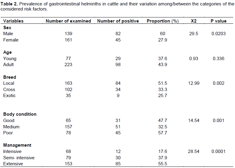
It was observed that the prevalence of gastrointestinal helminths had statistically significant difference association in relation to sex, breed, body condition and management system (P<0.05). However, it is also shown that there were no statistical association among the age groups (P>0.05) as indicated in Table 2.
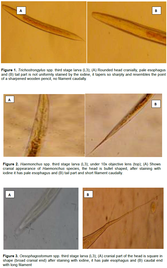
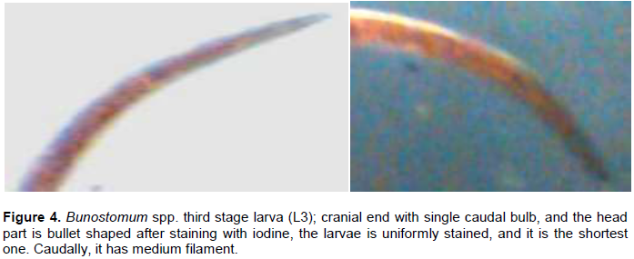
Morphological identification of L3 of most parasitic nematodes was based principally on examination of the caudal and cranial extremities, size of the larvae, presence or absence of filament, presence or absence of sheath, and also differentiation by staining property by lugolsiodine (Hansen and Perry, 1994). Based on these morphological characteristics, four nematode larvae belonging to genera Trichostrongylus, Haemonchus, Oesophagostomum and Bunostomum were recovered and identified as show in Figures 1 to 4.
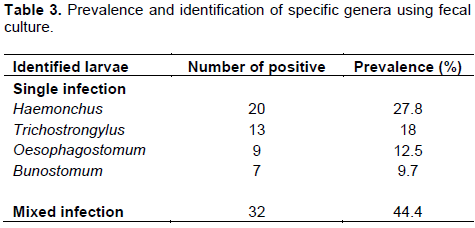
The pooled larval cultures showed that Haemonchus, Trichostrongylus and Oesophagostomum were the most common gastrointestinal nematodes found in the animals. Table 3 indicates that Haemonchus, Trichostrongylus and Oesophagostomum were the dominant genera among the cultivated larvae having 20 (27.8%), 13 (18%) and 9 (12.5 %), respectively. Mixed infection (poly infection) was more prevalent (32; 44.4%) than single infection (mono-infection).
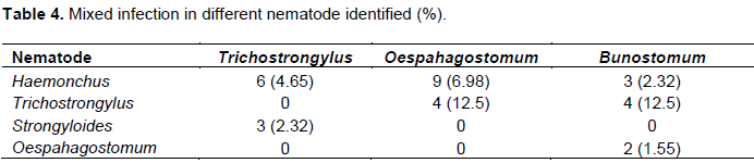
Mixed infection between different nematodes species was also observed and described as high prevalence of the Heamonchus, together with Oesophagostomum (6.98%) and Trichostrongylus (4.65%), and less prevalence of the Oesophagostomum together with Bunostomum (1.55%) as shown in Table 4.
DISCUSSION
Overall prevalence rate
The present study shows that 42.3% (127/300) of the cattle screened had a GIT helminth infection, indicating the high burden of GIT helminths among cattle in and around Bishoftu. This prevalence is comparable to that of Yimer et al. (2015) who reported 41.5% prevalence in Diredawa but lower than 50.08 and 61% in the East Showa Zone and Tulu district, West Harergae Zone reported by Telila et al. (2014) and Tulu and Lelisa (2016), respectively. The differences observed could be due to the variation in the periods or seasons in which the studies were conducted, climate and husbandry of the animals.
The current study overall prevalence of 28.6% for gastrointestinal nematode parasites in cattle is comparable to Birhanu (2011) in Gondar who reported 33.3%. It was observed that nematode infections were particularly high, as they accounted for 28.6% of the helminth burden. High nematode infection has a significant impact on livestock production since they result in reduced milk, meat, wool, hide products and stamina of working animals (Ekong et al., 2012), hence resulting in the reduction of production potentials such as decreased growth rate, weight loss in young growing calves, and late maturity of the animals (Swai et al., 2006).
The present finding disagree with the report of Epherem (2007) and Addisu and Berihu (2014) who reported 41.2% for Western Amhara Region and 49% for the West Arsi zone, respectively. These high prevalence values could be due to topography, season and climate that could favor the survival of parasitic stage, due to difference in management system of the study animals and breeds of these animals. Unlike the present study done on cattle of different breeds and from three management systems, those studies were conducted on cattle managed under extensive management, which could increase the degree of pasture contamination, leading to higher prevalence rates. In contrast to the present finding, Yehuelaeshet (2005) reported a lower prevalence value (11%) for Bahirdar. This difference in prevalence might be due to the differences in deworming practice, study design and ecology, season and husbandry system among the different studies (Singla, 1995).
Prevalence per helminth type
The highest prevalence of helminths was in the strongyle type, while other helminths parasites like cestodes were found to be the least prevalent. The result of the present study agrees with that of Telila et al. (2014) who reported a 41% prevalence of the strongyle type in East Showa zone, Central Ethiopia. In addition, this finding also agrees with Tulu and Lelisa (2016) who reported a 36.23% prevalence of the strongyle type egg in Tulo District, West Hararghe Zone. However, it disagrees with Etsehiwot (2004) and Derib (2005) who reported that trematodes are more prevalent than strongyle species. The high prevalence of strongyles may be due to the suitability of the climatic condition of Bishoftu for survival and transmission of these parasites.
The overall prevalence for trematode infection in this study is 11%. This finding is lower than the 60.42% reported for Andassa by Yenenehet al. (2012), 34.5% in Bahir Dar and its surroundings (Derib, 2005) and 52.53% for Jimma (Abebe et al., 2011). This variation could be associated with differences in geographical and/or climatic conditions and ecology since the presence of trematode infections is dependent on availability of the intermediate hosts. The only cestode observed in the cattle in the present study was Moniezia (2.6%) and the occurrence of parasite is very low as compared to other gastrointestinal parasites. This might be due to the fact that the climatic condition is not suitable for survival of the intermediate host (oribatid mite) and transmission of the parasite.
Infection rate among sexes
Statistically significant difference exists in the infection rate of gastro-intestinal helminth infection among sexes, with a higher prevalence rate in males (60%) than in females (27.9%). This is in agreement with Wondimu (2009), Birhanu (2011) and Tulu and Lelisa (2016) who found prevalence rates in males (45.86%) to be greater than that in females (41.4%). This might be due to males being more exposed to grazing areas than females. In the study area, females are used for milking purposes; as such, the owners do not allow them to graze on pastureland. However, this is in disagreement with previous study report that stated statistically non-significant difference among male and female cattle (P>0.05) (Tigist et al., 2012). In this study, a statistically significant difference was observed in GIT helminth infection in relation to body condition where a higher prevalence was recorded in poor body condition (57.7%) than good (47.7%) and medium body conditions (32.7%). This finding agrees with Tigist et al. (2012) but contradicts the findings of Regassa et al. (2006) who reported that body condition of the animal did not show significant association with prevalence of the parasite. This could be explained by the fact that loss of body condition in the study animals could be due to other factors, such as seasonal change of forageable feedstuff and the presence of other concurrent disease conditions.
Helminth infection and management system
The result of the present study showed significantly higher GIT helminth infection rate in extensively managed cattle (55.5%) than semi intensive (37.9%) and intensive (17.6%) managed cattle. This finding agrees with the reports of Keyyu et al. (2006) and Kabaka et al. (2013). This might be due to the fact that extensively managed cattle have more access to contaminated pasture.
Single and mixed infection
The pooled larval culture showed that Haemonchus, Oesophagostomum and Trichostrongylus were the most prevalent nematodes, with Haemonchus being present in 27.8% followed by Trichostrongylus (18%) and Oesophagostomum (12.5 %). The detection of Haemonchus species as the commonest infection in this study may suggest that these worms are seriously affecting the cattle in the study area. Similar report by Waruiru et al. (1998) indicated Haemonchus as the most prevalent species of gastrointestinal nematode in Kiambu District, Kenya. The overall prevalence of mixed infection (poly infection) was 32% which is characterized by two or more genera. The highest prevalence in this mixed infection with different genus is seen in Haemonchus with Oesophagostomum (6.98%).
CONCLUSION
The overall prevalence of gastrointestinal helminths parasite in the current study indicates that gastrointestinal helminthosis is an important health problem due to its high prevalence (42.3%) and occurrence of mixed infection. The phenomenon of mixed infection has been suggested to be an important cause of morbidity and reduced production in livestock. Furthermore, the immunosuppression of the host immune system by mixed infections increases host susceptibility to other diseases or parasites. Thus, it can be concluded that GIT helminths of cattle cause serious problems in livestock production in and around Bishoftu. Therefore, more studies on seasonal transmission pattern of all these parasites are required in order to design rational, economic and locally sustainable parasite control programs. Further studies that will help in taking obligatory preventive and control measures against parasitism as well as maximize the production, are suggested.
CONFLICT OF INTERESTS
The authors have not declared any conflict of interests.
REFERENCES
|
Abebe F, Behablom M, Berhanu M (2011). Major trematode infections of cattle slaughtered at Jimma municipality abattoir and the occurrence of the intermediate hosts in selected water bodies of the Zone. Journal of Animal and Veterinary Advances 10:1592-1597. |
|
|
Addisu B, Berihu H (2014). Study on prevalence of gastrointestinal nematodes and coccidian parasites affecting cattle in West Arsi Zone, Oromia Regional State, Ethiopia. Journal of Veterinary Science and Technology 5:207. |
|
|
Alsan M (2012). The effect of the tsetse fly on African development. National Bureau of Economic Research, 105 Massachusetts, Avenue, Suite 418, Cambridge, USA. |
|
|
Birhanu T (2011). Study on the prevalence of gastrointestinal nematodes infection of cattle in and around Gondar, North western Ethiopia. DVM thesis, Gondar University, Gondar, Ethiopia. |
|
|
Charles M (2006). Diagnostic veterinary Parasitology.3rdEdn.Stilous Elsevier Science. |
|
|
Central Statistical Agency (CSA) (2012). Central Statistical Agency of the Federal Democratic Republic Of Ethiopia. Agricultural Sample Survey of 2011/12 (2004 E.C).Volume II. Report on Livestock and Livestock Characteristics (Private Peasant Holdings), Central Statistical Agency, Addis Ababa, Ethiopia. |
|
|
Central Statistical Agency (CSA) (2013).Central Statistical Agency (CSA). Agricultural sample survey results for NNPR. Statistical report on livestock and livestock characteristics (private peasant holdings), Center of Statistical Agency, Volume II, Addis Ababa, Ethiopia. |
|
|
Derib Y (2005). Study on endoparasites of dairy cattle in Bahir Dar and its surrounding. DVM thesis, Faculty of Veterinary Medicine, Addis Ababa University, DebreZeit, Ethiopia. |
|
|
Ekong PS, Juryit R, Dika NM, Nguku P, Musenero M (2012). Prevalence and risk factors for zoonotic helminth infection among humans and animals-Jos, Nigeria, 2005–2009. Pan African Medical Journal 12:6. |
|
|
Epherem W (2007). Prevalence of Bovine GI helminths in selected Dairy farms of Addis Ababa. DVM Thesis, Jimma University, Jimma, Ethiopia. |
|
|
Etsehiwot W (2004). A Study on Bovine Gastro Intestinal Helminthes in Dairy Cows in and around Holota. DVM Thesis, Addis Ababa University, Debrezeit, Ethiopia. |
|
|
Foreit W (1999). In: Reference Manual of Veterinary Parasitology. 5thEdn, Wiley Blackwell. |
|
|
Frandson RD (1992). Anatomy and physiology of farm animals. Department of anatomy and neurobiology, collage of veterinary medicine and bio medic sciences, 5thEdn. Colorado state University, for Collins, Colorado. |
|
|
Getachew H, Guadu T, Fentahun T, Chanie M (2012). Small Ruminant Hydatidosis: Occurrence and Economic Importance in Addis Ababa Abattoir. Global Veterinaria 8(2):160-167. |
|
|
Gupta SK, Singla LD (2012). Diagnostic trends in parasitic diseases of animals. In: Veterinary Diagnostics: Current Trends. Gupta RP, Garg SR, Nehra V and Lather D (Eds), Satish Serial Publishing House, Delhi, pp. 81-112. |
|
|
Hansen J, Perry B (1994). The epidemiology, diagnosis and control of helminth parasites of ruminants. 2nd Edn. Nairobi, Kenya. |
|
|
Hendrix CM (1998). Diagnostic Veterinary Parasitology, 2nd Edn. Mosby, London. |
|
|
Kabaka WM, Gitau GK, Kitala PM, Maingi N, Venleeuwen JA (2013). The prevalence of gastro intestinal nematode infection and their impact on cattle in Nakuru and Makurweini districts of Kenya. Ethiopian Veterinary Journal 17:95-104. |
|
|
Keyyu JD, Kssuku AA, Msalilwa LP, Monrad J, Kyvsgaard NC (2006). Cross-Sectional prevalence of helminth infections in cattle on traditional, small-scale and large- scale dairy farms in Iringa District Tanzania. Veterinary Research Communications 30:45-55. |
|
|
Lancaster MB, Hong C (1987). Differentiation of third stage larvae of ovine Ostertagia type and Trichostrongylus species. Veterinary Record pp.120-503. |
|
|
Nicolson MJ, Butterworth MH (1986). A guide to condition scoring of zebu cattle. IntLivest Centre Africa, Addis Ababa, Ethiopia. |
|
|
Perry BD, Randolph TF, McDermott JJ, Sones KR, Thornton PK (2002). Investing in animal health research to alleviate poverty. International Livestock Research Institute (ILRI), Nairobi, Kenya, P 148. |
|
|
Rafiullah A, Abdul S, Sayyed Shabbir R, Muhammed S (2011). Prevalence of Gastrointestinal tract parasites in cattle of Khyber Pakhtukkhwa, Journal of Agricultural and Biological Science 9:6. |
|
|
Regassa F, Sori T, Dhuguma R, Kiros Y (2006). Epidemiology of Gastrointestinal Parasites of Ruminants in Western Oromia, Ethiopia. International Journal of Applied Research in Veterinary Medicine 4:51-57. |
|
|
Singh ST, Malhotra P, Singla LD (2014). Fatal natural infection with microfilariae of Setaria species in a cattle bull. Progress Research 9(1):355-356. |
|
|
Singla LD (1995). A note on sub-clinical gastro-intestinal parasitism in sheep and goats in Ludhiana and Faridkot districts of Punjab. India Veterinary Science Journals 19:61-62. |
|
|
Singla LD, Moudgil AD, Sood NK, Deshmukh S, Turkar S, Uppal SK (2014). A unique case report on Setaria species microfilariosis in adult cattle in Punjab (India). International Science Journal 1(2):1-3. |
|
|
Swai E, Mitui P, Mbise A, Kaaya E, Sank P (2006). Prevalence of gastrointestinal parasites infection in maasai cattle in Mygorongora District, Tanzania. LRRD. 18:1-11. |
|
|
Telila C, Abera B, Lemma D, Eticha E (2014). Prevalence of gastrointestinal parasitism of cattle in east Showa Zone, OromiaRegional State, Central Ethiopia. Journal of Veterinary Medicine and Animal Health 6:54-62. |
|
|
Thrusfield M (2005). Veterinary epidemiology.3rdEdn.Blackwell Publishers, London. |
|
|
Tigist A, Basaznew B, Mersha C (2012). Occurrence of Gastro Intestinal Nematodes of Cattle in and Around Gondar Town, Amhara Regional State, Ethiopia. Ethiopia. Acta Parasitology Globe 3(2):28-33. |
|
|
Tulu D, Lelisa K (2016). A Study on Major Gastro-Intestinal Helminths Parasites of cattle in Tulo District, West Hararghe Zone, South- Eastern Ethiopia. Journal of Veterinary Science and Animal Husbandry 3(2):1027. |
|
|
Van Wyk JA (2004). Morphological identification of nematode larvae of small ruminants and cattle simplified. Veterinary Parasitology 119:277-306. |
|
|
Wadhawa A, Tanwar RK, Singla LD, Eda S, Kumar N, Kumar Y (2011). Prevalence of gastrointestinal helminths in cattle and buffaloes in Bikaner, Rajasthan, India. Veterinary World 4(9):417-419. |
|
|
Waruiru RM, Mutune MN, Otieno RO (2005). Gastrointestinal parasite infections of sheep and goats in a semi-arid area of Machakos District, Kenya. Bulletin of Animal Health and Production in Africa 53(1):25-34. |
|
|
Wondimu A (2009). Study on prevalence of Gastrointestinal nematodes in Dairy cattle at Haramaya university dairy farm. DVM thesis, Addis Ababa University,Debzeit, Ethiopia. |
|
|
Yehuelaeshet D (2005). The Study on Endoparasite of Dairy Cattle in Bahir Dar and its surrounding. DVM thesis, Addis Ababa University, Debzeit, Ethiopia. |
|
|
Yeneneh A, Kebede H, Fentahun T, Chanie M (2012). Prevalence of cattle flukes infection at Andassa Livestock Research Center in north-west of Ethiopia. Veterinary Research Forum 3(2):85-89. |
|
|
Yimer M, Dinaol B, Mesfin A, Solomon S, Hassen B (2015). Prevalence of Gastrointestinal Nematode of Cattle in Selected Kebeles of Dire Dawa Districts Eastern Ethiopia. Advanced Biomedical Research 9(6):418-423. |
|
Copyright © 2024 Author(s) retain the copyright of this article.
This article is published under the terms of the Creative Commons Attribution License 4.0


