Full Length Research Paper

ABSTRACT
Avian hepatitis E virus (aHEV), clinically important in poultry industry, can cause death and reduce egg production of chickens, resulting in significant economic losses in the poultry industry. However, little is known about this aHEV infection in Burkina Faso. This study presents the results of distribution and characterization of aHEV in domestic and wild birds without clinical disease. In total 173 birds liver samples were collected from four Burkina Faso provinces, between February 2015 and June 2016. Reverse transcription polymerase chain reaction (RT-PCR) with aHEV specific degenerate primers was used to screen the presence of aHEV. RNA of aHEV was detected in 29 (16.8%) liver samples. Of these, the prevalence was diverse in different species of birds; the most frequent level was 35.3% in Numida meleagris, respectively followed by 23.5% in Gallus gallus domesticus, 13.3% in Streptopelia turtur, 13.3% in Columba livia, 6.7% in Anas platyrhynchos and 3.3% in Pternistis natalensis. The present study firstly revealed the prevalence of HEV infection in six species of birds in Burkina. It is therefore important to conduct further research on the impact on poultry mortality and egg production in our country.
Key words: Avian hepatitis E virus, zoonosis, birds, prevalence, Burkina Faso.
INTRODUCTION
Hepatitis E virus (HEV), known to have zoonotic potential (Pavio et al., 2010), is transmitted enterically, mainly through the consumption of contaminated food or water (Yugo and Meng, 2013). HEV is the causative agent of a self-limiting acute hepatitis, ranges from an asymptomatic to a severe course, as described in immune-compromised patients and pregnant women (Purdy and Sue, 2017; Zuin et al., 2017). The severity in pregnant women is reflected in a mortality rate reaching up to 10 to 30% compared with 0.5 to 4.0% in young adults (Ward et al., 2011). HEV is divided into two genera: Orthohepevirus with four species (A–D) and Piscihepevirus with one species (Spahr et al., 2018). Orthohepevirus A has at least 8 recognized genotypes of mammalian HEV. Orthohepevirus B consists of avian viruses and is divided into four proposed subtypes (I–IV) associated with geographical distribution (Sridhar et al., 2017).
Avian Hepatitis E virus (aHEV) was first isolated from chickens with big liver and spleen disease (BLSD) or hepatitis-splenomegaly (HS) syndrome. Phylogenetic analysis of the full or nearly complete genome of aHEV strains identified four different genotypes and showed a distant relationship to mammalian and swine HEVs (50 to 60% nucleotide sequence identity) (Smith et al., 2015). The aHEV genotype 1 has been described in Australia and Korea, genotype 2 in USA, genotype 3 in Europe and China, and more recently, genotype 4 in Hungary and Taiwan (Payne et al., 1999; Park et al., 2015; Wang et al., 2015; Moon et al., 2016; Zhang et al., 2016).
HEV infections are completely asymptomatic in many animal species, but it seems to have some pathogenic importance for chickens (Yugo et al., 2016). Besides the enlargement of spleen and liver, both ovarian regression and presence of serosanguinous abdominal fluid or clotted blood in the abdomen are commonly associated with the HS syndrome (Ritchie and Riddell, 1991; Payne et al., 1999; Haqshenas et al., 2001; Thiry et al., 2017). The disease mainly causes a decrease in egg production and an increase in mortality in birds (Sun et al., 2004; Peralta et al., 2009). However, aHEV can be detected in birds without symptoms as well (Yugo et al., 2016; Zhang et al., 2016; Zhang et al., 2017). The virus appears to spread easily within and between flocks via the fecal-oral route transmission (Yugo et al., 2016). Other routes of transmission, including aerosol, vertical, vector-borne, or mechanical carrier, have not been demonstrated in natural or experimental avian models (Meng, 2011).
Based on serological evidence, it appears that avian HEV is widespread in chicken flocks with seropositive rates of approximately 71% in the United States, 90% in Spain, 20% in Brazil and 57% in Korea (Kwon et al., 2012). The overall detection rate of avian HEV RNA in fecal samples was 62.9% in the United States (Gerber et al., 2015).
Human infection with aHEV has not been observed up to now as it was for swine HEV (Meng, 2010). However, aHEV exposure of human population have largely increase in relationship to the consumption of contaminated poultry eggs and meat, the use of poultry viscera as a culinary delicacy, and the handling of poultry (Hsu and Tsai, 2014). In addition to the already described capacity of the virus to recognize human hepatocyte (Hsu and Tsai, 2014), the existence of a yet unknown aHEV variant able to enter and infect human liver may have a critical public health implication in the future.
In West Africa, the status of avian HEV infection in chickens is largely unknown. Considering that aHEV infection is most prevalent and dangerous among birds, it is imperative to access the contribution of aHEV to poultry and wildlife in Burkina Faso. The aim of the present study were to determine the possible circulation of avian HEV both in domestic and wild birds without clinical symptoms in Burkina Faso.
MATERIALS AND METHODS
Sample collection
In total, 173 samples of different symptomless bird flocks (4 domestic bird species) or hunted animals (2 wild bird species) currently in food chain in Burkina, were collected between February 2015 and June 2016 from four Burkina Faso district (Figure 1): 34 Guinea fowls (Numida meleagris), 34 chicken (Gallus gallus domesticus), 30 mallards (Anas platyrhynchos), and 15 doves (Columba livia) for domestic flocks and 30 turtle dove (Streptopelia turtur), 30 natal francolins (Pternistis natalensis) hunted in the hunting areas of Burkina Faso.
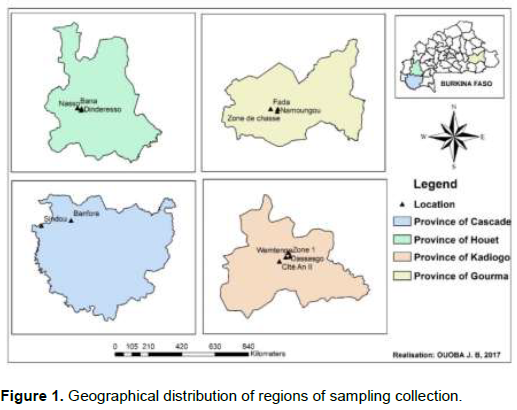
0.5 g of liver samples from each animal were collected and stored at -20°C in the RNAlater Buffer, until further use as source of HEV genomic RNA. Wild animals were samples in the provinces of Houet and Gourma where there are hunting areas. Kadiogo is in the center and does not have a hunting area, so no wild birds were taken in this area.
Samples RNA extraction and aHEV detection
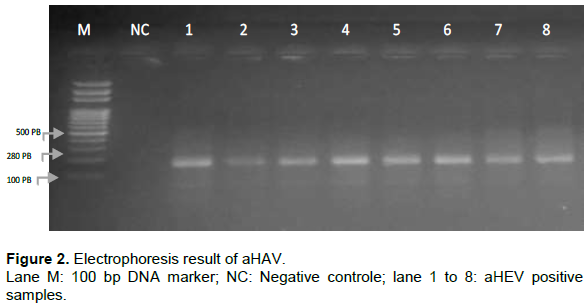
RNA extractions on the liver samples were performed using the SV total RNA isolation system kit (Promega, France). Extracts were subsequently used for detection of the partial capsid gene of aHEV using primers described previously (Bilic et al., 2009) in a reverse transcriptase polymerase chain reaction (RT-PCR). Briefly, external primers set Forw1_C-BLSV (5’-GGTATGGTTGATTTTGCCATAAAG-3’) and Rev1_C-BLSV (5’-GCTGCNCGNARCAGTGTCGA-3’) were used. The reverse transcriptase reaction and polymerase chain reaction were performed with the OneStep RT-PCR kit (Promega, France), according to manufacturer’s instructions under the following conditions: 50°C for 30 min; 95°C for 5 min; 45 cycles of 94°C for 30 s, 60°C for 30 s, and 72°C for 1 min, followed by a final elongation step of 72°C for 10 min. The negative control was water treated in the same way as the liver samples. Polymerase chain reaction (PCR) products with the expected size (280 bp) were revealed on a 1% agarose gel containing SybrGreen (Figure 2).
Statistical analysis
We performed the statistical analysis using R software version 2.13.0, through the package ‘Rcmdr’ version 2.5-1 (Fox et al., 2018). The differences in avian HEV RNA positivity between different variables (Locality and Species) were evaluated using logistic regression binomial. The best model was judged by Fisher's scoring algorithm. All tests were two-sided, and values of p < 0.05 were considered statistically significant. Odds ratios (ORs) and their 95% confidence intervals (95% CIs) were estimated to explore the strength of the association between aHEV positivity and the conditions investigated.
RESULTS
Avian HEV RNA were detected in 29 (16.8 and 95% CI [11.2 – 22.3]; p=2.2.10-16) of the 173 examined bird liver samples by RT-PCR (Tables 1 and 2). Of these, the prevalence was diverse in different birds species; the most frequent level was 35.3% (12/34, 95% CI [19.2 – 51.4]) in N. meleagris, followed by 23.5% (8/34, 95% CI [9.3 – 37.8]), in G. gallus domesticus; 13.3% (4/30, 95% CI [1.2 – 25.5]), in S. turtur; 13.3% (2/15, 95% CI [0 – 30.5]), in C. livia; 6.7% (2/30, 95% CI [0 – 15.6]), in A. platyrhynchos and 3.3% (1/30, 95% CI [0 – 9.8]) in P. natalensis. The highest proportions of positive samples were found in the domestic species 21.2% (24/113 95% CI [13.7 – 28.8], p=9.7.10-10) against 8.3% in wild birds (5/60, 95% CI, [1.3 – 15.3], p=1.1.10-10): Domestic birds had 4.8-fold higher risk than wild birds ([OR], 4.8; 95% CI, [1.8 – 15.9]; p=4.0. 10-3) (Table 2).
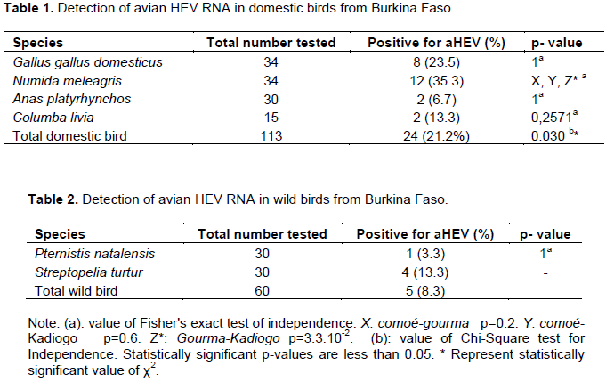
Comparison between domestic and wild birds within the area where both species were tested in sufficient numbers, show that the prevalence of N. meleagris (3/3 (100%)) is higher than that of S. turtur (4/30 (13.3%)) in Gourma (p˂ 0.01).
In addition, within a specie, the positive rates of avian HEV RNA in liver varied according different locations; thus, N. meleagris in the district of Gourma are more likely to be infected than those in province of Kadiogo (pË‚ 0.05). The total positive cases in a locality, without distinction of species, were respectively 18.2% (18/95 95% CI [10.6 – 25.8]) in the district of Kadiogo (p=1.42.10-9), 23.3% (7/30 95% CI [8.2 – 38.5]; p=3.5.10-3) in the district of Gourma, 6.2% (2/32 95% CI [0 – 14.6]; p=6.0.10-4) in the district of Houet, and 16.7% (2/12 95% CI [0 – 37.5]; p=2.1. 10-4) in the district of Comoé.
Thus, without distinction of species, in district of Kadiogo seems to have an approximately 1.85-fold higher risk than Comoé (odds ratio [OR], 1.8; 95% confidence interval [CI], [0.4 – 12.9]; p=0.4). Kadiogo had an approximately 0.7-fold lower risk than Gourma (OR, 0.7; 95% CI, [0.2 – 2.0]; p=0.5). Kadiogo had an approximately 5.4-fold higher risk than Houet (OR, 5.4; 95% CI, [1.4 – 35.8]; p=3.2.10-2).

DISCUSSION
Evidence of aHEV infection of poultry has been well documented from the United States, Canada, China, Australia, Israel, and several countries in Europe (Haqshenas et al., 2001; Swayne, 2003; Agunos et al., 2006; Guo et al., 2006; Peralta et al., 2009; Xiao et al., 2013; Zhang et al., 2017). This study represents the first report on the distribution and molecular characterization of avian Hepatitis E Virus in domestic and wild bird without clinical symptoms in Burkina Faso. The overall aHEV RNA prevalence was 16.8% (29/173) in six birds species sampled from four districts of Burkina Faso, which was lower than that in chickens in the United States (29.9%; Gerber et al., 2014), Brazil (20.0%; Billam et al., 2005), and Korea (28%; Kwon et al., 2012), by ELISA, in China (30.6%) by Reverse transcription-polymerase chain reaction (RT-PCR), and (35.1%; Sun et al., 2016) by ELISA. The low prevalence recorded in detecting avian HEV genome could be attributed to sampling of apparently healthy birds and alternatively to the primers used (Gerber et al., 2015), as the avian HEV genome shows a high variability (Sprygin et al., 2012). Besides, detection rate of aHEV RNA in the pooled fecal samples was 62.9% (39/62) (Gerber et al., 2015), hence fecal samples could be another samples source, suitable for the molecular detection of avian HEV.
This prevalence of aHEV RNA was diverse in different birds species; the most frequent level was 35.3% in N. meleagris, 23.5%, in G. gallus domesticus, 13.3%, in S. turtur, 13.3%, in C. livia, 6.7% in A. platyrhynchos and 3.3% in P. natalensis. The differences could be related to differences in ecological and geographical factors (Cong et al., 2014). Thus, the high rate of aHEV RNA showed in the domestic species (G. gallus domesticus, N. meleagris, A. platyrhynchos and C. livia), could be due the poultry were highly congested in livestock areas, feces likely serve as the main source for virus spread within the flock (Haqshenas et al., 2001; Saif et al., 2008; Ahmad et al., 2010; Meng, 2011; Yugo et al., 2016). Domestic birds could be most often subject to a re-infection, because proper sanitation conditions in the henhouse are lacking and bird drinking water can contain feces (Crespo et al., 2015). This results suggests the possibility of aHEV transmission from asymptomatic cases or repeated introduction through an unknown common source (Hsu and Tsai, 2014). Some studies have also shown that the high density of poultry increases the risk of disease transmission (Ricard and Marche, 1988). The low prevalence of aHEV RNA observed in wild birds (P. natalensis and S. turtur) also reflects that the congesting increases the likelihood of positive. Indeed, these birds live in liberty and are less congested compared to domestic birds. As for, A. platyrhynchos living in semi-liberty, the rate of positive sample (6.7%) was higher than in wild birds (Crespo et al., 2015). Burkina Faso is a developing country with low health and educational standards. Utilization of untreated bird feces for agriculture could increase the risk of virus dissemination, which in turn can infect wild birds. The high frequency of aHEV occurrence in bird livers in our country must be monitored to avoid an eventual outbreak. We have not investigated the source of aHEV infection in this study, but the role of wildlife in spreading the disease cannot be ignored (Crespo et al., 2015). The present study demonstrates the circulation of avian HEV in the domestic and wild birds without clinical symptoms, in Burkina Faso. This asymptomatic circulation of the virus in birds is of great interest and should be better monitored to avoid large epidemics. Thus we have to undertake studies on public health issues related to aHEV and the genetic diversity of aHEV inside the country.
ACKNOWLEDGEMENTS
J.B.O. received funding from the 3rd-cycle university scholarship program of the Embassy of France in Burkina Faso (http://www.burkina.campusfrance.org).
CONFLICT OF INTERESTS
The authors have not declared any conflict of interests.
REFERENCES
|
Agunos AC, Yoo D, Youssef SA, Ran D, Binnington B, Hunter DB (2006). Avian hepatitis E virus in an outbreak of hepatitis-splenomegaly syndrome and fatty liver haemorrhage syndrome in two flaxseed-fed layer flocks in Ontario. Avian Pathology 35(5):404-412. |
|
|
Ahmad T, Waheed Y, Tahir S, Safi SZ, Fatima K, Afzal MS, Qadri I (2010). Frequency of HEV contamination in sewerage waters in Pakistan. The Journal of Infection in Developing Countries 4(12):842-845. |
|
|
Bilic I, Jaskulska B, Basic A, Morrow CJ, Hess M (2009). Sequence analysis and comparison of avian hepatitis E viruses from Australia and Europe indicate the existence of different genotypes. Journal of General Virology 90(4):863-873. |
|
|
Billam P, Huang FF, Sun ZF, Pierson FW, Duncan RB, Elvinger F, Meng XJ (2005). Systematic pathogenesis and replication of avian hepatitis E virus in specific-pathogen-free adult chickens. Journal of Virology 79(6):3429-3437. |
|
|
Cong W, Meng QF, Shan XF, Sun WW, Qin SY, Zhang XX, Qian AD (2014). Seroprevalence and risk factors associated with hepatitis E virus infection in three species of pet birds in northwest China. Scientific World Journal 2014:296285. |
|
|
Crespo R, Opriessnig T, Uzal F, Gerber PF (2015). Avian Hepatitis E Virus Infection in Organic Layers. Avian Disease 59(3):388-393. |
|
|
Fox J, Bouchet-Valat M, Andronic L, Ash M, Boye T, Calza S, Chang A, Grosjean P, Heiberger R, Pour KK (2018). "Package 'Rcmdr'." http://cran.ma.imperial.ac.uk/web/packages/Rcmdr/Rcmdr.pdf |
|
|
Gerber PF, Trampel DW, Opriessnig T (2014). Identification and characterization of avian hepatitis E virus in 2013 outbreaks of hepatitis-splenomegaly syndrome in two US layer operations. Avian Pathology 43(4):357-363. |
|
|
Gerber PF, Trampel DW, Willinghan EM, Billam P, Meng XJ, Opriessnig T (2015). Subclinical avian hepatitis E virus infection in layer flocks in the United States. The Veterinary Journal 206(3):304-311. |
|
|
Guo H, Zhou EM, Sun ZF, Meng XJ, Halbur PG (2006). Identification of B-cell epitopes in the capsid protein of avian hepatitis E virus (avian HEV) that are common to human and swine HEVs or unique to avian HEV. Journal of General Virology 87(1):217-223. |
|
|
Haqshenas G, Shivaprasad HL, Woolcock PR, Read DH, Meng XJ (2001). Genetic identification and characterization of a novel virus related to human hepatitis E virus from chickens with hepatitis-splenomegaly syndrome in the United States. Journal of General Virology 82(10):2449-2462. |
|
|
Hsu IW, Tsai HJ (2014). Avian hepatitis E virus in chickens, Taiwan, 2013. Emerging Infectious Diseases 20(1):149-151. |
|
|
Kwon HM, Sung HW, Meng XJ (2012). Serological prevalence, genetic identification, and characterization of the first strains of avian hepatitis E virus from chickens in Korea. Virus Genes 45(2):237-245. |
|
|
Meng XJ (2010). Hepatitis E virus: animal reservoirs and zoonotic risk. Veterinary Microbiology 140(3-4):256-265. |
|
|
Meng XJ (2011). From barnyard to food table: the omnipresence of hepatitis E virus and risk for zoonotic infection and food safety. Virus Research 161(1):23-30. |
|
|
Moon HW, Lee BW, Sung HW, Yoon BI, Kwon HM (2016). Identification and characterization of avian hepatitis E virus genotype 2 from chickens with hepatitis-splenomegaly syndrome in Korea. Virus Genes 52(5):738-742. |
|
|
Park SJ, Lee BW, Moon HW, Sung HW, Yoon BI, Meng XJ, Kwon HM (2015). Construction of an infectious cDNA clone of genotype 1 avian hepatitis E virus: characterization of its pathogenicity in broiler breeders and demonstration of its utility in studying the role of the hypervariable region in virus replication. Journal of General Virology 96(5):1015-1026. |
|
|
Pavio N, Meng XJ, Renou C (2010). Zoonotic hepatitis E: animal reservoirs and emerging risks. Veterinary Research 41(6):46. |
|
|
Payne CJ, Ellis TM, Plant SL, Gregory AR, Wilcox GE (1999). Sequence data suggests big liver and spleen disease virus (BLSV) is genetically related to hepatitis E virus. Veterinary Microbiology 68(1-2):119-125. |
|
|
Peralta B, Biarnes M, Ordonez G, Porta R, Martin M, Mateu E, Meng XJ (2009). Evidence of widespread infection of avian hepatitis E virus (avian HEV) in chickens from Spain. Veterinary Microbiology 137(1-2):31-36. |
|
|
Purdy MA, Sue A (2017). The effect of phylogenetic signal reduction on genotyping of hepatitis E viruses of the species Orthohepevirus A. Archives of Virology 162(3):645-656. |
|
|
Ricard F, Marche G (1988). Influence de la densité d'élevage sur la croissance et les caractéristiques de carcasse de poulets élevés au sol. Annales de zootechnie. |
|
|
Ritchie SJ, Riddell C (1991). British Columbia. "Hepatitis-splenomegaly" syndrome in commercial egg laying hens. The Canadian Veterinary Journal 32(8):500-501. |
|
|
Saif YM, Glisson JR, McDougald LR, Nolan LK, Swayne DE (2008). Diseases of Poultry. Blackwell Publishing Ltd, 12th Edition pp. 443-452. |
|
|
Smith DB, Simmonds P, Study G, Jameel S, Emerson SU, Harrison TJ, Purdy MA (2015). Hepeviridae Consensus proposals for classification of the family Hepeviridae. Journal of General Virology 96(5):1191-1192. |
|
|
Spahr C, Knauf-Witzens T, Vahlenkamp T, Ulrich RG, Johne R (2018). Hepatitis E virus and related viruses in wild, domestic and zoo animals: A review. Zoonoses Public Health 65(1):11-29. |
|
|
Sprygin AV, Nikonova ZB, Zinyakov NG (2012). Avian hepatitis E virus identified in Russian chicken flocks exhibits high genetic divergence based on the ORF2 capsid gene. Avian Pathology 41(5):459-463. |
|
|
Sridhar S, Teng JL, Chiu TH, Lau SK, Woo PC (2017). Hepatitis E virus genotypes and evolution: emergence of camel hepatitis E variants. International Journal of Molecular Sciences 18(4):869. |
|
|
Sun Y, Du T, Liu B, Syed SF, Chen Y, Li H, Zhao Q (2016). Seroprevalence of avian hepatitis E virus and avian leucosis virus subgroup J in chicken flocks with hepatitis syndrome, China. BMC Veterinary Research 12(1):261. |
|
|
Sun ZF, Larsen CT, Dunlop A, Huang FF, Pierson FW, Toth TE, Meng XJ (2004). Genetic identification of avian hepatitis E virus (HEV) from healthy chicken flocks and characterization of the capsid gene of 14 avian HEV isolates from chickens with hepatitis-splenomegaly syndrome in different geographical regions of the United States. Journal of General Virology 85(3):693-700. |
|
|
Swayne DE (2003). Vaccines for list a poultry diseases: emphasis on avian influenza. Developments in Biologicals 114: 201-212. |
|
|
Thiry D, Mauroy A, Pavio N, Purdy MA, Rose N, Thiry E, de Oliveira-Filho EF (2017). Hepatitis E Virus and Related Viruses in Animals. Transboundary and Emerging Diseases 64(1):37-52. |
|
|
Wang X, Zhao Q, Dang L, Sun Y, Gao J, Liu B, Zhou EM (2015). Characterization of Two Novel Linear B-Cell Epitopes in the Capsid Protein of Avian Hepatitis E Virus (HEV) That Are Common to Avian, Swine, and Human HEVs. Journal Virology 89(10):5491-5501. |
|
|
Ward JWA, Koh HK (2011). World Hepatitis Day: a new era for hepatitis control. The Lancet 378(9791):552-553. |
|
|
Xiao CT, Gimenez-Lirola LG, Gerber PF, Jiang YH, Halbur PG, Opriessnig T (2013). Identification and characterization of novel porcine astroviruses (PAstVs) with high prevalence and frequent co-infection of individual pigs with multiple PAstV types. Journal of General Virology 94(3):570-582. |
|
|
Yugo DM, Hauck R, Shivaprasad HL, Meng XJ (2016). Hepatitis Virus Infections in Poultry. Avian Diseases 60(3):576-588. |
|
|
Yugo DM, Meng XJ (2013). Hepatitis E virus: foodborne, waterborne and zoonotic transmission. International Journal Environment Research Public Health 10(10):4507-4533. |
|
|
Zhang X, Bilic I, Marek A, Glosmann M, Hess M (2016). C-Terminal Amino Acids 471-507 of Avian Hepatitis E Virus Capsid Protein Are Crucial for Binding to Avian and Human Cells. PLoS One 11(4):e0153723. |
|
|
Zhang X, Bilic I, Troxler S, Hess M (2017). Evidence of genotypes 1 and 3 of avian hepatitis E virus in wild birds. Virus Research 228:75-78. |
|
|
Zuin M, Caserta C, Romano L, Mele A, Zanetti A, Cannatelli R, Battezzati PM (2017). Seroepidemiology of HEV and HAV in two populations with different socio-economic levels and hygienic/sanitary conditions. European Journal of Clinical Microbiology and Infectious Diseases 36(3):479-485. |
|
Copyright © 2024 Author(s) retain the copyright of this article.
This article is published under the terms of the Creative Commons Attribution License 4.0


