Full Length Research Paper

ABSTRACT
A cross-sectional study was conducted to determine the prevalence of mastitis in bovines and to, isolate and identify the major bacterial pathogens of lactating cows in six selected woredas of Bench Maji Zone of Southwestern Ethiopia from June 2017-October 2017. Three hundred eighty four lactating cows were examined for mastitis by combination of udder physical examination, California Mastitis Test and subsequent bacteriological isolation. During the study period, 116 (30.21%) cows had mastitis, of which 35 (30.17%) and 81(69.83%) showed clinical and subclinical mastitis, respectively. The prevalence rates of mastitis in cross breed and local breed cattle were found to be 71.43 and 28.65%, respectively. Based on parity, higher prevalence (45.45%) was recorded in cows which had greater than 5 parturitions and the lower prevalence (13.04%) was recorded in cows with 1-2 parturitions. Based on lactation stage, the prevalence was (45%) in late stage, followed by middle stage (36.60%) and early stage of lactation (13.85%). The prevalence rates of mastitis based on different age groups of lactating cows were found to be 48.78 , 30.54 and 18.52% in cows of greater than 8 years old, 4-8 years old and in cows less than 4 years old, respectively. Except parity, the other associated risk factors (breed, lactation stage and age group) had significant association (P<0.05) with the prevalence of mastitis in the study animals. Upon subsequent bacterial culturing, the quarter milk samples yielded three types of bacteria. Staphylococcus aureus (59.26%), Streptococcus agalactiae (38.27%) and Escherchia coli (2.47%) were the major isolates. In conclusion, the overall prevalence of mastitis in lactating cow of the study area was high and this suggests the need of improved hygienic practices and applies different methods for prevention and strategic control of the disease.
Key words: Bacteria, cows, Ethiopia, mastitis, prevalence.
INTRODUCTION
Ethiopia is believed to have the largest livestock population in Africa. This livestock sector has been contributing considerable portion to the economy of the country and still promising to rally round the economic development of the country. Among livestock, cattle play a significant socio-economic role in the livelihoods of the Ethiopian people. Livestock products (meat, milk, cheese and butter) and by-products supply animal protein that contributes to the improvement of the nutritional status of the people (CSA, 2015). Conversely, low annual per capita consumption of milk in Ethiopia (19 kg) revealed that current milk production in Ethiopia is insufficient to fulfill the requirements due to a multitude of factors (FAO, 2017).
Mastitis can reduces milk yield, increase culling rate, incur treatment cost and occasionally result in death from severe infection. Moreover, mastitis had been known to cause a great deal of loss or reduction of productivity, to influence the quality and quantity of milk yield and to cause culling of animals at an unacceptable age (Radostits et al., 2007).To increase milk production cross breeding of indigenous zebu cattle with exotic breeds particularly Holstein Friesian is widely practiced. This resulted in a larger portion of the dairy cattle population especially in urban areas to be with a high level of exotic blood. However, this market oriented dairy production in many African countries, is subjected to diseases of intensification including mastitis and reproductive disorders (Lemma et al., 2001).
Oviedo-Boyso et al. (2007) and Suriyasathaporn et al. (2000) revealed that mastitis is a multifactorial disease. As such, its incidence depends on exposure to pathogens, effectiveness of udder defense mechanisms and presence of environmental risk factors, as well as interactions between these factors. Seegers et al. (2003) indicated that mastitis has been described as the most common and costly disease in dairy production causing over 38% economic losses due to health problems. Many infectious agents have been identified as cause of mastitis in cattle. The most common organisms being Streptococcus agalactiae and Staphylococcus aureus whereas, environmental mastitis is associated with Coliforms and environmental Streptococci that are frequently found in the cow’s environment (Radostits et al., 2000; Quinn et al., 2002; Endale et al., 2016; Jafer et al., 2016; Belay and Tadele, 2017).
It is a serious problem in the dairy industry of Ethiopia (Mekonnen et al., 2005). Bovine mastitis is among the major health problem hindering dairy productivity in Ethiopia, which requires the development of methodologies of control program under the prevailing husbandry system (Fufa et al., 2013). However, the information about prevalence of the disease is inadequate. Such information is important when designing appropriate strategies that would help to reduce its prevalence and effects. From the economic point of view, mastitis especially the-subclinical form causes extensive economic losses that include reduction of milk yield, changes in the milk composition and reduction in milk as well as shortens the productive life span of the affected animals (Radostits et al., 2007).
Mastitis is one of the most important destructive infectious diseases of dairy cattle industry and it is considered of quite vital importance to the public health as it is associated with many zoonotic diseases in which milk acts as a vehicle for the infectious agents. Mastitis not only brings huge economic losses of dairy cow production, but it also cause public health and food safety. The safety of milk with respect to food born disease is a great concern around world this is especially true in developing countries like Ethiopia where the production of milk often take place under unsanitary conditions and consumption of raw milk which is typically produced in small dairy farm under unsatisfactory hygienic conditions is a common practice (FHR, 2006; Teshome and Tesfaye, 2016). Most of the studies in Ethiopia were carried out in Addis Ababa and its surrounding, which may not representative of other regions of the country (Almaw et al., 2009). In Bench Maji Zone, mastitis is commonly observed in dairy cattle. However, scientific data and literature is not available on the current status of mastitis in the targeted area. Therefore, the objectives of this study were to determine the prevalence of mastitis and to isolate and identify the most common bacteria associated with the subclinical and clinical mastitis of cows in study area.
MATERIALS AND METHODS
Description of the study area
This study was conducted in Bench Maji zone (BMZ) of Southwestern part of Ethiopia. The zone is found at a distance of about 561 km from Addis Ababa and 830 km from the regional capital Hawassa. Agro-ecologically, BMZ consists of 52% lowland (<1500 m above sea level (masl), 43% mid altitude (1500-2300 masl) and 5% highland (>2300 masl). The zone s found at 34°45’-36°10’ east and 5°40’-7°40’ north. The annual average temperature ranges from 15.1 to 27.5°C, while the annual rainfall ranges from 400 to 2,000 mm. The total cattle, sheep and goats population in the zone is about 334,502, 181, 203 and 93,952, respectively (CSA, 2016/2017).The study was conducted in six woredas of the zone namely Sheko, Guraferda, Debub Bench, Shey Bench, Semen Bench, Menitgoldia and Maji.
Study animals
The study was conducted on lactating local (indigenous zebu, Sheko) and cross breed cows that were managed under extensive, semi intensive and intensive farming system.
Study design
A cross-sectional study was carried out in June 2017 - October 2017 to investigate the prevalence of mastitis and to isolate and identify the most common bacteria associated with the subclinical and clinical mastitis of cows in study area.
Sampling method and sample size
Out of 10 woredas of the BMZ six woredas were selected purposively based on accessibility for transportation of milk samples and population of cattle. From each woreda, two kebeles were selected purposively and the household with at least one lactating cow was involved in the study. From each selected kebele, 32 lactating cows were selected by using simple random sampling method for CMT and bacteriological examinations. The sample size for the study is calculated based on the formula developed by Thrustfield (2007) for random sampling method. A 5% absolute precision and 95% confidence interval was used for determining sample size. Since there is no previous study on the prevalence of mastitis in the study areas, an expected prevalence of 50% was used to determine the maximum sample size.
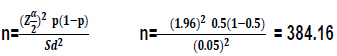

Where, P= is the expected prevalence, Sd = is standard deviation (desired absolute precision).
n = the total sample size
Accordingly, the calculated sample sizes was 384 samples.
Clinical examination
Physical examination for evidence of clinical mastitis was conducted in all lactating cows that were sampled in the study area. The udders of the selected cows were examined visually and by palpation for any presence of clinical mastitis. During examination, attention was paid to cardinal signs of inflammation, blindness, injuries, milk clots, symmetry, size, consistency of udder quarters and swelling (Radostits et al., 2007). A cow was considered to have clinical mastitis if it fulfilled at least two of the clinical findings, (1) pain reaction upon palpation, (2) changes in colour and consistency of milk (blood tinged milk, watery secretions, clots, pus) and (3) change in consistency of the udder (Lakew et al., 2009). Cows that did not have clinical mastitis were tested further for sub-clinical mastitis based laboratory investigation.
Milk sample collection and Laboratory investigation
According to Quinn et al. (2002) procedures of mastitis testing, the lactating cows’ milk samples were directly collected using universal sample collection bottles. The first 3-4 streams of milk were discarded. The collecting bottle was held as near horizontal as possible and by turning the teat to a near horizontal position and approximately 10 ml of milk were collected into the container. After collection, the sample was labeled and placed in ice box and transported to the Mizan Regional Veterinary Diagnostic Laboratory. The analysis was performed within two to three hours after sampling.
California mastitis test (CMT)
The CMT was conducted to diagnose the presence of subclinical mastitis (Quinn et al., 1999). Collected milk samples were poured in to four shallow cups in the CMT paddle and equal amount of CMT reagent was added to each cup and gentle circular motion was applied to the mixture on the horizontal plane. Based on the thickness of the gel formed by CMT reagent and milk mixture, test results were scored as 0 (negative), 1 (weak positive), 2 (distinct positive) and 3 (strong positive). Milk samples with test result of CMT 1 to 3, was classified as evidence of subclinical mastitis (Quinn et al., 1999; Radostits et al., 2007).
Bacterial isolation and identification
Bacteriological study was performed on milk samples from the sepositive CMT reactive and mastitis milk for culture. Identification of mastitis pathogens was carried out following microbiological procedures for diagnosis of bovine udder infection described in Quinn et al. (1999). One standard loop (0.01 ml) of milk was streaked on 7% blood agar. The inoculated plate was incubated aerobically at 37°C. The plates were checked for growth after 24, 48 and up to 72 h to rule out slow growing bacteria species. A milk sample was considered positive for mastitis pathogens if at least single colonies of a potential pathogen were detected and the positives were identified by biochemical tests. For primary identification, size, shape, color, hemolytic characteristics, Grams reaction and catalase production was used. For confirmation, biochemical tests were used after sub culturing isolated distinct colony on selective media. MacConkey agar (Oxoid) and Edward’s agar (Oxoid) were used to detect the most aerobic pathogens, enteric bacteria and Streptococci, respectively. Primary identification of Staphylococci was based on colony morphology, catalase test, Gram-staining morphology and differentiated from micrococci on the basis of the oxidative fermentative (OF) test carried out on semi-solid OF medium(Difco, Becton, Dickinson and Company, Franklin Lakes, NJ,USA). The Staphylococci were also tested for production of coagulase enzyme by the tube method as described by Quinn et al. (1994).
Isolates that produced Gram-positive cocci in clusters, and were catalase positive, glucose-fermentative, resistant to bacitracin and did not produce coagulase were identified as coagulase-negative staphylococci (CNS). S. aureus isolates were differentiated from other coagulase-positive staphylococci on the basis of mannitol fermentation on mannitol salt agar (Oxoid). The enteric bacteria were identified using colony morphology, oxidase test, lactose fermentation on MacConkey agar (Oxoid), indole production test, citrate utilization Quinn et al. (1999). Interpretation was made according to NMC (1990). The culture was considered negative if no growth occurs after 72 h of incubation and plates showing mixed and confluent growths, with no evidence of single discernible colonies, were not investigated further.
Data analysis
Data collected from the laboratory test and the questionnaire survey was recorded and coded in Microsoft excel spread sheets 2010 and analyzed using statistical data analysis of SAS version 9.10. The prevalence of mastitis was calculated as the number in study population testing positive divided by the total study units tested. The Chi-square (χ2) test was applied to determine existence of any association between the laboratory test positivity and the associated risk factors (such as breed type, parity, lactation stage and age of milking cows). For all analysis, a P-value of less than 0.05 was taken as significant.
RESULTS AND DISCUSSION
Prevalence of mastitis
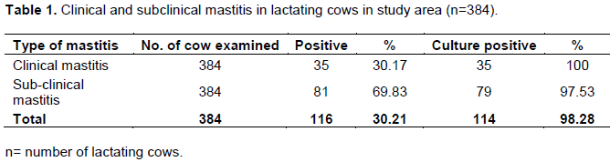
The overall prevalence of mastitis of cow level in the study areas were tested by using CMT and clinical inspection of the udder (Table 1). From the total 384 lactating cows examined during the study period, 116 (30.21%) cows had mastitis, of which 35 (30.17%) and 81(69.83%) showed clinical and subclinical mastitis, respectively. In the current study, the clinical and subclinical mastitis examined was higher than that reported by Alebachew and Alemu (2015) who found 21.2% clinical and 46.8% subclinical mastitis in selected commercial dairy farms in Addis Ababa. Jafer et al. (2016) also reported the minimum clinical mastitis (15.27%) and maximum subclinical mastitis (84.73%) in dairy farm of Dire Dawa City.
The present study showed an overall prevalence of 30.21% lower than that of Biffa et al. (2005) in and around Addis Ababa and Getahun (2006) in Haramaya who reported 38.9 and 36.9%, respectively. The difference in results could be due to variations in the distribution of mastitis risk, laboratory techniques, study design, climate, the level of management and animals studied. As indicted in the Table 1, all CMT positive samples were cultured for etiological agent identification. From 81 samples cultured, 79 were positive for known subclinical mastitis pathogens while all of the samples cultured from clinical mastitis were positive for mastitis.
The potential associated risk factors
Breed, parity, lactation stages and age had significant influence on the prevalence of bovine mastitis (P<0.05). The result showed that the prevalence of mastitis was significantly higher in cross breed (71.43%) than local breed of cows (28.65%) (Table 2). The effect of cross breed on the current prevalence of mastitis was relatively comparable with the reports of Jafer et al. (2016) in Dire Dawa city (71.1%). Compared to present study results, Belay and Tadele (2017) reported the lower prevalence in cross breeds (58.46%) and higher prevalence (38.2%) in local breeds in HoroGuduru Wollega Zone. In Ethiopia, many studies showed statistically significant difference in mastitis between local and cross breeds. Furthermore, cows with high milk yield is more susceptible to mastitis where as low-yielding cows tend to be more resistant (Biffa et al., 2005; Mekibib et al., 2010; Megersa et al., 2012; Moges et al., 2012). This may be due to genetic improvement for milk yield is accompanied by gradual decline in genetic resistance to mastitis (Radostits et al., 2008). Parity also showed an effect on the occurrence of mastitis. Higher prevalence (45.45%) was recorded in cows multiparous (greater than 5 parturition) and the lower prevalence (13.04%) was recorded in cows with first and second parity. Similarly, Alebachew and Alemu (2015) reported the higher prevalence (90.8%) in cows with 4-7 parturition and the lower prevalence (61.6%) in cows with 1-3 parturition in Addis Ababa.
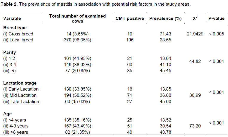
Lactation stage had association with the occurrence of mastitis were the prevalence was higher (45%) in late stage, followed by middle (36.60%) and early stages of lactation (13.85%). Belay and Tadele (2017) reported that the similar results based on the stage of lactation which was 34.21, 38.24 and 56.1% in the 1st, 2nd and 3rd trimester of lactation, respectively. The high prevalence of mastitis at late lactation might be due to an increased period of exposure of the udder during previous stages of early and mid-lactations.
There was a significant difference in prevalence between animals of different age of lactating cows (P<0.05). The highest prevalence (48.78%) was found in lactating cows of ages greater than 8 years, followed by cows of ages 4-8 years (30.54%) and the lowest prevalence (18.52%) was recorded in cows of ages less than 4 years. Correspondingly, Belay and Tadele (2017) reported that the highest prevalence (61.16%) of older cows (>9.5 years), followed by cows age 6-9.5 years (36.96%) and the lowest prevalence (34.15%) by cows age of 2.5-6 years. The high prevalence of the mastitis revealed in older animals might be due to the physiology of exhausted canal which is more dilated and remains partially open due to years of repeated milking. This facilitates the entrance of environmental and skin-associated microorganisms leading clinical or sub clinical mastitis. Blowey and Edmondson (2010) also reported the high occurrence of mastitis in older aged cows compared to young and adult cattle. This could be due to damage of teat canals in old animals facilitates access of bacteria into the mammary gland.
Identified and isolated major pathogens
As shown in Table 3, milk samples collected from 116 mastitis positive cows (35 clinical cows and 81 CMT positive subclinical cows) were cultured on blood agar. The dominant bacteria isolated were Staphylococcus species followed by Streptococcus species and other Gram negative enteric bacteria, of which Escherichia coli. By using further biochemical tests and selective media, three major strains of pathogenic bacteria namely S. aureus, S. Agalactiae and E. coli were found. Among three major pathogenic bacteria, S. aureus was the highest prevalent organism (59.26%); followed by S. Agalactiae (38.27%) and E. coli (2.47%).
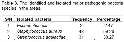
The prevalence of S. aureus in the present study was higher (59.26%) than early findings of Milne et al. (2002); Fufa et al. (2013) and Jafer et al. (2016) who reported 44.4% in Sebeta, 21.13% in Addis Ababa city and 48.4% in Dire Dawa, respectively. Likewise, this finding was disagreeing with the report of Bitew et al. (2010), Biruke and Shimeles (2015) who reported 20.3% in Bahir Dar and 45.1% in Addis Ababa, respectively. Present findings are comparable with the results of Endale et al. (2016) who reported 57.14% Staphylococcus species and 28.57% Streptococcus species in and around Sodo Town, Wolaita Zone, Ethiopia. The relative high prevalence of S. aureus in this study could be associated with the absence of post milking teat dipping, poor udder and teat washing before milking, poor hand milking practice and wide distribution of the organism inside the mammary gland and on the skin of teat and udder.
Available literature also showed that Staphylococcus species causing mastitis is the common and economically the greatest concern wherever dairy farming is practiced (Workineh et al., 2002; Fufa et al., 2013; Jafer et al., 2016). S. aureus has adopted to survive in the udder and established chronic and subclinical infection (Radostits et al., 1994). The isolation of S. aureus is of public health significance since it is a commonly recovered pathogen in outbreaks of food poising due to milk and milk product. This could be due to S. aureus is environmentally robust, surviving wide extremes of temperature and moisture. S. agalactiae (38.27%) was the second major pathogenic isolated in the study areas. This result was higher than the early findings of Kerro and Tareke (2003); Almaw (2004) and Bitew et al. (2010) who reported isolation rates of 13.1, 8.15 and 13.9%, respectively.
The justification given for S. aureus could also be a factor for S. agalactiae relative high isolation rate since both of them are contagious pathogens. The isolation of streptococcus species is of public health significance as it causes various gastrointestinal upset ranging from abdominal pain to diarrhea. Generally, the present study showed that contagious mastitis pathogens were the predominant isolated bacteria. This might be due to lack of effective udder and teat washing and drying, inter-cow hand washing and poor cleaning of milking area. Contamination of milkers’ hands, cloths and milking utensils leads to high spread of mastitis disease.
The present result also indicated that E. coli was the third predominant pathogens (2.47%) isolated in the study areas. This finding was much lower than the early findings of Iqbal et al. (2004) and Biruke and Shimeles (2015) who reported 18.6 and 40.7%, respectively.
However, it is comparable with the previous reports of Mekibib et al. (2010) at Holeta (4.6%) and Sori et al. (2005) in and around Sebeta (0.75%). The prevalence of E. coli is probably due to the fact that E. coli is the commonest environmental contaminants which are closely associated with hygiene. It becomes pathogenic whenever the hygienic conditions of the animal or environment become poor. In addition, the existence of high concentration of E. coli in milk also indicates the relatively poor quality of milk, related with substandard hygiene of the farm management.
CONCLUSION AND RECOMMENDATIONS
The overall 30.21% prevalence of mastitis at cow level was tested by using California Mastitis Test (CMT) and clinical inspection of the udder. Breed, parity, lactation stages and age have a significant influence on the prevalence of bovine mastitis (P<0.05). Increasing age, lactation stage, parity and poor management increased the risk of mastitis. The major pathogenic strains isolated were; S. aureus (59.26%), S. agalactiae (38.27%) and E. coli (2.47%). This indicates that mastitis caused by S. aureus is one of the major problems of dairy cows in milk production followed by S. agalactiae. The distribution of these bacterial pathogens in the herd indicates the economic impact of the disease. Beside the disease has economic importance it also to harm the health and well-being of human being. The professionals should apply different methods for prevention and strategic control of the disease and should be informed to the public about the relevance of pasteurization of milk before consumption to avoid food born infection and intoxication. There is a need of further study on drugs to which the bacterial are sensitive to use it used as primary choice to treat the disease in the study area.
CONFLICT OF INTERESTS
The authors have not declared any conflict of interests.
ACKNOWLEDGEMENTS
Sincere thanks are extended to the Mizan-Tepi University for supporting us with the financial budget required for the research work. Our special heartfelt gratitude and deepest appreciation goes to enumerators of each Woreda for their assistance, generous personal encouragement, willingness and cooperation during the period of data and samples collection. We extend our special thanks to Mizan Regional Veterinary Diagnostic Laboratory for allowing us to do the laboratory work in their Laboratory.
REFERENCES
|
Alebachew T, Alemu A (2015). Prevalence of bovine mastitis in lactating cows and its public health implications in selected commercial dairy farms of Addis Ababa. Global Journal of Medical Research: Global Veterinary Science and Veterinary Medicine 15(2):16-24. |
|
|
Almaw G (2004). A cross-sectional study of bovine mastitis in and around Bahir Dar and antibiotic resistance patterns of major pathogens. Addis Ababa University, Faculty of Veterinary Medicine DVM Thesis. |
|
|
Almaw G, Molla W and Melaku A (2009). Prevalence of bovine subclinical mastitis in Gondar town and surrounding areas, Ethiopia. Livestock Research Rural Development 21(7). |
|
|
Belay B, Tadele T. (2017). Epidemiology and financial impact of bovine mastitis in an animal production and research center and small holder dairy farms in Horo Guduru Wollega Zone, Western Ethiopia. Journal of Dairy Veterinary and Animal Research 5(4):8-9. |
|
|
Biffa D, Debela E, Beyene F (2005). Prevalence and risk factors of mastitis in lactating dairy cows in southern Ethiopia. International. Journal of Applied Research in Veterinary Medicine 3(3):189-198. |
|
|
Biruke D, Shimeles A (2015). Isolation and identification of major bacterial pathogen from clinical mastitis cow raw milk in Addis Ababa, Ethiopia. Academic Journal of Animal Diseases 4(1):44-51. |
|
|
Bitew M, Tefera A, Toles T (2010). Study on bovine mastitis in dairy farms of Bahir Dar town and its environs. Journal of Veterinary and Animal Advances 9:2912-2917. |
|
|
Blowey R, Edmondson P (2010). Mastitis Control in Dairy Herds (2ndedn) CAB. International, UK. |
|
|
Central Statistical Agency (CSA) (2015). Report on livestock and livestock characteristics (private peasant holdings). Agricultural Sample Survey. Volume V. Addis Ababa, Ethiopia pp.109-126. |
|
|
Central Statistical Agency (CSA) (2016/17). Report on livestock and livestock characteristics (private peasant holdings), Agricultural Sample Survey, Volume II. Addis Ababa, Ethiopia. |
|
|
Endale M, Eyob E, Addisu A, Naod T (2016). A Study on the prevalence of bovine mastitis and associated risk factors in and the surrounding areas of Sodo Town, Wolaita Zone, Ethiopia. Global Journal of Medical Research: Global Veterinary Science and Veterinary Medicine 16(2):16-24. |
|
|
Food and Agricultural Organization of the United Nations (FAO) (2017). Livestock production systems spotlight cattle sectors in Ethiopia. Africa Sustainable Livestock 2050. |
|
|
Food Hygiene Regulations (2006). A practical guide for milk producers to the food hygiene regulations, England 6 p. |
|
|
Fufa A, Gemechis F, Bekele M, Alemayehu R (2013). Bovine mastitis: prevalence, risk factors and bacterial isolation in small-holder dairy farms in Addis Ababa City, Ethiopia. Global Veterinaria 10(6):647-652. |
|
|
Getahun K (2006). Bovine mastitis and antibiotic resistance patterns of major pathogens in smallholder dairy forms in central high land of Ethiopia. MSc thesis: Debre-Zeit: Factory of Veterinary medicine, Addis Ababa university, Ethiopia. |
|
|
Iqbal M, Khan MA, Daraz B, Saddique U (2004). Bacteriology of mastitic milk and in vitro antibiogram of the isolates. Pakistan Veterinary Journal 24:161-164. |
|
|
Jafer K, Haimanot D, Hawi J, Tilahun Z, Girma K (2016). A Study on bovine mastitis, isolation and identification of staphylococcus species in Dairy Farms of Dire Dawa City, Eastern Ethiopia. Global Veterinaria 16(3):222-230. |
|
|
Kerro OD, Tareke F (2003). Bovine mastitis in selected areas of Southern Ethiopia. Tropical Animal Health Production 35:197-205. |
|
|
Lakew M, Tolosa T, Tigre W (2009). Prevalence and major bacterial causes of bovine mastitis in Asella, South Eastern Ethiopia. Tropical Animal Health and Production 41:1525-1530. |
|
|
Lemma M, Kassa T, Tegegene A (2001). Clinically manifested major health problems of crossbred dairy herds in urban and peri-urban production systems in the central high lands of Ethiopia. Tropical Animal Health and Production 33:85-89. |
|
|
Megersa B, Manedo A, Abera M, Regassa A, Abunna F (2012). Mastitis in lactating cows at Hawassa town: prevalence, risk factors, majorbacterial causes and treatment response to routinely used antibiotics. American-Eurasian Journal of Scientific Research 7(2):86-91. |
|
|
Mekibib B, Furgassa M, Abuna F, Megersa B, Regassa A (2010). Bovine mastitis: prevalence, risk factors and major pathogens in dairy farms of Holleta town, Central Ethiopia. Veterinary World 9(3):397- 403. |
|
|
Mekonnen H, Workineh S, Bayleyegne M, Moges A, Tadele K (2005). Antimicrobial susceptibility profile of mastitis isolates from cows in three major Ethiopian dairies. Medicine Veterinary 176(7):391-394. |
|
|
Milne MH, Barrett DC, Fitzpatrick JL, Biggs AM (2002). Prevalence and an etiology of clinical mastitis on dairy farms in Devon. Veterinary Record 158:241-243. |
|
|
Moges N, Hailemariam T, Fentahun T, Chanie, Melak A (2012). Bovine mastitis and associated risk factors in small holder lactating dairy farms in Hawassa, Southern Ethiopia. Global Veterinaria 9(4):441-446. |
|
|
National Mastitis Council (NMC) (1990). Microbiological procedures for the diagnosis of bovine udder infection.3rd. Arlington, Va, USA: National Mastitis Council Inc. |
|
|
Oviedo-Boyso J, Valdez A, Cajero-Juárez M, Ochoa-Zarzosa A, Meza-López-Meza JE, Patino A, Baizabal-Aguirre VM (2007). Innate immune response of bovine mammary gland to pathogenic bacteria responsible for mastitis. Journal Infection 54:399-409. |
|
|
Quinn PJ, Carter ME, Markey B Carter GR (1994). Clinical veterinary microbiology, Wilfe Publishing, London pp. 95-101. |
|
|
Quinn PJ, Carter ME, Markey B, Carter GR (1999). Clinical veterinary microbiology. Harcourt Publishers Ltd.: London. |
|
|
Quinn PBK, Markey ME, Carter, Donnelly WJC, Leonard FC and Maguire D (2002). Veterinary microbiology and microbial diseases.1st Published Blackwell Science 1td. Radostits OM, Blood DC, Gay CC (1994). Veterinary Medicine: A text book of the diseases of cattle, sheep, pigs, goats and horses. 8th ed. Bailliere Tindall: London 8:563-613. |
|
|
Radostits OM, Blood DC, Gay CC (1994). Veterinary Medicine: A text book of the diseases of cattle, sheep, pigs, goats and horses. 8th ed. Bailliere Tindall: London 8:563-613. |
|
|
Radostits OM, Gay CC, Blood DC, Hinchlif KW (2007). Mastitis In: Veterinary medicine 9thed., Harcourt Ltd, London pp. 174-758. |
|
|
Radostits OM, Gay GC, Blood DC, Hinchillif KW (2000). Mastitis In: Veterinary Medicine, 9th Edition, Harcourt Limited, London pp. 603- 700. |
|
|
Radostits OM, Gay CC, Hinchcliff KW, Constable PD (2008).Veterinary Medicine; Text books of the disease of Cattle, Sheep, Pigs, goats and Horses.(10thedn), Elsevier, UK. pp. 673-680. |
|
|
Seegers H, Fourichon C, Beaudeau F (2003). Production effects related to mastitis and mastitis economics in dairy cattle herds. Veterinary Research 34(5):475-491. |
|
|
Sori H, Zerihun A, Abdicho S (2005). Dairy Cattle Mastitis In and Around Sebeta, Ethiopia International Journal of Applied Research in Veterinary Medicine 3:332-338. |
|
|
Suriyasathaporn W, Schukken YH, Nielsen M, Brand A (2000). Low somatic cell count: a risk factor for subsequent clinical mastitis in dairy herd. Journal of Dairy Science 83:1248-1255. |
|
|
Teshome G, Tesfaye A (2016). Physicochemical properties and microbial quality of raw cow milk produced by smallholders in Bench Maji-Zone, Southwestern Ethiopia. Food Science and Quality Management 55:55-62. |
|
|
Thrusfield M (2007). Veterinary epidemiology 3rd edition, Blackwell Publishing. |
|
|
Workineh S, Bayleyegn M, Mekonnen H, Potgieter LND (2002). Prevalence and an etiology of mastitis in cows from two major Ethiopian dairies. Tropical Animal Health and Production 34:19-25. |
|
Copyright © 2024 Author(s) retain the copyright of this article.
This article is published under the terms of the Creative Commons Attribution License 4.0


