
ABSTRACT
Hip dysplasia in cats appears to have a similar prevalence as that seen in dogs especially with large breeds of cats like the Maine coon. However, cats do not commonly show signs of lameness like dogs do. This study was to determine the prevalence of hip dysplasia in domestic shorthair breed of cats in three municipal districts in Accra, Ghana. Twenty (12 males and 8 females) adult (2.3 ± 0.8 years) shorthair breed of cats of both gender weighing 1.7 ± 0.4 kg were included in this study. They were sedated with xylazine and atropine injection prior to radiography. Extended and frog-leg ventro-dorsal radiographs were taken using a manual processing x-ray machine. Norberg angles (NA), joint space scores and subjective morphological alterations for each hip were assessed. Comparisons were made for the NA scores within gender, between gender, between the right and left hips using Independent t-test. The relationship between the joint space scores and weight, age or gender of the cats was assessed. The morphological changes were also assessed. Mean manual Norberg angle measurements for 19 cats were 93.67 ± 1.9° and mean digital Norberg angle measurements for 19 cats was 102.35 ± 1.1°. One cat with complete cranio-dorsal luxation, due to history of preceding accident had manual Norberg angle of 38° and digital Norberg angle of 45°. Two cats displayed mild subluxation of the hip joint. There was no considerable difference in the NA or joint space measurements. This study found a ten percent prevalence of hip dysplasia in domestic shorthair breed of cats in the three Municipal Districts in Accra, Ghana. Both manual and digital methods for measuring the NA provided results which were within similar ranges.
Key words: cats, hip dysplasia, radiographs, prevalence, shorthair, domestic.
INTRODUCTION
Hip dysplasia is an abnormal development of the hip joints affecting humans, large and small animals. It can occur unilaterally but it is mostly bilateral. It presents with varying degrees of laxity of the joint ligaments, abnormal changes of the femoral head and acetabulum leading to secondary osteoarthritis (Allan and Davies, 2018). Hip dysplasia is an inherited disease of dogs (Alexander, 1992; Smith et al., 1990). Gierson (2012) proposed a lack of well-defined genetic association of hip dysplasia in cats however, studies by Low et al. (2019) showed a correlation between older and heavier cats having the tendency of severe feline hip dysplasia affecting both limbs and younger and lighter cats having mild conditions which often affect one hip. In cats, joint laxity may not play the same role in the pathogenesis of hip dysplasia as in dogs due to the shallow acetabulum usually found in cats. Subluxation of the femoral head is not consistently seen and there is minimal remodeling of the femoral head (Keller et al., 1999). The condition affects all breeds of dogs and it appears to be higher in large breeds compared with small breeds of dogs. The occurrence among breeds can be from as high as 70.5% in Bulldogs and 48.2% in St. Bernards to as low as 1.9% in Borzois (Corley and Keller, 1993). Even though hip dysplasia occurs in toy breeds of dogs as well as cats, their affected hips rarely produce the bony changes commonly seen in heavier dogs (Brinker et al., 2006). Hip dysplasia can occur in any breed of cat. The occurrence in domestic shorthair cats was 10.4%; derived from a mixed breed population of 899 cats (Keller et al., 1999). However, it is as high as 24.9% percent in purebred cats like the Maine Coon (Loder and Todhunter, 2017).
Hip dysplasia in companion animals can be screened qualitatively by identifying abnormal radiographic changes on the acetabulum, the head or neck of the femur (Flückiger, 2007). The commonly used screening methods include the Orthopaedic Foundation for Animals (OFA), British Veterinary Association/Kennel Club (BVA/KC) and Federation Cynologique Internationale (FCI). These methods describe the tightness or laxity of the hip joint and deviations from the ideal ball and socket conformations; these may include mild to severe osteoarthritic changes (Dennis, 2012). Computed Tomography (CT), Magnetic Resonance Imaging (MRI) and ultrasonography are other less commonly used methods of diagnosis (Phillip and Harry, 2012; Guillot et al., 2012). Common objective measurements used in the assessment of coxofemoral joints for presence or absence of hip dysplasia include the Norberg angle (NA) and the Distraction index (DI) (Allan, 2000). Norberg angle is used to measure hip joint laxity in research studies and used as a selection criterion in the canine registry databases of the prevalence of hip dysplasia by breed (Tomlinson and Johnson, 2000). The NA is measured when a line drawn from the cranial acetabular edge to the center of the respective femoral head intersects a line drawn between the two femoral heads. The NA varies with the depth of the acetabulum and the amount of coxofemoral laxity observed on the radiograph. The NA measurement can be carried out manually or digitally. The DI is a unit less index of passive coxofemoral joint laxity that is calculated from compression and distraction radiographs of the coxofemoral joints. Low DI values correspond with low radiographic signs of degenerative joint changes.
Some studies have shown that the DI values are a more reliable predictor of degenerative joint disease (DJD) in hip dysplasia than the Ortolani sign, Norberg angle, or OFA measurement of hip conformation (Smith et al., 1993, 1995). Hip dysplasia and subsequent osteoarthritis are clinical problems in cats, however, the prevalence in domestic shorthair breed of cats in Accra, Ghana is not yet known. The purpose of this study was to determine the prevalence of hip dysplasia in the domestic shorthair breed of cats in Accra, Adenta and Ledzokuku municipal districts of Accra, Ghana and to identify which measuring method (manual or digital) provided a more reliable result.
MATERIALS AND METHODS
This study was approved by the institute of Animal Care, Use and Research Ethics Committee of the University of Ibadan (UI-ACUREC/19/0050) and informed consent forms were administered on all the owners. Twenty, 12 males and 8 females, apparently healthy client-owned domestic shorthair adult cats with mean age of 2.3 ± 0.8 years and mean weight of 1.7 ± 0.4 kg were included in this study. They were patients presented at The Vet Place, Achimota in Accra with no previous history of musculoskeletal problems for the past year. They had no history of castration or spaying.
The cats were sedated with intramuscular injections of 0.5 mg/kg of xylazine (Xylazine 2% Inj®, Dutch Farm International BV, Holland) and 0.04 mg/kg of atropine (Atrowell®, Wellona Pharma, India). An extended and frog-leg ventrodorsal view of the hip joint were taken for each cat using a manual processing X-ray machine (TUR® manual processing X-ray machine, Dresden). The extended and frog-leg ventrodorsal radiographs were qualitatively graded as dysplastic or normal based on the Orthopaedic Foundation for Animals scoring system. Qualitatively determined subluxations without any signs of osteoarthritis were graded as mild hip dysplasia (Flückiger, 2007).
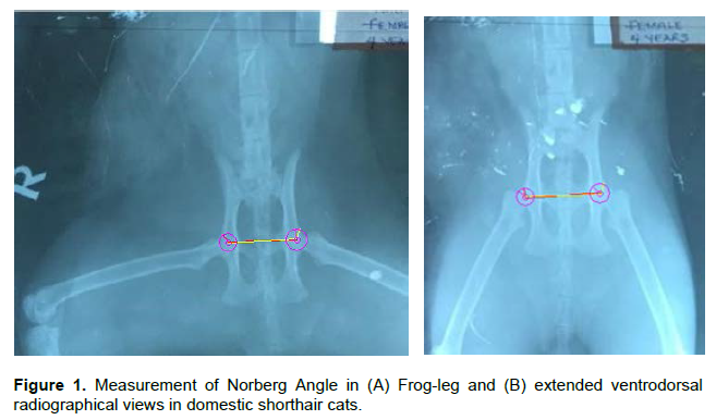
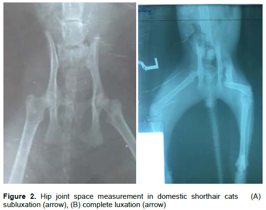
The radiographic images were also analyzed using the Norberg angle measurements for the right and left hips. A line was drawn from the cranial acetabular rim of the right hip to the center of the femoral head of the right limb. This line then transects a line drawn between the two femoral heads. This was repeated for the left hip. The angle measured is the Norberg angle (Peiffer et al., 1974). As shown in Figure 1, digital Norberg angle measurements were made with Digimizer®. The joint space for each hip was measured using a digital Vernier caliper from the cranial acetabular edge to the head of the femur facing it. Subluxation and complete luxation of the hip joint is presented in Figure 2.
Data was presented as mean and standard deviation. The Pearson’s correlation test was done to determine any relationship between the coxofemoral joint space scores with mean age, weight and gender. The Independent t-test was used to define statistically significant differences in the measurements between Norberg angle scores between gender, within gender, between right hip joints and between left hip joints as well as between manual and digital Norberg angle scores. A P-value of <0.05 was significant.
RESULTS
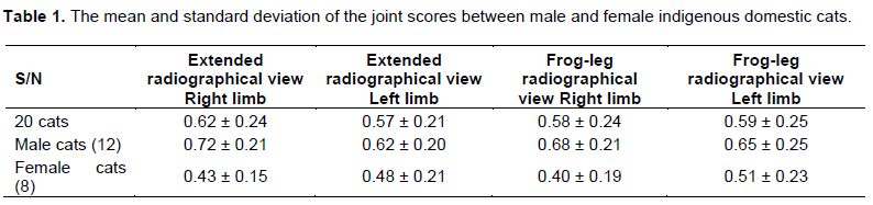
The results of this study revealed a 10% (2/20) prevalence of hip dysplasia in domestic shorthair breeds of cats of the Accra, Adentan and Ledzokuku municipal areas of Accra, this excludes the cat with complete luxated hip due to an automobile accident. The mean and standard deviation of the joint scores between male and female indigenous domestic cats are shown in Table 1. There was no considerable difference between the measurements of the hip joint space scores.
The Pearson’s correlation coefficient between coxofemoral joint scores and gender (-0.035), weight (0.191) and age (0.136) show there was a weak negative association between coxofemoral scores and gender, though it was not significant (0.888). There was a positively weak association between coxofemoral scores with weight (0.420) and age (0.568), though they were not statistically significant. The P-value was set at P <0.05.
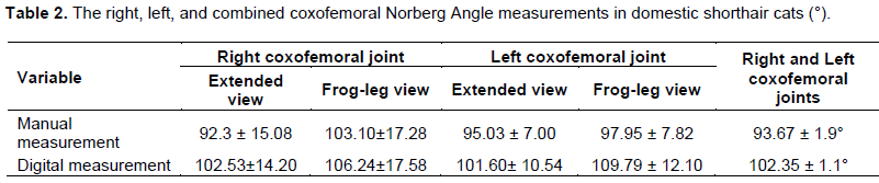
The acetabulum, head and neck of the femur were assessed for any deviation from normal congruency and appearance using the orthopaedic Foundation for Animals scoring system. One cat had a luxated right hip joint. Two cats had a subluxated hip joint each. The mean and standard deviation of the Norberg angle for the right, left and combined hip joints were shown in Table 2. There was no significant difference (P < 0.05) in the manual and digital methods of measuring the hip joint in both the extended and frog-leg radiographs.
DISCUSSION
The results of this study revealed two cases of subluxation of the hip joint without any osteoarthritic changes to the bones. This may be attributed to their low body weight which does not cause remodeling of the coxofemoral joint, this is in agreement to the findings of Brinker et al. (2006) that hip dysplasia in toy breeds of dogs and cats rarely produce bony changes in the affected hips. There was no significant difference when comparing the measurements for each pair of hips. The variables (Norberg angle scores) derived manually and digitally for all the cats were also similar. This indicates that both left and right hip are evenly predisposed to hip dysplasia and agrees with findings by Low et al. (2019).
Allan and Davies (2018), explained that radio-graphically, joint space widening is not easily identified in small animals; this may be because dogs and cats cannot be X-rayed in a standing position or due to inconsistencies between the direction of the X-ray beam and the joint space. This is similar to results obtained in this study. The joint space scores were compared with regards to gender, age, and weight. This study showed the joint space values were all less than 1 mm apart from the luxated limb. This may be the beginning phase of hip dysplasia in contrast to chronic hip conditions where the joint space capsule can be as thick as 5-7 mm in a bid to stop femoral luxation (Morgan et al., 2000).
The variables (Norberg angle scores) derived were analyzed using the Independent t-test. The mean values for Norberg angles using manual measurements in this study was 93.67 ± 1.9°; and 102.35 ± 1.1° using the digital measurement, which agrees with studies by Ana et al. (2018); whose studies revealed cats with perfect congruency had mean Norberg angles of 102.6°. This study also agrees with findings of 84 ± 10° to 95 ± 5° by Langenbach et al. (1998) and between 89.75° to 93.02° by Milken (2007) for dysplastic hips. It also agreed with studies by Kimura et al. (2012) whose studies showed a Norberg angle of 84.96 ± 6.78° for dysplastic hips and 97.46 ±11.52° for normal hips. However, the luxated hip in this study had a value of 38° (manual measurement) and 45.862° (digital measurement). This also agrees with studies by Clarke and Bennet (2006) which show low angles for dysplastic or luxated hips. There was no influence of gender on the variables derived. This is similar to results in dogs and agrees with studies by Keller and Corley (1996), Ana et al. (2018) and Loder and Todhunter (2017) but did not conform with results obtained in studies by Hayes et al. (1979) and Koeppel and Ebner (1990) who proposed hip dysplasia was more prevalent in females.
In conclusion, the results of our study showed there was no advantage of the digital method of measurement over the manual method. Both can be used to derive reliable values for the detection of hip dysplasia in cats. The actual incidence of hip dysplasia in indigenous domestic cats in Accra may well be higher than this study shows; hence, there is need for further research with larger population sample in this subject area.
CONFLICT OF INTERESTS
The authors have not declared any conflict of interests.
AKNOWLEDGEMENT
The authors sincerely appreciate the support of Dr. Selom Tettey and the entire staff of The Vet Place, Achimota, Accra Ghana.
REFERENCES
|
Alexander JW (1992). The pathogenesis of canine hip dysplasia. Veterinary Clinics of North America: Small Animal Practice 22(3):503-511. |
|
|
Allan GS (2000). Radiographic features of feline joint diseases. Veterinary Clinics of North America: Small Animal Practice 30(2):281-302. |
|
|
Allan G, Davies S (2018). Radiographic sSigns of jJoint dDisease in dDogs and cCats. Textbook of Veterinary Diagnostic Radiology pp. 403-433. |
|
|
Ana CVH, Danilo M, Ana CSS, Paula F, José FI (2018). Incidence of hHip dysplasia in dDomestic cCats - sStudy with 86 aAnimals. Biomedical Journal of Scientific and Technical Research 9(1). |
|
|
Brinker D, Piermattei G, Flo's C (2006). Handbook of small animal orthopedics and fracture repair. Fourth Edition, Elsevier Inc, United States of America pp. 475-511. |
|
|
Clarke SP, Bennett D (2006). Feline osteoarthritis: a prospective study. Journal of Small Animal Practice 47(8):439-45. |
|
|
Corley EA, Keller GG (1993). Hip dDysplasia: A pProgress rReport and uUpdate. Columbia, Missouri: Orthopedic Foundation of Animals (suppl) |
|
|
Dennis R (2012). Interpretation and use of BVA/KC hip scores in dogs. In Practice 34(4):178-194. |
|
|
Flückiger M (2007). Scoring radiographs for canine hHip dDysplasia -The big three organisations in the world. European Journal of Companion Animal Practice 17(2):135 -140 |
|
|
Gierson J (2012). Hips, elbows and stifles: common joint diseases in the cat. Journal of Feline Medicine and Surgery 14(1):23-30. |
|
|
Guillot M, Moreau M, d'Anjou MA, Martel-Pelletier J, Pelletier JP and Troncy E (2012). Evaluation of osteoarthritis in cats: novel information from a pilot study. Veterinary Surgery 41(3):328 -335. |
|
|
Hayes HM, Wilson GP and Burt JK (1979). Feline hip dysplasia. Journal of the American Animal Hospital Association 15(4):447-448. |
|
|
Keller GG, Corley EA (1996). Hip dysplasia: Orthopedic Foundation for Animals: Data on the Maine Coon Cat. Maine Coon Breeders and Fanciers Association Scratch Sheet P 18. |
|
|
Keller GG, Reed AL, Lattimer JC, Corley EA (1999). Hip dysplasia: a feline population study. Veterinary Radiology and Ultrasound 40(5):460-464. |
|
|
Kimura T, Kimura S, Okada J, Suzuki S, Kitanaka T (2020). Retrospective radiographic study of degenerative joint disease in cats: Prevalence based on orthogonal radiographs. Frontiers in Veterinary Science 7:138. doi.org/10.3389/fvets.2020.00138 |
|
|
Koeppel E, Ebner J (1990). Hip dysplasia in the cat [article in German]. Kleintierpraxis 35:281-298 |
|
|
Langenbach A, Giger U, Green P, Rhodes H, Gregor TP, LaFond E and Smith G (1998): Relationship between degenerative joint disease and hip joint laxity by use of distraction index and Norberg angle measurements in a group of cats. Journal of the American Veterinary Medical Association 213(10):1439-1443 |
|
|
Loder RT, Todhunter RJ (2017). Demographics of hip dysplasia in the Maine Coon cat. Journal of Feline Medicine and Surgery 20(4):302-307. |
|
|
Low M, Eksell P, Högström K, Olsson U, Audell L and Ohlsson A (2019). Demography, heritability and genetic correlation of feline hip dysplasia and response to selection in a health screening programme. Scientific Reports Nature Research 9(1):1-9. |
|
|
Milken VM (2007). Estudoradiográfico comparative da dysplasia coxofemoral entre gatos da raçapersa e semraçadefinida. Tese (DoutoradoemMedicinaVeterinária) - Faculdade de Medicina Veterinária e Zootecnia da UNESP, Botucatu pp. 29-40. |
|
|
Morgan JP, Wind A, Davidson AP (2000). Hereditary bone and joint diseases in the dog: osteochondroses, hip dysplasia, elbow dysplasia. Hannover, Germany: Schlutersche pp. 109-208. |
|
|
Peiffer RL, Young WO, Jr, Blevins WE (1974). Hip dysplasia and pectineus resection in the cat. Feline Practice 4:40-43. |
|
|
Phillip W, Harry S (2012). Conditions of the feline pelvic region. In Practice 34(9):498-511. |
|
|
Smith GK, Biery DN, Gregor TP (1990). New concepts of coxofemoral joint stability and the development of a clinical stress-radiographic method for quantitating hip joint laxity in the dog. Journal of American Veterinary Medical Association 196(1):59-70. |
|
|
Smith GK, Gregor TP, Rhodes WH, Biery DN (1993). Coxofemoral joint laxity from distraction radiography and its contemporaneous and prospective correlation with laxity, subjective score, and evidence of degenerative joint disease from conventional hip-extended radiography in dogs. American Journal of Veterinary Research 54(7):1021-1042 |
|
|
Smith GK, Popovitch CA, Gregor TP, Shofer FS (1995). Evaluation of risk factors for degenerative joint disease associated with hip dysplasia in dogs. Journal of the American Veterinary Medical Association 206(5):642-650, |
|
|
Tomlinson JL, Johnson JC (2000). Quantification of measurement of femoral head coverage and Norberg angle within and among four breeds of dogs. American Journal of Veterinary Research 61(12):1492-1500. |
|
Copyright © 2024 Author(s) retain the copyright of this article.
This article is published under the terms of the Creative Commons Attribution License 4.0


