Full Length Research Paper

ABSTRACT
A cross-sectional study was conducted from November, 2013 to April, 2014 in pastoral areas of Oromia and Somali regional states to determine the prevalence of brucellosis in small ruminants and assess associated risk factors. The multistage sampling technique was used on total population in the selected district during the study period. A total of 420 serum samples were collected from 129 sheep and 291 goats in extensive management system, with no previous vaccination history. Of 420 sera examined, 36 (8.5%) were positive to Rose Bengal plate test (RBPT). The sera screened positive by RBPT were retested using complement fixation test (CFT) and among 36 sera sample tested, 15 (3.6%) were positive for brucella antibodies. The prevalence of brucellosis among sheep and goats was found to be 2 (0.48%) and 13 (3.09%), respectively. The results of the present study showed that there was no significant difference in seroprevalence to Brucella antibodies and species, sex and age of the animals examined (p > 0.05). The occurrence of brucellosis among small ruminants in selected districts could pose productivity and reproductive problem in addition to public health risk. Thus, implementing control measures and raising public awareness on prevention methods of brucellosis should be suggested.
Key words: Brucellosis, complement fixation test, Ethiopia, Rose Bengal plate test, pastoral areas, small ruminant.
INTRODUCTION
Ethiopia is one of the developing countries with domestic small ruminant population estimated to be 26.1 million sheep and 21.7 million goats (Community-supported Agriculture (CSA), 2006). Small ruminants are the chief source of cash income to small holders (EPAIAT, 2003; Akbarmehr and Ghiyamirad, 2011). This is because sheep and goats provide rapid cash turn over (Corbel Center for food security and Public Health and OIE, 2009; Godfroid et al., 2011). Most of the sheep and goat populations in Ethiopia are raised under pastoral conditions. These small ruminants and their milk/meat products represent an important export commodity, which significantly contributes to the National economy. There is also a growing export market for sheep and goats meat in the Middle Eastern Gulf states and some African countries. At optimum off take rates, Ethiopia can export 700,000 sheep and 2 million goats annually, and at the same time supply 1,078,000 sheep and 1,128,000 goats for the domestic market (Alemu and Markel, 2008).
Even though the animals contribute much to the National economy, its development is hampered by different constraints. The most important constraints to small ruminant productions are poor management system, low genetic endowment and widespread endemic diseases including parasitic infestation, viral and bacterial diseases. Among many factors that limit economic return from small ruminants, reproductive diseases including brucellosis are the major disease constraints found in pastoral areas (International Livestock Research Institute (ILRI), 2006).
Brucellosis is a highly contagious and important zoonotic disease which particularly impedes international trade (Refai, 2002). It is caused by different species of the genus Brucella, a small, gram negative, non-motile, non spore forming, rod shaped (Coccobacilli) bacteria (Amenu et al., 2010; Kaoud et al., 2010) that are pathogenic for a wide variety of animals and also for humans (Mantur and Amarnath, 2008). In animals, it mainly affects reproduction and fertility, reduces the survival of newborns and diminishes milk yield. In human beings, the symptoms of disease are weakness, joint and muscle pain, headache and undulant fever (McDermott and Arimi, 2006).
It is an important disease of both livestock and people in sub-Saharan Africa (Radostits et al., 2008). The disease has much significance due to its transmission between animals and human through animal products and by products (OIE, 2004). Brucella melitensis (biovars 1, 2 or 3) is the main causative agent of caprine and ovine brucellosis and it is highly pathogenic for humans causing one of the most serious zoonoses in the world (OIE, 2008; Ragassa et al., 2009). All infected tissues, cultures and potentially contaminated materials should therefore be handled with great care (Alton et al., 1975).
Despite being endemic in many developing countries, brucellosis remains under diagnosed and under-reported. Furthermore, since brucellosis is an important cause of veterinary morbidity and mortality, the disease can also cause important economic losses in developing countries (Radostits et al., 2008). Even though the disease is endemic in the country, especially in pastoral areas, very limited researches have been done on small ruminant brucellosis. Hence, the study was designed to determine the seroprevalence of small ruminant brucellosis and assess potential risk factors in the selected districts of Oromia and Somali regional states of South Eastern Ethiopia.
MATERIALS AND METHODS
Study areas
The study areas were the pastoral areas located in Somali and Oromia National states, where most of the pastoralists of Ethiopia live. Borana Zone is found at 724 km South of Addis Ababa. It is one of the Oromia Regional States located at 3° 36’ North 3° 43’ East. The total human population of the district (Woredas) is estimated to be 43,837 (CSA, 2011) whereas small ruminant population was sheep (100, 261) and goat (99,201) (CSA, 2006). The average annual rainfall ranges from 400 to 700 mm. The mean daily temperature is 25 to 44°C. Livelihood of the people is largely dependent on livestock and livestock products, subsequent food and water shortage for the settled and mobile population of the area. Whereas, Somali districts are found around 820 km East of Addis Ababa, Somali regional states of Ethiopia, part of Liban Zone and located in coordinates 4° 25’ North, 41° 25’ East. The zone is bounded by the confluence of the Ganale Dorya with Dawa river by Afder Zone on the South West by Somalia and on the South by Kenya. The altitude of Woreda ranges from 200 to 1000 m above sea level, with an average daily temperature of 29 to 45°C. The Woreda has a total human population of 37,404, sheep (380,030) and goats (436,099). The rainfall pattern can be characterized as erratic, unpredictable and unreliable, with average rainfall of 200 mm. The livelihood is largely dependent on livestock and livestock products, with subsequent food and water shortage for the settled and mobile population of the area. Elevation of this Woreda ranges from 500 to 1500 m above sea level, with average daily temperature of 28 to 44°C. The total human population of these districts accounts for 66,495, with sheep (180,000) and goats (420,000).
Study animals
Animals (ovine and caprine) of both sexes, different age groups greater than six months and no history of vaccination against brucellosis, kept under the extensive whereas, small ruminants which were diseased and who have had history of vaccination against brucellosis were excluded.
A cross-sectional study was conducted from November, 2013 to April, 2014 in the study areas to determine the sero-prevalence of small ruminant brucellosis and assess potential risk factors for the transmission and spread of the disease. The cross sectional study design measures all variables on participants at same point in time and prevalence of the disease but not the incidence.
Sampling and sample size determination
Multistage sampling technique was used according to Dohoo et al. (2003) in the survey of small ruminant (sheep and goat) brucellosis. The peasant association (PA) was considered as primary unit, the herds as secondary units and individual animals as tertiary units. Sheep and goat herd in 8 PAs from four districts (2 kebeles per District) were sampled during the study based on the livestock population of each district. In order to determine the desired sample size, there were no previous reports of prevalence in the districts. The average expected prevalence rate was assumed to be 50% for the area within 95% confidence intervals (CI) at 5% desired accuracy as stated by Thrusfield (2007) formula:
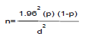
Where n = sample size; p = expected prevalence; d = desired level of precision (5%). However, the sample size was 420 to increase the representativeness of the samples to the wider population. Hence, n = 420 goats and sheep were sampled by considering 10% non respondent rate. Sampling was proportionally distributed based on the total small ruminant population in the study districts and accessibility to road for peasant association (PAS).
Sampling procedures
Blood samples were collected from a total of 420 study animals in the study areas during the study period, while laboratory analysis of specimens was made in National Animal Health Diagnostic and Investigation Center (NAHDIC). Essential materials that were used for sample collection and transportation were offered by the research institute. The blood samples were collected from the jugular vein of the animals aseptically. About 5 to 7 ml of blood was collected from sheep and goats through sterile vacutainer test tube and venoject needle. Immediately, each animal was tagged and the respective blood samples were labeled accordingly. This blood was let down to clot for about 2 to 3 h in room temperature then the clotted blood samples were stored at 4°C till serum extraction, usually within 24 h. Then, sera were extracted and dispensed into cryovials in NAHDIC and serum storage was made at -20°C. Then each serum samples were subjected to the laboratory test through the OIE (2004) recommended diagnostic tool.
Serological test procedures
RBPT was performed in NAHDCI on all sera samples collected as per the procedure described by Alton et al. (1975) and OIE (2004). The antigen was obtained from Institute Pourquier, Montpellier, France. The test was conducted in National Animal diagnosis and Investigation center (NADIC) in Sebeta Veterinary laboratory. The interpretation of the results was done according to the degree of agglutination.
Complement fixation test (CFT)
Sera samples found positive by RBPT were further tested by CFT at NAHDCI, Sebeta, Ethiopia, according to the protocol described in OIE Manual (2004). The CFT is the test approved by the World Organization for Animal Health (OIE) as the definitive test for further confirmation.
The data collected in the field were entered into a computer on a Microsoft Excel spreadsheet. Statistical analysis (multivariate logistic regression) was performed using ‘Statistical package for the social sciences’ (SPSS), version 20. Categorical variables (species, sex, age and area) were expressed in percentages. The prevalence proportion was calculated as the number of animals testing positive by the RBPT/CFT, divided by the total number of animals tested. The association between each risk factor and the outcome variable were assessed using the Chi-square test. For all analyses, a p-value of less than 0.05 was taken as significant.
Before any attempt to collect data, the protocol was approved by Institutional Review Board (IRB) of School of Veterinary Medicine, College of Medical and Health Sciences, Wollega University. Official permission was also obtained from animal owners and Agricultural Administration Office of the districts (Woredas). Moreover, the guideline was also used.
RESULTS
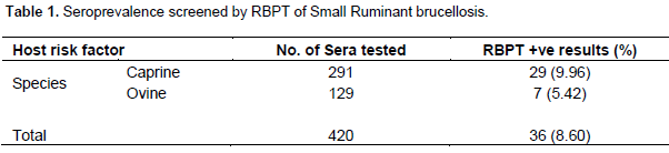
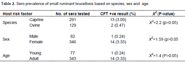
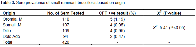
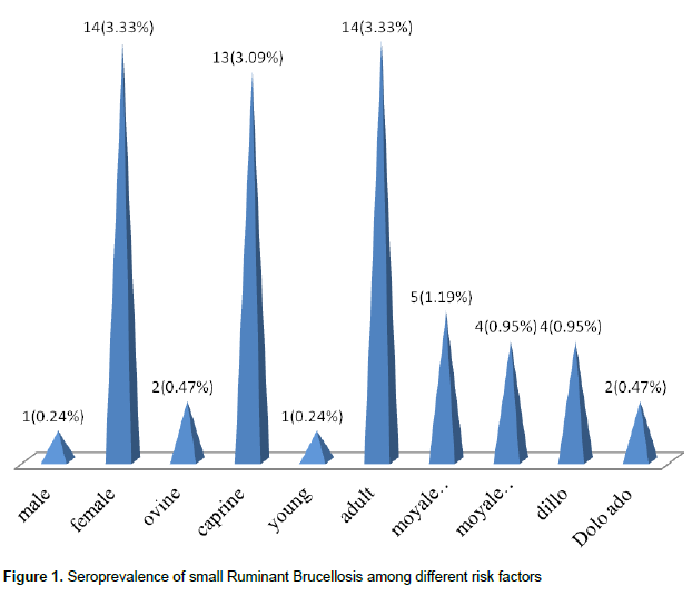
Out of 420 small ruminant sera tested, 36 (8.60%) sera were positive by RBPT. Among these, sheep (ovine) and goat (caprine) account for 7 (5.42%) and 29 (9.96%), respectively (Table 1). The results were further confirmed by CFT, where 15 (3.6%) were positive for small ruminant brucellosis during the study period (Tables 2 and 3). In the present study, statistical analysis of the data showed that there was no significant difference between the brucellosis and potential host risk factors (species, sex and age) of the examined animals (p > 0.05). However, significant difference was observed between the disease and origin of the animals (p < 0.05) (Table 3). The prevalence of small ruminant brucellosis in various origins was indicated (Figure 1).
DISCUSSION
The present study indicated the overall seroprevalence of small ruminant brucellosis in Oromia and Somali regional states to be 36 (8.60%) by the RBPT and 15 (3.6%) CFT. The result revealed a moderate prevalence and natural transmission of Brucella organisms in the study area. The finding was in line with the previous studies conducted by Omer et al. (2000) in Eritrea who reported, 3.8% in goat and in imported sheep by Refai (2002) in Iran who reported 3%. However, the result was higher than the result reported in Borena by Teshale et al. (2006), with 1.09% sheep in Yabello; Megersa et al. (2010), with 1.56% in Borena; Bekele et al. (2011), with 3.2% in Somali. On the other hand, the finding was lower than that reported by Wesinew et al. (2013), with 4.8% and Ashenafi et al. (2007), with 11.6% in Afar region, in the low lands of Ethiopia; Waghela (1976), with 6.01% in sheep, 6.01% in goat in Kenya; El-Ansary et al. (2001), with 14.2% in sheep, 16.2% in goat in Sudan. This difference could be due to various factors such as differences in diagnostic assay, sampling technique, study area and sample size used.
Higher prevalence was observed in goats (3.09%) than in sheep (0.47%). This finding is lower than the reports of PFE (2004), with 14.2% sheep, 16.72% in goat and Benkirane (2006), with 7.2% in sheep and 5.29% in goat. Goats are at higher risk of acquiring Brucella infection than sheep. This may be due to the greater susceptibility of goats to Brucella infection and also excreting the organism for a long period, unlike sheep; this reduces the potential for disease spread among sheep flocks.
Little difference was also recorded in the prevalence of brucellosis between adults and young animals. The prevalence in adult age is higher (3.3%) than young age (0.24%). It has been reported that brucellosis is essen-tially a disease of sexually mature animals (El-Ansary et al., 2001). Sexually mature and pregnant animals are more prone to Brucella infection and brucellosis than sexually immature animals of either sex (Walker, 1999). On the other hand, it is also true that younger animals tend to be more resistant to infection and frequently clear an established infection, although latent infections can occur. This might be due to the fact that sex hormones and erythritol, which stimulate the growth and multiplica-tion of Brucella organisms, tend to increase in con-centration with age and sexual maturity (Walker, 1999).
In this study, there was significant difference between the disease and origin of the animals (p < 0.05). Higher prevalence was found in Moyle Oromia (1.16%) and low prevalence was found in Dolo Ado (0.93%). This may be due to the difference in animal management system, sample size, presence of carrier animal in the origin of the region and population density of small ruminant in the area. Brucellosis is, therefore, well entrenched across the entire regions of Oromia and Somali. This might be attributable to the use of similar animal production and management systems throughout the districts of the region as well as similar agro-ecological conditions. Moreover, unrestricted animal movements may have enhanced the spread of infection, such as: Movements of animals in search of pasture and water/nomadic move-ment within the animal in search of feeding across the countries, trade within and between zones and districts, the mixing of animals at marketplaces and watering points. Accordingly, Quinn et al. (1999) found that the prevalence of small-ruminant brucellosis was higher, at points such as river and grassing land, especially during the dry season, in the regions, zones and district where there was frequent mixing of flocks.
The study indicated that RBPT which is based on B. abortus antigen was less sensitive in detecting antibodies against Brucella melitensis (Yibeltal, 2005). Almost half of the sera were found to be tested positive for anti-Brucella antibodies by RBPT and negative by CFT. This could be due to cross-reactions between Brucella and other bacteria which share similar epitopes. It might also be due to variations in animal management and production systems.
CONCLUSION
The study revealed that brucellosis is a widespread and well-established infection among goats and sheep in the study areas. The sero-prevalence of brucellosis was higher in goats than sheep, as well as female, adult aged and animals within dense and large herd size. It could be concluded that the positive animal can be a potential risk factors to the free disease animals in the areas, unless the management system is improved. Thus, the author recommend that the ongoing veterinary extension program for the community should be strengthened in order to effectively control the animal movement for successful prevention and control of the disease.
ACKNOWLEDGEMENTS
The authors would like to thank Wollega University and NAHDIC research institute for logistic and financial support. Administrations of Oromia and Somali Pastoral areas of Ethiopia, animal owners and all individuals for technical aid during the study period are highly acknowledged.
CONFLICT OF INTEREST
Authors declare that there are no conflicts of interests.
REFERENCES
| Akbarmehr J, Ghiyamirad M (2011). Serological survey of brucellosis in livestock animals in Sarab City (East Azerbaijan Province), Iran. Afr. J. Microbiol. Res. 5(10):1220-1223. | ||||
| Alemu Y, Markel R (2008). Sheep and Goat Production Handbook for Ethiopia, Ethiopia Sheep and Goat Productivity Improvement Program (ESGPIP). Addis Ababa, Ethiopia. P 2-6. | ||||
| Alton GG, Jones MJ, Lois M, Peitz DE (1975). Serological methods. In Laboratory Techniques in brucellosis 2nded. World health organization, Geneva. pp. 64-124. | ||||
| Amenu K, Thys E, Regassa A, Marcotty T (2010). Brucellosis in in Arsi-Negele District, Ethiopia, Prevalence in Ruminants and People's Behavior towards Zoonoses. J. Trop. Anim. Health Prod.28:205-210. | ||||
|
Ashenafi F, Teshale S, Ejeta G, Fikru R, Laikemariam Y (2007). Distribution of brucellosis among small ruminants in the Pastoral region of Afar, Eastern Ethiopia. Faculty of Veterinary Medicine, Addis Ababa University, Debre Zeit, Ethiopia and National Animal Health Research Center, Sebeta, Ethiopia. Rev. Sci. Technol. 26 (3):731-739. Pubmed |
||||
|
Bekele M, Mohammed H, Tefera M, Tolosa T (2011). Small ruminant brucellosis and Community Perception in Jijiga District, Somali Regional State and Eastern Ethiopia. J. Trop. Anim. Health Prod. 43:893-898. Crossref |
||||
| Benkirane A (2006). Ovine and caprine Brucellosis. World distribution and control/eradication strategies in West. Department MIMC, IAV Hassan II, BP 6202, Rabat-Institute, Rabat, Morocco. | ||||
| Central Statistical Agency (CSA) (2006). Agricultural sample survey Report on Livestock and livestock characteristics. Statistical bulletin, Addis Ababa, Ethiopia. | ||||
| Central Statistical Agency (CSA) (2011). Agricultural sample survey Report on Livestock and livestock characteristics, Addis Ababa, Ethiopia. Stat. Bull. 505 (2):23-46. | ||||
| Corbel Center for food security and Public Health and OIE (2009). Brucellosis. Available at http://www.cfsph.iastate.edu/Factsheets/pdfs/brucellosis.pdf | ||||
| Dohoo L, Martin W, Stryhn H (2003). Veterinary Epidemiologic Research. AVC Inc., University of Prince Edward Island, 550 University Avenue, Charlottetown, Prince Edward Island, Canada. pp. 335-360. | ||||
|
El-Ansary EH, Mohammed BA, Hamad AR, Karom AG (2001). Brucellosis among animals and human contacts in eastern Sudan. Saud. Med. J. 22 (7):577-579. Pubmed |
||||
| Ethiopian Participatory Applied Assessment Team (EPAIAT) (2003). Impact assessment of community-based animal health workers in Ethiopia: initial experience with participatory approach and method in Afar and North Wollo. EPAIAT, Addis Ababa. | ||||
|
Godfroid J, Schulz HC, Barbier T (2011). Brucellosis at the animal/ ecosystem/ human interface at the beginning of the 21st century. Prev. Vet. Med. 102(2):118-131. Crossref |
||||
| International Livestock Research Institute (ILRI) (2006). Domestic animal genetic resources information system (DAGRIS), Addis Ababa. Available at http://dagris.ilri.cgiar.org | ||||
| Kaoud A, Zaki M, El-Dahshan R, Nasr A (2010). Epidemiology Brucellosis among Farm Animals. Nat. Sci.8:190-197. | ||||
| Mantur G, Amarnath K (2008). Brucellosis in India, a review. J. Biol. Sci. 3:539-547. | ||||
|
McDermott B, Arimi SM (2006). Brucellosis in Sub-Saharan Africa: epidemiology, control and impact. J. Vet. Microbiol. 90:111-134. Crossref |
||||
|
Megersa B, Biffa D, Abunna F, Regassa A, Godfroid H (2010). Seroprevalence of brucellosis and its contribution to abortion in cattle, camel, and goat kept under pastoral management in Borana, Ethiopia. Trop. Anim. Health Prod.43:651-656. Crossref |
||||
| National Meteorological Services Agency (NMSA) (2013). Monthly report on temperature and Rainfall Distribution for Eastern Ethiopia Metrological Office, Addis Ababa, Ethiopia. | ||||
|
Omer MK, Skjerve E, Holstad G, Woldehiwet Z and Macmillan AP (2000). Prevalence of antibodies to Brucella spp. in cattle, sheep, goats, horses and camels in the state of Eritrea; influence of husbandry systems. J. Epidem. Infect. 25(2):447-453. Crossref |
||||
| Pastoralist Forum Ethiopia (PFE) (2004). Background to the Ethiopian livestock industry. In Proc. 3rd National Conference on Pastoral Development in Ethiopia: Pastoralism and sustainable pastoral development, 23-24 December Addis Ababa, Ethiopia. pp. 78-79. | ||||
| Quinn PJ, Carter ME, Markey B, Carter GR (1999). Clinical Veterinary Microbiology, 1st ed. Mosby, Edinburgh. pp. 261-267. | ||||
| Radostits OM, Gay CC, Blood DC, Hinchcliff KW (2000). Veterinary medicine: a textbook of the diseases of cattle, sheep, pigs, goats and horses, 8th edn. WB Saunders Ltd, Oxford. pp. 867-882. | ||||
| Radostits OM, Blood DC and Gay CC (2008). Veterinary Medicine, 9th edn. WB Saunders Co, Philadelphia. | ||||
| Ragassa G, Mekonnen D, Yamuah L, Tilahun H, Guta T and Smits HL (2009). Human brucellosis in Traditional pastoral communities in Ethiopia. Int. J. Trop. Med. 4:59-64. | ||||
|
Refai M (2002). Incidence and control of brucellosis in the-Near East region. J. Vet. Microbiol. 90(1-4):81-110. Crossref |
||||
| Teshale S, Muhie Y, Dagne A, Kidanemariam A (2006). Sero prevalence of small ruminant brucellosis in selected districts of Afar and Somalia postural areas of Eastern Ethiopia and the impact of husbandry practice. Rev. Med. Vet. 157:557-563. | ||||
| Thrusfield M (2007). Veterinary Epidemiology 3rd ed. UK: Blackwell Scientific Ltd. pp. 182-198. | ||||
| Waghela S (1976). Animal brucellosis in Kenya: a review. Bull. Afr. Anim. Health Prod. 24(1):53-59. | ||||
| Walker RL (1999). Veterinary Microbiology. Blackwell Science, Cambridge, Massachusetts. pp. 196-203. | ||||
| Wesinew A, Moti Y, Hailu D, Tadele T (2013). Control and prevention of brucellosis in small ruminants: time for action. J. Vet. Res. 170:97-98. | ||||
| World Organization for Animal Health (OIE) (2004). Manual of Stan-dards for Diagnostic Tests and Vaccines. 4th ed, Paris. pp 475-481. | ||||
| World Organization for Animal Health (OIE) (2008). World organization for animal health. Manual of standards for diagnostic tests and vaccine. Office of international des epizooties, Paris. | ||||
| World Organization for Animal Health (OIE) (2009). Ovine and Caprine Brucellosis, Brucella melitensis: Manual of Diagnostic Tests and Vaccines for Terrestrial Animals. Office International des Epizooties, Paris. | ||||
| Yibeltal M (2005). A seroprevalence study of small ruminant brucellosis in selected sites of the Afar and Somali regions, Ethiopia. DVM thesis, Faculty of Veterinary Medicine, Addis Ababa University, Debre Zeit, Ethiopia. | ||||
Copyright © 2024 Author(s) retain the copyright of this article.
This article is published under the terms of the Creative Commons Attribution License 4.0


