ABSTRACT
The spermiogram and biometrical observations on the testes and epididymis of 20 adult Uda rams were investigated. The study was aimed at providing information on the reproductive potentials of Uda rams in Ibadan, and thus improved evaluation of breeding soundness in rams. The average age of the rams was 26.40±7.39 months, and their mean body weight was 39.80±3.00kg. The lengths of the right testes (14.34±1.24) and epididymis (20.50±2.94) as well as the circumference of the right testes (19.08±2.46) were significantly higher than their left counterparts (12.86±2.49, 18.50±3.36 and 16.40±4.35, respectively). The longer testes had a higher number of sperm cell abnormalities (23.43±4.97) compared to the shorter testes (15.03±6.91). Progressively, decreasing number of abnormalities was observed from the testes down the length of the epididymis from the head to the tail. The sperm cell abnormality that occurred most was the curved tail while the rudimentary tail was the least observed. The age was positively and significantly correlated with the body weight (r= 0.59, P<0.01) as well as most of the right testicular and epididymal parameters. The right testicular and epididymal volume was positively correlated with sperm motility of right caput epididymis (r= 0.82, P<0.05), and some other spermiogram parameters than the left which did not show any positive correlation. In conclusion, higher correlation observed between age and testicular measurements than with body weight implies that age is an important factor for selection of the Uda breed of ram.
Key words: Testes, epididymis, semen, Uda, ram, Ibadan.
The Uda breed of ram (Ovis aries) is one of the four distinct indigenous breeds of ram in Nigeria, and is popularly known as the black and white or brown and white found in the Sudano-Sahelian vegetational region (Ibrahim et al., 2012). Uda is an excellent mutton producer and is highly esteemed during Moslem holidays, on local markets and for export (Devendra and McLeroy, 1982). The mammalian testes are reliable predictors of spermatozoa production (Ibrahim et al., 2012). The two major functions of the testes are production of spermatozoa through a process called spermatogenesis (Blood et al., 2007), and the production of the male sexual hormones called androgens. Investigations of testicular biometrics are important for various aspects of reproduction such as puberty characterization, sexual maturity determination, evaluation of spermatogenesis, monitoring the normality of the testis and gauging potential sperm production (da Silva Santos et al., 2013).
Biometric parameters such as scrotal circumference, testicular weight and testicular length are vital measurements in the andrological evaluation of a breeding animal (Omar, 2016). Among these parameters, scrotal circumference is often used because it is easy to measure, and displays a high correlation with body weight and reproductive capacity, particularly sperm production but should not be used in isolation for the selection of breeders (da Silva Santos et al., 2013). A complete andrological evaluation (a breeding soundness examination), including an evaluation of semen quality, should be performed to certify the reproductive capacity of a male (da Silva Santos et al., 2013).
The epididymis is an extremely convoluted structure that is closely attached to the dorsal part of the lateral surface of the testes and functions in storage, maturation and absorption of sperm cells (Ibrahim et al., 2012). Although, research has been done on Uda breed of ram, there is dearth of information on the andrological study as regards semen quality of Uda rams found in Ibadan. This study will provide information about the morphological changes of spermatozoa during epididymal transit, breeding soundness, fertility potential and correlation between the testicular and epididymal biometrics in Uda ram. This will therefore be of great importance to animal breeders and artificial insemination programmes as well as add knowledge to the existing information on some sheep breeds.
The study was conducted on 20 apparently healthy Uda rams presented for slaughter at Bodija abattoir located in Ibadan North local Government Area of Oyo state, Nigeria on geographic grid reference of longitude 3°54′39′′ E and latitude 7°25′35′′ N. Prior to slaughter, the rams weighed 39.80±3.00 kg, and were aged 26.40±7.39 months. Immediately the animals were slaughtered, the intra-scrotal testes were maintained at a warm condition (37°C) and transferred to the laboratory within 10 min. Each testicle with attached epididymis was weighed using a micro-sensitive electronic weighing balance (Lark) to the nearest 0.01 g, and the volume was based on Archimedes principle of water displacement evaluated from the volume of water they replaced. The testes collected were separated from the epididymis and weighed individually. The detached right and left epididymides were also weighed individually. The testicular circumference, testicular lengths and epididymal lengths were measured using a flexible metric tape and recorded.
Semen was obtained from all 40 testicles (20 pairs) from the parenchyma of the testis and from the caput, corpus and cauda epididymis through incisions about 1 cm long made with a scalpel blade. The semen samples were analysed to determine the percentage sperm motility, viability and morphological characteristics as described by ZemjÄnis (1962). Percentage motility was evaluated by adding a drop of semen to a drop of 2.9% buffered sodium citrate (Analar, England) on a warm glass slide covered with a glass slip and viewed at a magnification of x40. Only sperm cells moving in a unidirectional motion were included in the motility rating, while sperm cells moving in circles or backward direction were discarded (ZemjÄnis, 1962). Percentage viability was done by staining one drop of semen and one drop of warm Eosin-Nigrosin stain (Qualikems, New Delhi) on a warm slide. A thin smear was then made out of the mixture of semen and stain. The smear was air-dried and observed under the microscope. The ratio of the in vitro dead sperm cells was observed and it is based on the principle of Eosin penetrating and staining the dead autolysing sperm cells whereas viable sperm cells repel the stain (ZemjÄnis , 1962).
A drop of semen was placed with two drops of Wells and Awa stain (BDH, England). The semen and stain were thoroughly mixed together, and a smear was made on another slide. The smear was dried and viewed under light microscope (Olympus CX21, Tokyo Japan) starting from lower magnification to higher magnification. The presence of abnormal cells out of at least 400 sperm cells from several fields on the slide was noted (ZemjÄnis, 1962). The mean and standard deviation of the values of the parameters for the right and left testes, and epididymides was calculated. The T-test was done to compare the parameters of the right and left testes and epididymides. The morphological characteristics of the spermatozoa from the right and left testes and epididymides were also compared using Analysis of variance (ANOVA). Simple correlation of the testicular and epididymal parameters was also calculated.
The mean age and body weight in the present study were recorded as 26.40±7.39 months and 39.80±3.00 kg, respectively. Table 1 showed the means of right and left epididymal and testicular weight, testicular weight, epididymal weight, testicular length, epididymal length, testicular circumference and testicular and epididymal volume. Significant difference was not observed between the mean values of the right and left weight of epididymis and testes, testicular weight, epididymal weight and testicular and epididymal volume of the right testes and epididymis. Significant difference was observed between the mean values of the right and left testicular length, epididymal length and testicular circumference. Table 2 shows the spermiogram of the Uda rams. Significant difference was observed for the motility values between the right and left epididymal head and body while for other motility values, no significant difference was observed. Significant difference was not observed for the mean sperm livability values between the right and left testes, epididymal head, epididymal body and epididymal tail.
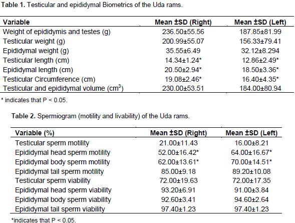
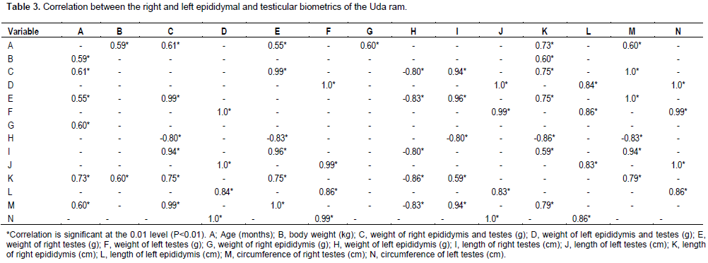
Table 3 shows that Uda rams age was positively and significantly correlated with the body weight (r = 0.59) as well as with most of the right testicular and epididymal biometrics such as the weight of right epididymis and testes (r = 0.61), weight of right testes (r = 0.55), weight of right epididymis (r = 0.60), length of right epididymis (r = 0.73) and circumference of right testes (r = 0.60). The weight of the left epididymis was negatively correlated with the weight of the right epididymis and testes (r = -0.80), weight of the right testes (r = -0.83), length of right testes (r = -0.80), length of right epididymis (r = -0.86) and circumference of right testes (r = -0.83) at P<0.05 (Table 3). From Table 4, the right testicular and epididymal volume was positively correlated (P<0.05) with motility of right testes (r=0.45), motility of right caput epididymis (r=0.82), motility of right corpus epididymis (r=0.61), motility of right cauda epididymis (r=0.71), motility of left cauda epididymis (r=0.71) and sperm viability of right caput epididymis (r=0.80). The left testicular and epididymal volume was not correlated with other spermiogram parameters except a negative correlation with sperm viability of left corpus epididymis (r=-0.92, P<0.05).
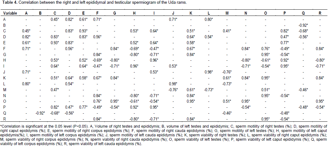
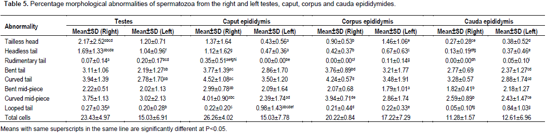
The sperm viability of right testes was positively correlated with motility of right testes (r=0.51), motility of right caput epididymis (r=0.64), motility of right corpus epididymis (r=0.58), motility of right cauda epididymis (r=0.67), motility of left testes (r=0.84), sperm viability of right corpus epididymis (r=0.61), sperm viability of right cauda epididymis (r=0.84), sperm viability of left testes (r=0.95) and sperm viability of left cauda epididymis (r=0.84). The motility values of the right testes, right caput epididymis, right cauda epididymis, left testes and left caput epididymis were positively correlated (P<0.05) with their corresponding sperm viability values. From Table 5, it was also observed that the longer testis (right) had a higher number of sperm cell abnormalities compared to the shorter one (left) with the rudimentary tail abnormality being the least observed, and the curved tail abnormalities being the most observed. The number of sperm cell abnormalities progressively decreased from testes down the length of the epididymis (from caput to cauda).

The mean body weight of 39.80±3.00 kg reported for Uda rams in this study is higher than those reported by Hassan et al. (2009) in Bangladesh native rams. All the testicular and epididymal biometrics were higher than those reported by Gemeda and Workalemahu (2017) in bucks. The testes weight, epididymal weight and length were lower while testicular length and epididymal and testicular volume were higher than those reported in Uda rams by Ibrahim et al. (2012)
The testes weight recorded were higher than those reported in Balami and Yankassa rams (Ibrahim et al., 2012), West African Dwarf rams (Ahemen and Bitto, 2007) but lower than values reported for Dorper rams by Besta (2006). The testicular lengths reported in this study were higher than those reported by Hassan et al. (2009) in rams. The testicular length and epididymal, and testicular volume recorded were higher than those reported in Balami and Yankassa rams (Ibrahim et al., 2012). The epididymal weight recorded was similar to values reported for Dorper rams (Besta, 2006), higher than values reported for West African Dwarf rams (Ahemen and Bitto, 2007) and Yankassa rams but lower than values reported for Balami rams (Ibrahim et al., 2012). The epididymal length recorded was lower than those reported in Balami and Yankassa rams (Ibrahim et al., 2012).
Sperm motility in the right testes was higher than the left though not statistically significant which is similar to the finding of Ajani et al. (2015). It is often expected that the longer epididymis should have higher motility as more space will encourage maturity of the spermatozoa in the epididymis, but this was not so in the Uda rams used for this study as there was significant increase in the motility of spermatozoa in the head (caput) and body (corpus) of the left epididymis (the shorter) compared to the right (the longer). The significant correlation between the age and body weight as well as most of the right testicular and epididymal biometrics is similar to the findings of Koyuncu et al. (2005) in Kivircik lambs. However, in contrast to the findings of Koyuncu et al. (2005) in which body weight was more closely correlated with testicular measurements than age, this study showed higher correlation between age and testicular measurements than with body weight. Significant correlations were found between the paired testicular weight and all testicular dimensions and size. This was similar to the findings of Abdou et al. (1978) and Koyuncu et al. (2005). Correlation between the testicular circumference and testicular length is similar to the findings of da Silva Santos et al. (2013) in buffalo.
Testicular length could provide a useful estimate of testicular growth because of its high correlations with the other testicular measurements. This is in agreement with the findings of Land and Carr (1975), Kumi-Diaka et al. (1985), Koyuncu et al. (2005), Hassan et al. (2009), Ibrahim et al. (2012) and Omar (2016). The number of sperm cell abnormalities progressively decreased from testes down the length of the epididymis (from caput to cauda). This observation may be linked to the effectiveness of epididymal function across the length of the epididymis. The curved tail and curved mid-piece abnormalities occurred most. This is similar to the finding of Ajani et al. (2015) with observation of curved tail as the mostly occurring abnormality. In conclusion, higher correlation observed between age and testicular biometrics than with body weight implies that age is an important factor for selection of the Uda breed of ram.
The authors have not declared any conflict of interests.
REFERENCES
|
Abdou MS, Hassun TM, El-Sawaf SA (1978). Testicular and epididymal sperm numbers and related parameters in the developing Awassi ram. Aust. J. Biol. Sci. 31(3):257-266.
Crossref
|
|
|
|
Ahemen T, Bitto II (2007). Sperm production rate, Gonadal andextragonadal sperm reserves of the West African Dwarf rams in Makurdi. Proc. of the 32nd Annu. Conf. of Nig. Soc. for Anim. Prod. pp. 99-101.
|
|
|
|
|
Ajani OS, Oyeyemi MO, Olusoji MJ (2015). Correlation between age, weight, scrotal circumference and the testicular and epididymal parameters of Red Sokoto bucks. J. Vet. Med. Anim. Health. 7(5):159-163.
Crossref
|
|
|
|
|
Besta N (2006). Effect of different dietary energy levels on productive and reproductive traits in Dorper rams. (Doctoral dissertation, University of the Free State).
|
|
|
|
|
Blood DC, Studdert VP, Gay CC (2007). Saunders Elsevier, St. Louis,Missouri, USA, P 2172. Available at:
View
|
|
|
|
|
da Silva Santos PR, Andrighetto C, Jorge AM (2013). The correlation between age, body weight and testicular parameters in Murrah buffalo bulls raised in Brazil. J. Reprod. Dev. 59(1):14-17.
Crossref
|
|
|
|
|
Devendra C, McLeroy GB (1982). Goat and sheep production in the tropics. Longman.
|
|
|
|
|
Gemeda AE, Workalemahu K (2017). Body Weight and Scrotal-Testicular Biometry in Three Indigenous Breeds of Bucks in Arid and Semiarid Agroecologies, Ethiopia. J. Vet. Med. 2017: 5276106.
Crossref
|
|
|
|
|
Hassan MR, Pervage S, Ershaduzzaman M, Talukder MA (2009). Influence of age on the spermiogramic parameters of native sheep. J. Bangladesh Agric. Uni. 7(2):301-304.
|
|
|
|
|
Ibrahim AA, Aliyu J, Ashiru RM, Jamilu M, Ibrahim A, Aliyu J, Ashiru M, Jamilu M (2012). Biometric study of the reproductive organs of three breeds of sheep in Nigeria. Int. J. Morphol. 30(4):1597-1603.
Crossref
|
|
|
|
|
Koyuncu M, Kara Uzun S, Ozis S, Duru S (2005). Development of testicular dimensions and size, and their relationship to age and body weight in growing Kivircik (Western Thrace) ram lambs. Czech J. Anim. Sci. 50(6):243-248.
Crossref
|
|
|
|
|
Kumi-Diaka J, Adesiyun AA, Sekoni V, Ezeokoli CD (1985). Scrotal dimensions and ejaculate characteristics of three breeds of sheep in tropical Nigeria. Theriogenology 23(4):671-677.
Crossref
|
|
|
|
|
Land RB, Carr WR (1975). Testes growth and plasma LH concentration following hemicastration and its relationship with female prolificacy in sheep. J. Reprod. Fertil.45(3):495-501.
Crossref
|
|
|
|
|
Omar CA (2016). Study of some testicular dimensions and their relationship to body weight in Karadi ram lambs. Assiut Vet. Med. J. 62(150):31-38.
|
|
|
|
|
ZemjÄnis R (1962). Diagnostic and Therapeutic Techniques in Animal Reproduction. Williams & Wilkins. Available at:
View
|
|