Full Length Research Paper

ABSTRACT
Myiasis is the infestation of living tissues of human and animals with larvae of flies in the Order Diptera. This study determined the gross and histopathological lesions due to Cordylobia anthropophaga infestations of Dogs in Kitui County, Kenya. Four dogs identified and confirmed to have canine cutaneous myiasis (CCM) infestation were purchased for the study. All the study animals were clinically examined for skin lesions characteristic of CCM and euthanized for complete necropsy examination and histopathological sample collection. Tissue sections were collected from areas with lesions of CCM namely; skin, skeletal muscles and regional draining lymph nodes. They were preserved in 10% buffered formalin and were processed and analyzed using standard methods. Grossly, all dogs had poor body condition, patches of alopecia, emaciated and had the pathognomonic lesions for canine cutaneous myiasis (CCM) characterized by multifocal coalescent nodular lesions (harboring Cordylobia anthropophaga larvae) with 3 mm diameter central pore (furuncles) and erythematous base. Some had serous, hemorrhagic or purulent discharges from the furuncles that matted surrounding hairs. Lesions were distributed mainly on the ventral abdomen, axilla, flanks, legs, perineum and external reproductive organs. Examination of the skin revealed that the furuncular lesions extended throughout the skin thickness from epidermis to the sub-cutis and underlying pale skeletal muscles. There was regional lymphadenopathy in affected body regions. Histopathology confirmed the main lesions in all the dogs were parasitic granulomas, congested blood vessels in the surrounding tissues and eosinophilic lymphadenitis. The granulomas were located in the skin and the underlying skeletal muscles. The lesions were characterized by centrally located parasite, surrounded by connective tissue and heavy infiltration with inflammatory cells predominantly eosinophils. Draining lymph nodes had eosinophilic lymphadenitis. The reported pathological lesions resulted in unthriftiness, alopecia, lethargy and anorexia due to pain and stress resulting to emaciation and possible death. There is need to eradicate the etiological agent as it affects the wellbeing of dogs in the study area.
Key words: Canine, Cordylobia anthropophaga, Myiasis, gross pathology, histopathology, Kitui County.
INTRODUCTION
Myiasis is a disease condition which occur at various anatomical body sites namely; skin, eyes, nose, urogenital, ears, blood, intestines and brain, where the dipteran larvae feed on the host`s liquid body substances, ingested food, dead or living tissue (Zumpt, 1965; Hosni et al., 2019). Cutaneous myiasis is classified into three broad categories based on clinical manifestation of the disease in the cutaneous system: furuncular, creeping and wound myiasis (Spradbery, 2002; Faramarzi et al., 2009). Myiasis is more often globally found in domestic animals in the tropics especially in developing countries where it is associated with poor animal welfare (Yanuartono et al., 2019). It is mainly predisposed by low level of animal hygiene, cage and environment hygiene (Yanuartono et al., 2019). In Kitui County, canine cutaneous myiasis (CCM) is predisposed by lack of proper housing and poor hygiene measures (Kamuti et al., 2022). Myiasis causing flies are attracted by skin injuries, urine and feces in the animals` environment. Female flies deposit eggs in the skin wounds or on sleeping areas especially beddings, sand or straws (Ogo et al., 2012). The eggs hatch and resulting larvae penetrate the animal skin after coming into direct contact from the environment or beddings where it feeds on dead or living tissues and body fluids of the host animal (Stevens and Wall, 2001). The typical clinical presentation of CCM includes multiple erythematous, nodular (furunculoid) lesions which ooze serous fluid or pus and clinical signs such as lethargy, anemia, unthriftness, emaciation and alopecia (Johnson et al., 2016; Henok, 2017). The nodular lesion mostly harbors one diptera larvae but multifocal coalescent cutaneous nodular lesions can also be found on affected body sites (Abebe, 2017). Earlier, Ogo et al. (2009) reported the occurrence and preference of Cordylobia anthropophaga larvae in dogs in Nigeria. Mugachia (2018) observed skin lesions suggestive of CCM in Kitui County while Kamuti et al. (2022) described the etiological agent, C. anthropophaga causing these lesions in dogs in Kitui County.
Globally, publications on the type of pathological lesions associated with CCM infestation and their distribution in the dog`s body are unavailable. This study was carried out to determine the gross and histopathological lesions associated with cutaneous myiasis in dogs in Kitui County.
MATERIALS AND METHODS
Source of canine cutaneous myiasis infested dogs
Dogs were sourced from Kitui County as the area was endemic with CCM as reported by Mugachia (2018). The CCM in Kitui County had been confirmed to be caused by C. anthropophaga larvae infestation (Kamuti et al., 2022).
Description of the study dogs
Ethical clearance for the use of the dogs in this study was sought and approved by Biosafety, Animal Use and Ethics committee of the Faculty of Veterinary Medicine, University of Nairobi (UoN), REF: FVMBAUEC/2021/303). Four dogs with clinical cutaneous myiasis (CM) were purchased from owners in households in Kitui Central sub-county, Kitui County. These dogs had clinical manifestation of CCM infestation as reported by Mugachia (2018) and Kamuti et al. (2022). The dogs were transported alive in cages to the Department of Veterinary Pathology, Microbiology and Parasitology, Faculty of Veterinary Medicine, UoN for euthanasia, routine necropsy and tissue sampling for histopathological examination.
Necropsy examination of the dogs
Physical examination was done to determine type, size, color and location of external lesions caused by CCM infestation in the dogs. They were noted and recorded.
The dogs were thereafter sedated one at a time using Xylazine HCl at a dosage rate of 1.1mg/kg body weight intramuscular route and then put into general anesthesia using a combination of Xylazine HCl and Ketamine 5% at a dosage rate of 1.1mg/kg body weight and 2.2 mg/kg body weight respectively. They were euthanized by intra-cardiac injection of 30 ml of Lignocaine HCL 2%.
Necropsy technique was carried out as described by Mcdonough and Southard (2017). The location, distribution, size (length and diameter of the furuncular central pore), extent, shape and color of the furuncular skin lesions in the dog carcasses were noted, measured and recorded. The underlying tissues (subcutis and muscles) were examined for gross changes (Caswell and Plattner, 2012). Tissues for histopathology were collected from affected skin, skeletal muscles and draining lymph nodes and preserved in 10% buffered formalin.
Histopathology examination
Tissues fixed in 10% buffered formalin from study dog carcasses were processed for histopathology examination using routine standard procedures and stained with hematoxylin and eosin (H&E) as described by Comanescu et al. (2012).
Histopathology slides were then examined and photomicrographs taken using an Olympus digital microscope with an inbuilt camera (Olympus CX21 microscope).
RESULTS
Gross lesions
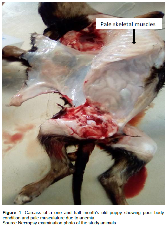
All the four dogs had poor body condition, rough hair coats, hair matted with serous, sero-hemorrhagic or purulent discharges, pale mucus membranes of the eye conjunctiva and generalized pale skeletal muscles indicative of anemia (Figure 1).
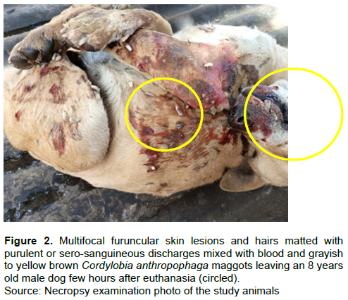
The pathognomonic skin lesions for CCM recorded in all the dog carcasses were multifocal nodular skin lesions with a central pore (furuncle). The furuncles were distributed on the ventral abdomen, lateral and medial sides of the thighs, perineum and tail, lateral sides of the chest (flanks), sternum, neck, skin of the ears, vulva mucosa, scrotal skin and the inter digital spaces of the fore and hind limbs.
The furuncular lesions were nodular, erythematous at the base (approximately 1 cm diameter) and had a central pore (approximately 3mm diameter). The hairs around the furuncles were raised. Most furuncles discharged freely and matted the hair around them with serous, purulent or sero-hemorrhagic exudates from the opened discharging pore at the center of the lesions. On application of digital pressure at the base of the furuncles, the lesions oozed serous fluid or pus mixed with blood.
Yellow-brown maggots also emerged from the squeezed furuncles to the skin surfaces (Figure 2). There were scarred, depressed tissue patches of scattered alopecia in areas where the lesions had already healed after the maggots had dropped from the host especially on ventral abdomen, flanks and lateral sides of the thighs.
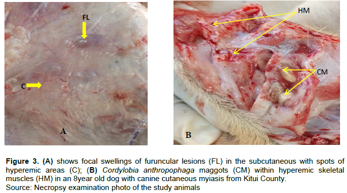
On observation of the skin, the furuncular lesions extended throughout the skin thickness from epidermis to the sub-cutis. They were characterized by firm consistency, edema and hyperemia (Figure 3a). The underlying skeletal muscles were bright red due to hyperemia (Figure 3b). The draining lymph nodes especially the pre-scapular and popliteal lymph nodes in the affected regions were enlarged and edematous on cut longitudinal sections (regional lymphadenopathy) in all the four dogs necropsy examination was done.
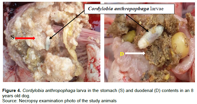
There were C. anthropophaga maggots mixed with stomach and duodenal contents (pseudomyiasis) without notable tissue reaction on the cardiac sphincter, stomach and duodenal mucosae in one of the dogs (Figure 4). There were no significant lesions in other body organs, systems and tissues.
Histopathology lesions
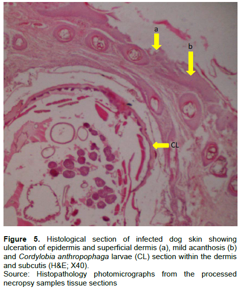
Microscopically, there was ulceration of the epidermis and superficial dermis and thickening of the epidermal layer (acanthosis) of the affected skin. Cross sections of Cordylobia anthropophaga larva were found within the dermis and subcutis (Figure 5).
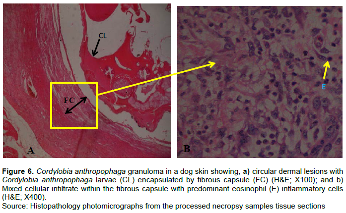
Multifocal circular cavities with C. anthropophaga larvae were observed in the dermis and in the subcutaneous tissue of the skin (Figure 6a). There was also dilatation of adjacent blood vessels with red blood cells. The dermal circular lesions (parasitic granulomas) were surrounded by a fibrous capsule with a mixed inflammatory cellular infiltrate with mononuclear cells and numerous eosinophils as the predominant inflammatory cells (Figure 6b).
There was destruction of the underlying subcutaneous skeletal muscles which showed edema and necrosis. There was proliferation of fibroblasts and subsequent deposition of eosinophilic collagen fibers mixed with inflammatory cellular infiltrates with predominant eosinophils.
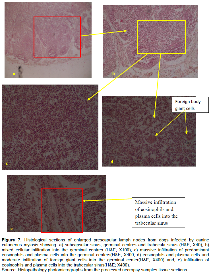
Regional lymph nodes draining affected areas had massive infiltration of eosinophils and plasma cells within the sub-capsular sinus, germinal centers and trabecular sinuses extending to the medullary sinuses. The lymph nodes also had reactive germinal centers with moderate infiltration of foreign body giant cells into the germinal centers (Figure 7).
DISCUSSION
These study findings confirmed that Cordylobia anthropophaga maggot infestation in dogs is associated with significant pathological lesions. The pathognomonic gross lesions associated with CCM in study dogs were multifocal nodular skin lesions with a 3mm diameter central pore (furuncular lesions). These lesions were limited to the skin, sub-cutis and the underlying subcutaneous muscles. These findings concurred with previous work by Leschnik et al. (2008) who reported that C. anthropophaga larvae infestation produced lump like subcutaneous lesions with a central hole in the skin of human and animals. The furuncular skin lesions were distributed on the ventral abdomen, lateral sides of the thighs, perineum, tail, flanks, sternal region, neck, skin of the ears and the interdigital spaces of both the fore and hind limbs.
Most of these body parts get in contact with the ground when the dog is lying. These findings were in agreement with those of Singh (2020) who reported that ventral body parts of dogs like vulvar lips and perianal region are more vulnerable to the attack of flies because these regions frequently come in contact with the ground while the dog is sitting and become soiled with faeces and the dipteran maggots infest and irritate host animals. The base of the furuncular lesions was erythematous, and oozed serous, purulent or hemorrhagic fluid freely or on application of digital pressure. The hairs around the furuncular lesions were matted by the discharges. The base of the furuncular lesions in the sub-cutis was congested and there was also edema in the affected body regions.
Lesions where larva had left the skin were scarred with firm consistency due to healing by fibrosis resulting in multifocal patches of alopecia. Some of this is as reported by Johnson et al. (2016) that anemia, unthriftness, alopecia, lethargy, emaciation and anorexia as the main clinical signs in dogs affected by canine cutaneous myiasis in the Greater Accra region in Ghana. However, these authors did not describe the other gross lesions. Pseudomyiasis was also another finding in this study. This could have occurred as dogs tried to bit infected areas after itching hence, swallowed the maggots.
Some larvae were still active crawling freely on the carcasses whereas others were dead and firmly attached to the furuncular lesions. The present study findings were in agreement with previous work by Johnson et al. (2016). The histopathological skin lesions associated with CCM were ulceration of the epidermis and superficial dermis and mild acanthosis at points of entry of C. anthropophaga larvae into the host animals. There were multifocal circular cavities harboring the C. anthropophaga larvae extending from the dermis to the subcutis layer of the skin. There was hyperemia of the blood vessels adjacent to the furuncular cavities. C. anthropophaga larvae formed parasitic granulomas surrounded by fibrous capsule infiltrated with a mixed cellular infiltrate of mononuclear and eosinophils. Unlike Hausdorfer et al. (1993) who reported that infestation by C. anthropophaga larvae caused dermatitis characterized by neutrophils and eosinophils inflammatory cells. Subcutis muscular changes of necrosis and healing by fibrosis where the maggots had left the host are also reported in this study.
There are no previous detailed reports on the gross and microscopic pathological lesions associated with CCM, elsewhere as reported in this study.
CONCLUSION AND RECOMMENDATION
Cordylobia anthropophaga infestation caused significant gross and microscopic pathological lesions in the affected dogs. Increased awareness of risk factors associated with CCM in Kitui County is recommended to ensure wellbeing of dogs and other vulnerable animals.
CONFLICT OF INTERESTS
The authors have not declared any conflicts of interests.
ACKNOWLEDGEMENTS
The authors would like to thank Mr. J. K. Mulwa, Mr. K. I. Mwendwa, Mr. M. M. Masika, Mr. M. Muthangya, Mr. R. M. Kiteme and Mr. J. Nzuki of Kitui County Directorate of Veterinary Services for their technical assistance while collecting data in the field. Special thanks to Mr. D.G. Muriithi and the late Mr. J.G. Mukiri of the Department of Veterinary Pathology, Microbiology and Parasitology, UoN for their technical support in processing tissue samples.
REFERENCES
|
Abebe HT (2017). Survey on prevalence of canine cutaneous myiasis in some selected kebeles of Dire Dawa city administration. BioRxiv: Survey on prevalence of canine cutaneous myiasis in some selected kebeles of Dire Dawa City administration | bioRxiv |
|
|
Caswell J, Plattner B (2012). Gross pathology description and interpretation. Department of Pathobiology, University of Guelph. |
|
|
Comanescu M, Annaratone L, DÀrmento G, Cardos G, Sapino A, Bussolati G (2012). Critical Steps in Tissue Processing in Histopathology. Recent Patents on DNA and Gene Sequences 6:22-32 |
|
|
Faramarzi A, Rasekhi AR, Kalantari, M, Hatam GR (2009). Chrysomya bezziana as a causative agent of human myiasis in Fars Province, Southern Iran. Iranian Journal of Arthropod-Borne Diseases 3(1):60-63. |
|
|
Hausdorfer SS, Bourlond A, Pirard C (1993). Histopathological aspects of myiasis. Dermatology 186(4):298-300. |
|
|
Hosni ME, Kenawy MA, Nasser MG, Al-AShaal SA, Rady MH (2019): A brief review of myiasis with special notes on the blow flies' producing myiasis (F.: Calliphoridae). Egyptian Academic Journal of Biological Sciences 11(2):25-32. |
|
|
Johnson SA, Gakuya DW, Mbuthia PG, Mande JD, Afakye K, Maingi N (2016). Myiasis in dogs in the Greater Accra Region of Ghana. Vector Borne Zoonotic Diseases 16(1):54-57. |
|
|
Kamuti NM, Mbuthia PG, Waruiru RM, Githigia SM, Keya EA (2022). Prevalence, Etiology, and Risk Factors Associated with Occurrence of Canine Cutaneous Myiasis in Kitui County, Kenya. Veterinary Medicine International, volume. 2022, ArticleID 5699060, 9 pages, 2022. |
|
|
Leschnik M, Lowenstein M, Edelhofer R, Kirtz G (2008). Imported non-endemic, arthropod-borne and parasitic infectious diseases in Austrian dogs. The Middle European Journal of Medicine 120(4):59-62. |
|
|
Mcdonough SP, Southard P (2017): Necropsy guide for dogs, cats, and small mammals. Front matter - necropsy guide for dogs, cats, and small mammals - Wiley online library |
|
|
Mugachia JC (2018). Daily Nation. Seeds of Gold: Vet on Call: These flies will eat your animals alive | Nation. |
|
|
Ogo NI, Onovoh E, Ayodele DR, Ajayi OO, Chukwu CO, Sugun M, Okeke IO (2009). Cutaneous canine myiasis in the Jos metropolis of Plateau State, Nigeria, associated with Cordylobia anthropophaga. Veterinarski Arhiv 79(3):293-299. |
|
|
Ogo NI, Onovoh E, Okubanjo OO, Galindo RC, De la Lastra JP, De la Fuente J (2012). Molecular identification of Cordylobia anthropophaga Blanchard (Diptera: Calliphoridae) larvae collected from dogs (Canis familiaris) in Jos South, Plateau State, Nigeria. Onderstepoort Journal of Veterinary Research 79(1). |
|
|
Singh A (2020). Canine Myiasis and its Causal Agents in India. Journal of Biomedical Research & Environmental Sciences 1(5):150-153 |
|
|
Spradbery JP (2002). A manual for the diagnosis of the screwworm fly, Fisheries and Forestr. (2nd edition). Australia: Department of Agriculture. |
|
|
Stevens JR, Wall R (2001): Genetic relationship between blowflies (Calliphoridae) of forensic importance. Forensic Science International 120:116-123: |
|
|
Yanuartono Y, Nururrozi A, Indarjulianto S, Raharjo S, Purnamaningsih H (2019). Myiasis in ruminants: Diagnosis, Therapy and Prevention Management. Journal of Tropical Animal and Veterinary Science 9(2):67-73. |
|
|
Zumpt F (1965). Myiasis in man and animals in the old world. A textbook for physicians, veterinarians and zoologists. Butterworth & Co. (Publishers) Ltd. 88, Kingsway, London W.C.2. UK. |
|
Copyright © 2024 Author(s) retain the copyright of this article.
This article is published under the terms of the Creative Commons Attribution License 4.0


