ABSTRACT
A cross sectional study was conducted in and around Kemissie, Dawa Cheffa District to estimate the prevalence and identify possible risk factors for the disease in the study area. Animal identification based on age, sex, breed and body condition score and management system of the study animals were taken into account followed by coprological examination for the presence of the developmental stages of parasites or parasite in naturally infected cattle. For this purpose, four hundred and five randomly selected animals were examined during the study period out of which 75 (18.5%) were found positive for schistosomiasis based on fecal examination. Variation together with age and sex of animals did not show statistical significance (P>0.05) in the disease occurrence. However, the prevalence of the disease was highest in young animals (23.14%), followed by adults (17.61%) and least in old animals (15.49%). On the other hand, statistical significant association (P<0.05) was seen with the variation of breeds, body condition score and management systems in the disease occurrence. In conclusion, relatively moderate prevalence (18.5%) was recorded in and around Kemissie and based on the results obtained, recommendations were forwarded.
Key words: Cattle, coproscopy, Kemissie, prevalence, Schistosomiasis.
Animal production has been considered as the main component of agricultural development in most parts of sub-Saharan Africa. Like in many developing countries of the region, domestic animals play a crucial role in Ethiopia. They provide food in the form of meat and milk, and non-food items such as draft power, manure and transport services as inputs into food crop production, and fuel for cooking. Livestock are also a source of cash income through sales of the above items, animal hides determine social status within the community. Ethiopia is known for its high livestock population, being the first in Africa and tenth in the world (Gebrecherkos and Berihun, 2012). The recent livestock population estimate shows that the country has about 52.1 million heads of cattle, 24.2 million sheep, 22.6 million goats and 44.9 million poultry (MoARD, 2013).
Trematode parasitism is one of the biggest problems reducing ruminant productivity around the world and skins. Furthermore, they act as a store of wealth and (Vercruysse and Claerebout, 2001). In Ethiopia, the potential of livestock sector is not efficiently and fully exploited due to several constraints like malnutrition,traditional management practice, poor genetic makeup and prevailing diseases. Among the prevailing diseases in the country, trematodes are one of the main parasitic problems of cattle and other ruminants (Fromsa et al., 2011).
These parasitic diseases are found in vast water lodged and marshy grazing field, a condition anticipated to be ideal for the propagation and maintenance of the intermediate host snails and hence the high prevalence of trematode infection (Solomon and Abebe, 2007).
Among these trematode infections, Schistosomiasis (also known as Bilharzias) is economically important worldwide. It is a chronic and debilitating tropical disease caused by adult blood flukes (parasitic trematode worms) of the genus, Schistosoma that deposit eggs in blood vessels surrounding the bladder or gut of infected mammalian hosts (McManus et al., 2009).
It is an endemic disease of cattle in Africa and Asia while at least 165 million cattle are infected with bovine Schistosomiasis worldwide. Animal Schistomes are mainly restricted to tropical and sub tropical areas (Upadhayay, 2005). It has a restricted distribution, which is found commonly in northern, eastern, southwestern and central parts of Ethiopia (Habtamu and Mariam, 2011). Routine diagnosis of visceral Schistosomiasis relies heavily on observation of clinical signs and fecal examination for eggs of the parasite. It is a zoonotic disease affecting both humans and livestock. The growing interest in organic, outdoor and aquaculture farming are likely to present higher risk of infection (Chhabra and Singla 2009).
The blood flukes are grouped under the genus Schistosoma (McCauley et al., 1984) and the family Schistosomatidae. Schistosomes under this group are elongate, unisexual and dimorphic trematodes, which inhabit the blood vessels of their hosts. Members of Schistosomatidae show morphological and physiological peculiarities which set them apart from all other trematodes. Firstly, they are dioecious, the male bearing the female in a ventral canal, gynaecophoric canal and secondly they live in the blood stream of warn-blooded hosts, being the only trematodes to do so (Smyth, 2005). One of the most important in animals is Schistosoma bovis which occurs in the portal and mesenteric veins of cattle, sheep and goats in Central, East and West Africa, the Mediterranean area and in the Middle East (Soulsby, 2006).There are three main species of Schisotomes infecting humans, Schistosoma mansoni, Schistosoma japonicum which inhabit the mesenteries around the intestine and S. haematobium, which are found in the venules surrounding the bladder (Upadhayay, 2005). Although, little or no overt clinical signs may be seen over a short period, frequent Schistosome infections, in nthe long term, cause significant losses to the herd (Ravindran et al., 2007).The disease is characterized by its chronic nature and affects the productivity and reproduction performances; and predisposes animals to other diseases (McCauley et al., 1984). Within the last five years, no work has been done indicating the current situation of bovine schistosomosis in the study area. Therefore, the aims of this study were to estimate the current prevalence of bovine schistosomosis and to identify major risk factors associated with the disease in the study area.
Study area
The study was conducted to determine the prevalence of bovine Schistosomosis in and around Kemissie, Dawa Cheffa District from September 5, 2011 to May 10, 2012, Oromo Nations Zone of the Amhara Regional state, Northeastern Ethiopia. It is located at 325 km North of Addis Ababa. The approximate geographical location of the area is between 10°01’ to 11° 25’ N and 39° 41’ to 40° 24’ E. The altitude of the area ranges from 1000 to 2500 m above sea level and the maximum temperature is 33°C and the minimum temperature of the area is about 12°C. The mean annual rainfall of the area ranges from 600 to 900 mm. The area has a long heavy rainy season from June to September and short rainy season from March to May (OZARD, 2006).
Study population
The study was conducted in local Zebu and cross breeds between exotic and local breeds of cattle in the study area that was managed under extensive and semi-intensive management systems in the study area. The study population comprises of cattle at different age (De launta and Habel, 1986) and sex category. Four peasant associations (PAs) were selected purposively for their accessibility (convenience) of transportation and nearness to towns. These PAs were Kemissie, Shekla, Woledi and Tuche, all of which are under the same climatic and geographical features (OZARD, 2006). All the local breeds were often kept out-doors grazing on a marshy land and watered on natural rivers whereas most of the crossbreeds were kept in-door especially during milking periods where there is sufficiently gathered feed resources but watering is usually out-door especially during the dry season where it is difficult to fetch from a distant area for animals to drink. During sampling, deworming history of individual animals was obtained from the owners and recently dewormed animals were excluded from being sampled.
Study design
The study design for this particular study was cross sectional. The sampling method used was simple random sampling. To determine the sample size, an expected prevalence of 50% was taken into consideration since there was no previous study conducted recently within the last five years in the study area. The desired sample size for the study was calculated by the formula given by Thursfield (2005) with 95% confidence interval and 5% absolute precision and it was 384. However, to increase the precision level, 405 animals were selected randomly from the selected PAs of the study area.
Study methodology
Sample collection and transportation
About 10 g (Foreyt, 2001) of fresh fecal samples were collected per
rectum from each cattle using sterile rectal plastic gloves and placed in a clean plastic container and each sample was clearly labeled with animal identifications, such as body condition score (Nicholson and Butterworth, 1986), sex, age (De launta and Habel, 1986), breed and management systems for each animal on a data recording format.
Coprological examination
Coproscopy was used to determine positivity of the animals for the disease. Fecal samples collected from the study animals directly from the rectum were stored in a clean universal bottle containing 10% formalin and labeled separately. A modified simple sedimentation technique (Gupta and Singla, 2012) was employed. Each sample was repeatedly examined more than four times recommended by Habtamu and Mariam (2011) to observe the eggs of the parasites by using a light microscope.
Data analysis
Data were entered and stored in a Microsoft Excel spread sheet program and were coded for analysis. SPSS version17.0 software, data were analyzed. Chi-square (χ2) test was used to determine the variation in the infection and prevalence among ages, sexes, breeds, body condition score and management systems. Statistical significance was set at P<0.05 to determine whether there are significant differences between the parameters measured between the groups.
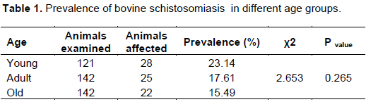
Out of 405 fecal samples examined, an overall prevalence of 18.5% was observed as 75 samples were found positive for schistosomiasis in the study area. Slightly higher prevalence of the disease was seen in young cattle (23.14%) followed by adults (17.61%) and least in old age (15.49%) as shown in Table 1. However, the prevalence of schistosomiasis iid not vary significantly (p>0.05) among the age groups.
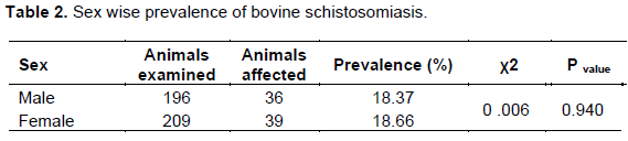
Prevalence of Schistosomiasis in female and male animals was 18.66 and 18.37%, respectively. However, no significant difference (P>0.05) was observed between sexes (Table 2).
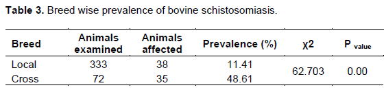
Prevalence of the disease in the cross breeds (48.61%) was higher, which showed higher variation from local breeds (11.41%). The result of statistical analysis revealed a significant difference (P<0.05) between breeds of animals (Table 3).
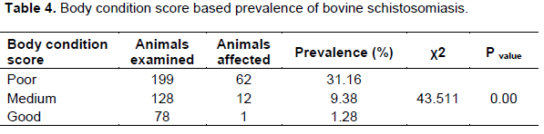
Prevalence of schistosomiasis in poor body condition animals (31.16%) was found to be highest. However, the prevalence in animals with medium and good body condition was 9.38 and 1.28%, respectively. A significant difference (P<0.05) in prevalence was observed in body condition of the study animals (Table 4).
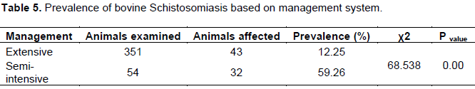
The prevalence of the disease in animals under semi-intensive management system (59.26%) was higher. However, the prevalence of the disease in animals under extensive management was only 12.25%. The result of statistical analysis revealed a significant difference (P<0.05) in each management system (Table 5).
An overall prevalence of 18.5% bovine schistosomiasis was recorded based on coprological investigation. This result agrees with previous reports of Getachew et al. (2006) in Kemissie who reported similar prevalence of 17.2% as in the present case. This may be because both studies were conducted in the same study area. However, the result of the present study was not in agreement with the previous reports by Ameni et al. (2001) who reported higher prevalence (28%) in Kemissie. This may be due to climatic changes such as repeated draught leading to drying of natural habitats of the intermediate host, snail and the larval stages of the parasite may not be reached by infective stages and may decrease their population leading to decrease in the prevalence of the disease in the area. Hansen and Perry (1993) reported that schistosomiasis is closely associated with large permanent water bodies such as ponds, lakes and marshy pastures; and according to Urquhart et al. (1996), it may be due to development of resistance for the disease when animals are repeatedly exposed to the infection in the study area. The prevalence of the disease was also not in agreement with the previous reports in different areas by different authors; Habtamu and Mariam (2011), 37.3% in Bahir Dar; 43% in Bati (Getachew et al., 2006), 57.3% in India (Ravindran et al., 2007). This might be due to the differences in temperature, moisture, humidity, availability of large permanent water bodies and soil that might favor multiplication and survival of the intermediate host, snails. Hansen and Perry (1993) reported that a key determinant in the epidemiology of bovine schistosomiasis is the relative abundance of the intermediate hosts and their ability to develop and survive in the environment and associated with large permanent water bodies. It was also reported by Urqhart et al. (1996) that Schistosoma species are totally dependent on water as a medium for infection in both the intermediate host and final host.
Prevalence of the disease in different age groups has values of 23.14% in young, 17.61% in adults and 15.49% in old animals with a non-statistical difference (P > 0.05). However, the result of the study indicates that the prevalence of the disease decreases as age increases. This may be because young animals have lower immunity when compared with adult and older cattle. According to the reports by Habtamu and Mariam (2011), calves are traditionally weaned at about 1.5-2 years and then allowed to graze with adult cattle. In addition to this, adult and old group of cattle are left to graze on the field where cercaria infection is high, furthermore in this group of animals, acquired immunity is not established, hence egg shed in these animals is high. There was statistically non-significant difference (P > 0.05) between the sexes categories indicating that it has no effect on the prevalence of the disease. This may be because exposure of both sex groups in similar pasture lands and watering points is the same and eventually developing the disease equally (Habtamu and Mariam, 2011).
A significant difference (P < 0.05) was observed between breeds in the study animals. High prevalence was recorded in cross breed cattle (48.61%) than in local breeds (11.41%). This wide gap may be due to local breeds acquired a high degree of immunity as a result of repeated natural exposure to the disease for a longer period. The main manifestation of immunity was suppression of worm fecundity (Cheng, 1986). It was also reported that local cattle that naturally acquired infections are capable of reducing egg production. Furthermore, there is difference in natural or innate immunity between indigenous and cross breed of cattle (Habtamu and Mariam, 2011).
The results of this study indicated that there is a significant difference in body condition scoring (P<0.05). Infection rates in poor body condition animals were significantly highest (31.16%), moderate in medium (9.38%) and least in animals having good body condition (1.28%). Based on the reports of Urqhart et al. (1986) and Hansen and Perry (1993), the disease causes anorexia and emaciation which leads to weakening of the immune status of the animal that leads to low immune response to the parasite and predisposes animals to other diseases McCauley et al. (1984). This indicates the importance of the disease in causing poor body condition (emaciation).
An attempt was also made to analyze the prevalence of the management system of the animals. The prevalence of the disease in animals that were kept under semi-intensive management system was higher (59.26%) than animals kept under extensive management system (12.25%). The result of statistical analysis revealed that there is a significant difference (P < 0.05) in the management system. This may be because extensively managed animals acquired a high degree of immunity as a result of repeated natural exposure to the disease (Cheng, 1986). However, animals kept in semi-intensive management system become susceptible to the infection as they have no acquired immunity to withstand the disease.
CONCLUSION AND RECOMMENDATIONS
The result of this study indicated that schistosomiasis is a moderately prevalent bovine infection in the study area. Even though its prevalence seems lower, it has higher economic importance due to loss of productivity and mortality in animals and it is a zoonotic disease. However, it is evident that proper evaluation of the epidemiology of the disease is absent in the study area recently. The major risk factors associated with the disease on this finding were management system, body condition score and cattle breeds. However, sex and age were not that much significant in this study. However, the prevalence of the disease was highest in young cattle followed by adults and least in older age animals. The result of the present study showed that there is a reduction in its prevalence when compared with previous reports in the study area. However, micro dams constructed for irrigation purpose may be threatening factors to increase its prevalence again in the future in the area. In the area, no measures being taken to control the disease and no drugs available in veterinary clinics even though the disease is endemic.
Therefore, detailed studies should be conducted on the epidemiology of the disease to expand and implement disease investigation and control strategy. Young aged animals should be kept at home or the weaning time of calves should be extended until they become mature or until their immunity develops well. Fencing water bodies is supposed to be practiced to reduce water contamination with fluke eggs. Applications of molluscicide ought to be used to reduce snail population at seasons where the number of the population increases in the area.
The authors have not declared any conflict of interests.
REFERENCES
|
Ameni G, Erko B, Bogale T (2001). Preliminary study on the major bovine trematode infection around Kemissie, Northeastern Ethiopia and treatment trial with praziquantel. Bull. Anim. Health. Prod. Afr. 49:62-67.
|
|
|
|
Cheng TC (1986).General Parasitology, 2nd edition, Academic Press, Medical University of South Carolina, Charlton. pp. 332-342.
|
|
|
|
|
Chhabra MB, Singla LD (2009). Food-borne parasitic zoonoses in India: Review of recent reports of human infections. J. Vet. Parasitol. 23(2):103-110.
|
|
|
|
|
De launta A, Habel RE (1986). Applied Veterinary Anatomy. W.B. Saunders Company. USA.
|
|
|
|
|
Foreyt WJ (2001). Veterinary Parasitology Reference Manual, 5th edition, Black well,Lowa State University Press, Washington DC, USA. P 3.
|
|
|
|
|
Fromsa A, Meharenet B, Mekibib B (2011). Major Trematode Infections of Cattle Slaughtered at Jimma Municipality Abattoir and the Occurrence of the IntermediateHosts in Selected Water Bodies of the Zone. J.Anim. Vet. Adv. 10(12):1592-1597.
Crossref
|
|
|
|
|
Gebrecherkos, Berihun A (2012). Prevalence of bovine fascilosis in municipal Abattoir of Adigrat, Tigray, Ethiopia. Mekelle University College of Veterinary Medicine, Mekelle Ethiopia.
|
|
|
|
|
Getachew T, Tesfu K, Berhanu E, Legesse W, Ahmed A, Nega B, Girmay M (2006). Pilot control of fasciolosis and related animal fluke infections by the use of Endod and reduced morbidity: I pre-intervention studies. Ethiop. vet. J. 10(1):67-70.
|
|
|
|
|
Gupta SK, Singla LD (2012). Diagnostic trends in parasitic diseases of animals. In: Veterinary Diagnostics: Current Trends. Gupta RP, Garg SR, Nehra V and Lather D (Eds), Satish Serial Publishing House, Delhi. pp. 81-112.
|
|
|
|
|
Habtamu A, Mariam SW (2011). Repeated simple sedimentation technique and prevalence of bovine schistosomosis in selected sites of Bahir Dar woreda. Ethiop. Vet. J.15(1).
Crossref
|
|
|
|
|
Hansen J, Perry B (1993). The Epidemiology, Diagnosis and Control of Helminth Parasites of Ruminants, 2nd edition, published by the ILRAD, Nairobi, Kenya. pp. 40-41.
|
|
|
|
|
McCauley EH, Majid AA, Tayeb A (1984). Economic evaluation of the production impact of bovine schistosomiasis and vaccination in the Sudan.Preven. Vet. Med. 2(6):735-754.
Crossref
|
|
|
|
|
McManus DP, Li Y, Gray DJ, Ross AG. (2009). Conquering 'snail fever': schistosomiasis and its control in China. Exp. Rev.Anti-infect.Ther. 7(4): 473-485.
|
|
|
|
|
Ministry of Agriculture and Rural Development(MoARD)(2013).Major challenges and Achievements in Ethiopian Livestock production.
|
|
|
|
|
Nicholson MJ, Butterworth MH (1986). A Guide to Body Condition Scoring in Zebu Cattle. International Livestock Center for Africa (ILCA). Addis Ababa, Ethiopia.
|
|
|
|
|
OZARD (2006). Annual Report, Oromya Zone Agricultural and Rural Development Department, Amhara Regional State, Kemmisse. P 19.
|
|
|
|
|
Ravindran R, Lakshmanan B, Ravishankar C, Subramanian H (2007). Research note. Visceral schistosomiasis among domestic ruminants slaughtered in Wayanad, South India. Southeast Asian J. Trop.Med. Public Health 38(6):1008.
|
|
|
|
|
Smyth JD (2005). Animal Parasitology. Cambridge University Press.3rd edition. London. P 236.
|
|
|
|
|
Solomon W, Abebe W (2007). Effects of a strategy antehelmtic treatment intervention bovine fasciolosis. A conducted in facilities in endemic area in north western Ethiopia. Vet. J. 11(2):59-68.
|
|
|
|
|
Soulsby EJL (2006). Helminths, Arthropods, and Protozoas of Domestic Animals. 7th edition. London. P 72.
|
|
|
|
|
Thrusfield M (2005). Veterinary Epidemiology.2nd edition, UK, Blackwell Sci. pp. 228-247.
|
|
|
|
|
Upadahayay AKU (2005). Textbook of Preventive Veterinary Medicine. 1st edition. pp. 380-381.
|
|
|
|
|
Urquhart GM, Armour J, Duncan JL, Jennings FW (1996). Veterinary helminthology: Veterinary Parasitology. New York, Churchill: Livingstone Inc. pp. 114-116.
|
|
|
|
|
Vercruysse J, Claerebout E(2001). Treatment vs non-treatment of helminth infections in cattle: defining the threshold. Vet. parasitol. 98(1):195-214
Crossref
|
|