Full Length Research Paper

ABSTRACT
A study on incidence of lumpy skin disease (LSD) was conducted at 20 exports-oriented cattle feedlots found in Adama district in Central Ethiopia between June and December, 2011. The 11,189 bulls in the 20 exports-oriented feedlots were clinically examined and evaluated for LSD, its incidence, mortality and morbidity. The overall incidence and mortality rate of LSD in cattle feedlots was 6.1 and 1.8% respectively. Statistically significant difference was observed among sites of feedlots operations in incidence (χ2 = 251.4, df = 5, p < 0.05) and mortality rate (χ2 = 167.9, df = 5, p < 0.05). The overall case fatality of the disease was 30% with significant difference among all feedlot operation sites (χ2 = 326.7, df = 5, p < 0.05). Majority of the affected population was observed with variable degree of lameness that was accompanied with edema of limbs with the progression of the disease. The nodules became necrotic, and eventually a deep scab formed (sit-fast). The results of this study indicated that the complex epidemiological situation of the disease in Ethiopia needs more detailed investigation if improved vaccine-based control is to be achieved effectively and efficiently.
Key words: Cattle, Central Ethiopia, feedlots, lumpy skin disease.
INTRODUCTION
Lumpy skin disease (LSD) is a serious skin disease of cattle caused by lumpy skin disease virus (LSDV). It causes acute to sub-acute systemic disease characterized by mild to severe symptoms including disseminated appearance of skin lesions, 2 to 5 cm in diameter and lymphadenopathy, accompanied by high fever, which can sometimes exceed 41°C (Babiuk et al., 2008). The virus causes significant economic losses in cattle industry due to reduction in milk production, decreased weight gain, abortion, decrease draft power, temporary or permanent sterility, damaged hides and deaths thus decreasing their commercial value, (Rovid, 2008; Gari et al., 2011).
Furthermore, restrictions to the global trade of live animals and animal products, costly control and eradica-tion measures such as vaccination campaigns as well as the indirect costs because of the compulsory limitations in animal movements cause significant financial losses (Tuppurainen and Oura, 2012). In outbreaks of the disease, the morbidity rate varies widely depending on the immune status of the hosts and the abundance of mechanical arthropod vectors and usually ranges from 3 to 85%. In general, mortality rate is low (1 to 3%) but may sometimes reach 40% (Coetzer, 2004; Tuppurainen and Oura, 2012).
LSD is mechanically transmitted by different types of biting and blood-feeding arthropods (Kitching and Mellor, 1986; Chihota et al., 2001; Magori-Cohen et al., 2012). This disease is mainly spread to new areas by infected animals, but it could also be transmitted by contaminated hides and other products. Outbreaks can be eradicated by quarantine, depopulation of infected and exposed animals, proper disposal of carcasses, cleaning and disinfection of the premises and insect control (Rovid, 2008). In endemic countries, the control of LSDV can be achieved through the use of attenuated live virus vaccines (Brenner et al., 1992).
LSD is now endemic in most of sub-Saharan Africa, parts of North Africa and has been reported from the Middle East (Gari et al., 2010; Salib and Osman, 2011; Body et al., 2012; Tuppurainen and Oura, 2012). Currently, LSD outbreaks occur almost in all agro-ecological zones of Ethiopia (Gari et al., 2008; Gari et al., 2010; Gari et al., 2011). Since the export of live cattle from Ethiopia is currently largely feedlot-based, the occurrence of LSD in the feedlots certainly has affected access of the country to international markets. Therefore, the objective of this study was to determine the incidence of lumpy skin disease and associated risk factors in export-oriented cattle feedlots at Adama District in Central Ethiopia.
MATERIALS AND METHODS
Study area
The study was conducted in Adama district (Nazret/Nazareth) (Figure 1) situated in East Shewa Zone of Oromia regional state located between 8°33' N, 39°16′ E, 8.55° N, and 39.27° E at an elevation of 1,712 m above sea level South-East of Addis Ababa. It is about 99 km away from the nation’s capital Addis Ababa, on the famous and ever busy port highway of Ethiopia. It is a bowl-like sinking sight surrounded by small hills just in the heart of the great East Africa Rift Valley. The mean annual temperature varies between 18°C and 30°C and its mean annual rainfall is 410 to 820 mm. Natural vegetations grown in Adama district are grouped under the Acacia wood land and savannah vegetation.
Study design and subject
A longitudinal study design was implemented in this study. During the study period, 11,189 bulls found in 20 exports-oriented feedlots were followed for the occurrences of new cases of LSD. The bulls were originated from Borena pastoral area and they are on finishing stage for export in Adama districts. The bulls aged 3 to 5 years vaccinated earlier for LSD were examined. Bulls in each selected feedlots were closely monitored for three months for progression of the disease. Cohorts of sick bulls were isolated to observe clinical signs until recovery or death. The diagnosis of the disease was made on the basis of clinical signs suggestive of LSD.
Clinical examination
During the study period, all bulls were carefully examined for presence of characteristic clinical signs of LSD on skin and mucus membrane. Suspected cases of LSD were restrained in a crush pen and examined for physical status, temperature, superficial lymph node and skin lesions according to Radostits et al. (2007). In each feedlot, animals manifesting the characteristic signs of LSD like nodules on the skin and mucous membranes, rise in temperature were considered as infected. All cases of LSD diagnosed among feedlots, veterinarian and/ or bull attendant were further clinically examined by research team. Endemically occurring skin diseases of cattle were taken into consideration to rule-out the differential diagnoses while the clinical examination was conducted.
Data analysis
Data was classified, filtered, coded using MS Excel and were analyzed using descriptive statistics. Chi-square test was used for comparison of incidence and morbidity rate. Multivariable logistic regression analysis was used to assess the association between incidence and mortality rate with potential risks factors identified. For all analyses confidence level was held at 95% and P < 0.05 was set for significance. Statistical analyses were conducted using STATA v 12.
RESULTS
Observational clinical signs amongst the studied cattle
From the total of 11,189 bulls observed, 681(6.1%) bulls showed clinical signs and lesions suggestive of LSD. Affected animals showed severe clinical signs charac-terized by generalized skin nodules, enlarged peripheral lymph nodes and edema of the dependent parts (brisket and forelegs). Majority of the affected population was observed with variable degree of lameness that accompanied edema of limbs. With progression of the disease the nodules became necrotic, and eventually a deep scab formed (sit-fast) (Figure 2).
An evaluation of mortality and morbidity rates of LSD
From the total of 11,189 bulls, 681(6.1%) and 204 (1.8%) bulls were found affected and dead with LSD, respect-tively. Statistically significant difference in incidence (χ2 = 2.514, df = 5, p < 0.05) was observed among the sites of feedlots operations, with the highest rate in Koshe (11.9 %) and lowest in Dera (1.4%) (Table 1). Mortality rates were also significantly different (χ2 = 1.68, df = 5, p < 0.05) among all the sites of operations, the highest observed in Koshe (4.8%) and lowest in Dera(0.5%). The overall case fatality of the disease was 30% with significant difference among all feedlot operation sites (χ2 = 326.7, df = 5, p < 0.05) (Table 2). The highest case fatality was recorded in Koshe (40.5%), and the lowest case fatality recorded in Jogo (15.8%).

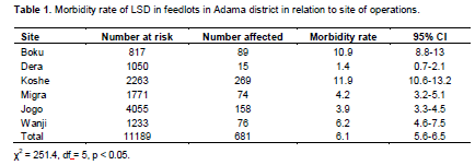
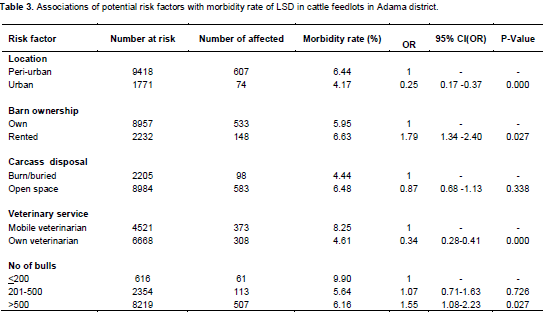
Table 3 shows the associations of potential risk factors with infection of LSD among the bulls in the feedlots based on multivariate logistic regression analysis. The analysis showed that bulls kept in peri-urban sites were more likely infected by LSD than in urban (odd ratio (OR) = 4). Fattening of bulls in rented barn also increased the risk of LSD infection compared to privately owned barn (OR = 1.79). Provision of veterinary service with mobile veterinarians was found to be associated with LSD infections compared to veterinary service provision by own veterinarian (OR = 2.9). Bulls housed more than 500 bulls per barn were more likely at risk of LSD infection than bulls kept less than 200 and between 200 and 500 bulls per barn with odd of 1.55 and 1.45, respectively. Caracas disposal methods were not significantly associated with LSD occurrence.

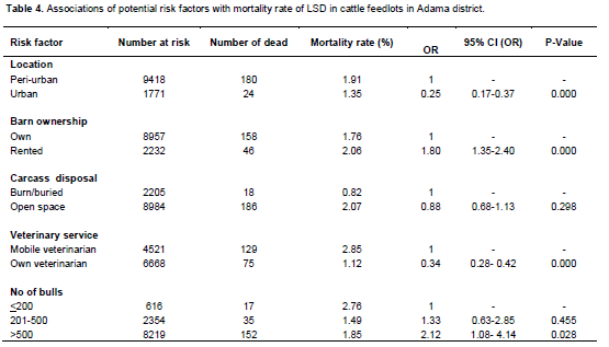
The results of multivariate logistic regression analysis of potential risk factors on LSD mortality are presented in Table 4. According to the analysis, fattening of bulls at peri-urban sites increased the risk of death (OR = 4) due to LSD compared to urban sites. Bulls that finished in rented barn were also more likely at risk of death than privately owned barn (OR = 1.80). Furthermore, the risk of LSD death increased when veterinary services were provided by mobile veterinarians compared to own veteri-narian (OR = 2.94). The bulls that were more than 500 in number per barn were more likely at risk of LSD deaths than those kept at less than 200 and between 200 and 500 bulls per barn with odd of 2.12 and 1.59, respec-tively. Carcass disposal methods were not significantly associated with LSD death.
DISCUSSION
The presence of characteristic skin nodules and outstanding features of the LSD are best indicative of the occurrence of LSD at affected feedlots. LSD infection causes a variable degree of clinical and pathological outcomes. The clinical signs observed in this study such as generalized skin nodules, edema of the dependent parts, ocular discharge and lameness were similar with the clinical signs documented elsewhere (Coetzer, 2004; Brenner et al., 2006; Kumar, 2011; Gari et al., 2010; Salib
and Osman, 2011; Body et al., 2012).
The recurrent LSD outbreaks in feedlots during the rainy season might be linked with the optimum season for the development of insect population (Kitching and Mellor, 1986; Chihota et al., 2001; Gari et al., 2010). LSD vaccines (Neethling strains) were used in the feedlots as part of Sanitary and phytosanitray (SPS) requirements and rules and regulations of animal quarantine. However, vaccination of bulls started a week after the last bull purchased entered the feedlots and were registered by the quarantine service, with a time gap that was sufficient for transmission of the disease to susceptible bulls.
Furthermore, the disease was spread among the vaccinated bulls in the feedlots during epizootic period regardless of vaccination. The problems of vaccine failure and re-infection of vaccinated animals has been also reported by other authors (Hunter and Wallace, 2001; Kara et al., 2003; Brenner et al., 2009; Gari et al., 2010; Kumar, 2011). None of the isolation pens for LSD infected bulls was insect proof with very high probability of viral transmission through mechanical vectors (Kitching and Mellor, 1986; Chihota et al., 2001). It is likely that du-ring LSD outbreak in feedlots, optimal conditions for the spread of LSDV were created through the presence of high numbers of susceptible animals in combination with the poor biosecurity practice and the high abundance of insect vectors.
The magnitude of LSD occurrence varied across sites of feedlots operation during the study period. The ob-served LSD incidence at animal level found in this study was 6.1% which is in close agreement with previous findings from Nekemt area of 7.02% (Regassa, 2003) and 8.1% reported by Gari et al. (2010) in different agro-ecological zones of Ethiopia. Mortality (1.8%) observed during this outbreak was similar with previous reports by Salib and Osman (2011) in Egyptian cattle. Furthermore, the ob-served mortality rate in the present study is in agreement with the previous reports of 2.12% in Ethiopia by Gari et al. (2010). The significant differences observed between the morbidity, mortality and case fatality rates in the six sites might be attributed to variations in availability of suitable condition for the presence of blood-feeding ar-thropods (Ali et al., 1990; Gari et al,. 2010) and difference in biosecurity practice used in the feedlots (Tuppurainen and Oura, 2012).
Fattening of bulls in peri-urban sites was a significant risk factor for LSD infection and death. This might be due to the fact that in peri-urban area there is relatively higher vegetation converge than in urban site which could support biting fly population. Furthermore, in peri- urban site, small holder farmers’ cattle grazed around the feedlots areas could increase the risk of LSD infection and death in peri-urban than in urban areas (Gari et al., 2010). Fattening of bulls in rented barn also increased the risk of LSD infection and death. This could be due to the fact that in rented barns, there is higher probability of feedlot operators’ turnover. This might compromise the biosecurity level of the barn by disposing risky material in the barn and lower probabilities of disinfecting or thoroughly cleaning of the barn between the batches. These in turn increase the probability of the virus trans-mission between the batches since LSDV is remarkably stable and surviving for long periods at ambient temperature, especially in dried scabs (Rovid, 2008).
Provision of veterinary service with mobile veterinarian was found to be significant risk factor for LSD infections and death compared to veterinary service provision by own veterinarian. This might be due to the fact that there is higher probability of using contaminated equipments between feedlots by mobile veterinarian. Therefore, if proper needle hygiene was not practiced, needles contaminated with virulent LSDV during the actual vaccination and treatment procedure serve as vehicle for transmission of the virus (Carn and Kitching, 1995; Magori-Cohen et al., 2012; Tuppurainen and Oura, 2012). Furthermore, mobile veterinarians are less equipped than business owner; there might be higher probability of failure in one or more steps of the cold chain of vaccine. As indicated by Tuppurainen and Oura (2012), lack of appropriate storage facilities for storage of vaccine resulted in inactivation of vaccine because of exposure to direct sunlight or high environmental temperatures during the vaccination process.
Bulls that held more than 500 bulls per barn were more likely at risk of LSD infection and death than those kept in less than 200 and between 200 and 500 bulls per barn. This might be associated with increase opportunity for transmission of the virus by arthropod vectors between bulls. In this study, carcass disposal methods were not significantly associated with LSD infection and death. This might be due to the fact that dead bodies disposed in shallow pits nearby feedlots or throw in open air where insects have easy accesses to disposed carcasses and thus facilitating the transmission of the virus to neighboring feedlots likewise.
CONCLUSION
The present study indicates that LSD outbreak occurred in the cattle feedlots and resulted in bull mortality and morbidity which affect livelihood of business owners and have major threat to national economies as they tend to affect the international trade. The results of this study, indicates that the complex epidemiological situation of the disease in Ethiopia needs more detailed investigation for improved vaccine-based control for it to be achieved efficiently.
ACKNOWLEDGMENT
The authors are grateful to the Faculty of Veterinary Medicine, Addis Ababa University for funding this research. Feedlots operators are highly appreciated for their all-round cooperation during LSD outbreak investigation.
CONFLICT OF INTEREST
The authors declare that they have no conflicts of interest.
REFERENCES
|
Ali AA, Attia EH, Selim A, Abdul-Hamid YM (1990). Clinical and pathological studies on lumpy skin disease in Egypt. Vet. Rec. 127: 549-550. Pubmed |
||||
|
Babiuk S, Bowden TR, Boyle DB Wallace DB, Kitching RP (2008). Capripox viruses: an emerging worldwide threat to sheep, goats and cattle. Transbound. Emerg. Dis. 55:263-272. Crossref |
||||
| Body MK, Singh P, Hussain MH, Al-Rawahi A, Al-Maawali M, Al-Lamki K, Al-Habsy S (2012). Clinico-histopathological findings and PCR based diagnosis of lumpy skin disease in the Sultanate of Oman. Pak. Vet. J. 32(2):206-210. | ||||
| Brenner JD, David A, Avraham U, Klopfer-Orgad I, Samina, Peleg BA (1992). Experimental infection with local lumpy skin disease virus in cattle vaccinated with sheep pox vaccine. Isr. J. Vet. Med. 47:17-21. | ||||
| Brenner JM, Haimovitz E, Oron Y, Stram O, Fridgut V, Bumbarov L, Kuznetzova Z, Oved A, Waserman S, Garazzi S, Perl D, Lahav N, Edery and Yadin H (2006). Lumpy skin disease (LSD) in a large dairy herd in Israel. Isr. J. Vet. Med. 61:73-77. | ||||
|
Brenner J M, Bellaiche E, Gross D, Elad Z, Oved M, Haimovitz A, Wasserman O, Friedgut Y, Stram V, Bumbarov L, Yadin H (2009). Appearance of skin lesions in cattle populations vaccinated against lumpy skin disease: statutory challenge. Vaccine 27:1500-1503. Crossref |
||||
|
Carn VM, Kitching RP (1995). An investigation of possible routes of transmission of lumpy skin disease virus (Neethling). Epidemiol. Infect. 114:219-226. Crossref |
||||
|
Chihota CM, Rennie LF, Kitching RP, Mellor PS (2001). Mechanical transmission of lumpy skin disease virus by Aedes aegypti (Diptera.: Culicidae). Epidemiol. Infect. 126:317-321. Crossref |
||||
| Coetzer JAW, (2004). Lumpy skin disease. In: Coetzer JAW, Justin RC (eds.), Infectious Diseases of Livestock 2nd Ed. Oxford University Press, Cape Town, South Africa. pp. 1268-1276. | ||||
|
Gari G, Bonnet P, Roger F, Waret-Szkuta A (2011). Epidemiological aspects and financial impact of lumpy skin disease in Ethiopia. Prev. Vet. Med. 102 (4):274-283 Crossref |
||||
|
Gari G, Waret-Szkuta A, Grosbois V, Jacquiet P, Roger F (2010). Risk factors associated with observed clinical lumpy skin disease in Ethiopia. Epidemiol. Infect. 138:1657-1666. Crossref |
||||
|
Gari G, Biteau-Coroller F, LeGoff C, Caufour P, Roger F (2008). Evaluation of indirect fluorescent antibody test (IFAT) for the diagnosis and screening of lumpy skin disease using Bayesian method. Vet. Microbiol. 129:269-280. Crossref |
||||
|
Hunter P, Wallace D (2001). Lumpy skin disease in Southern Africa: a review of the disease and aspects of control. J. South Afr. Vet. Assoc. 72:68-71. Crossref |
||||
|
Kara PD, Afonso CL, Wallace DB, Kutish GF, Abolnik C, Lu Z, Vreede FT, Taljaard LCF, Zsak A, Viljoen GJ, Rock DL (2003). Comparative sequence analysis of the South African Vaccine strain and two virulent field isolates of Lumpy Skin Disease Virus. Arch. Virol. 148: 1335-1356. Pubmed |
||||
|
Kitching RP, Mellor PS (1986). Insect transmission of capripoxvirus. Res. Vet. Sci. 40:255-258. Pubmed |
||||
|
Kumar SM (2011). An outbreak of lumpy skin diseases in Holstein dairy herd in Oman. A clinical case report. Asian J. Anim. Vet. Adv. 6(8): 851-859. Crossref |
||||
|
Magori-Cohen R, Louzoun Y, Herziger Y, Oron E, Arazi A, Tuppurainen E, Shpigel N, Klement E (2012). Mathematical modelling and evaluation of the different routes of transmission of lumpy skin disease virus. Vet. Res. 43:1. Crossref |
||||
| Radostits OM, Gay CC, Hinchcliff KW and Constable PD (2007). Veterinary Medicine: A textbook of diseases of cattle, horses, sheep, pigs and goat. 10th Ed. WB Saunders Co., Philadelphia, USA. | ||||
| Regassa C (2003). Preliminary study of major skin diseases of cattle coming to Nekemt Veterinary Clinic. Unpublished DVM Thesis, Addis Ababa University. | ||||
| Rovid SA (2008). Lumpy Skin Disease. The center for food security and public health, Iowa State University. College of Veterinary Medicine. Available at: http://www.cfsph.iastate.edu/DiseaseInfo/disease.php?name=lumpy-skin-disease&lang=en Accessed April 2012. | ||||
| Salib FA, Osman AH (2011). Incidence of lumpy skin disease among Egyptian cattle in Giza Governorate, Egypt. Vet. World 4(4):162-167. | ||||
|
Tuppurainen ESM, Oura CAL (2012). Review: Lumpy skin disease: An Emerging Threat to Europe, the Middle East and Asia. Transbound. Emerg. Dis. 59:40-48. Crossref |
||||
Copyright © 2024 Author(s) retain the copyright of this article.
This article is published under the terms of the Creative Commons Attribution License 4.0


