Full Length Research Paper

ABSTRACT
A cross-sectional study was employed to estimate the prevalence of skin diseases in small ruminant and risk factors associated to its occurrence in Gamo Gofa zone from July, 2012 to April, 2014. The study areas were clustered into two agro-ecological zones; lowland and highland area. A total of nine hundred (450 sheep and 450 goats) were examined. Detailed physical examinations and systemic examinations, followed by skin scraping and laboratory tests were carried out to diagnose skin diseases. The Pearson’s chi-square (χ2) test was used to assess the degree of association between skin diseases and risk factors. The overall prevalence was found to be 42.33% (381/900). Significantly higher prevalence (p<0.05) of small ruminant skin disease was observed in goats (52.22%) than sheep (38.66%). Furthermore, the study also revealed significantly higher prevalence (p<0.05) in unvaccinated (42.92%) than vaccinated (29.52%) group of animals. The occurrence of skin diseases was statistically significantly associated with age and sex of animals. The prevalence was higher in males (30.38%) than females (22.49%), and in young age groups as compared to adults. The external parasites identified include manges (Sarcoptic, Chorioptic, Psoroptic and Demodectic), ticks (Ambyloma varigatum, Rhipicephalus evertisi evertisi and Boophilus decloratus), lice (Bovicola species and Linognathus species) and sheep ked (Melophagus ovinus). Viral infections, predominantly of pox virus infection were noted in sheep (10.44%) and goat (13.11%) and contagious ecthyma 2.44% on sheep and 2.00% on goats. The overall prevalence of viral disease showed significant association (p<0.05) with vaccination history and age of the study animals. The high prevalence of skin disease on small ruminant has shown there is urgent need for its strategic prevention and control, as skin and hides represent the second major export commodity of the country. It is recommended that external parasite control should be strategically designed and technologically verified in local context.
Key words: Ethiopia, external parasites, Gamo Gofa, goat, sheep, prevalence.
INTRODUCTION
Small ruminants contribute 35 and 14% of meat and milk consumption, respectively in Ethiopia (Kebede, 2013). Hide and skin export has got the largest share of animal products next to live animal export and skin is the most important item in generating foreign currency, next to coffee (Kumsa et al., 2012). Ethiopia supplies a wide range of both processed and semi-processed sheep and goat skins to the world market (Kebede, 2013). Whereas hides and skins account for 12 to 16% of the total value exports in Ethiopia (Tefera, 2012). The current utilization of hides and skins is estimated to be 48% for cattle hide, 75% for goat skin and 97% for sheep skin with the expected off take rate of 33% and 75% for sheep, goat and cattle, respectively (Berhe, 2009; Yacob et al., 2008 and Tefera, 2012). In Ethiopia, hides, skins, leather and leather products are the most widely traded agro-based livestock commodities with an estimated value of over US$100 billion/year and continues to conspicuously exhibit a huge unexplored potential (Mekonnen et al., 2013). Even though small ruminants are important components of Ethiopian farming system, their contribution to food production, rural income and export income are far below the expected potential (Mekonnen et al., 2013). This is because small ruminant production not only in Ethiopia but in most of the developing countries is constrained by complemented effects of prevailing diseases, subclinical parasitism, poor feeding and managements (Abadi, 2000; Singla, 1995; Tefera, 2012; Yacob et al., 2008; Yacob, 2013). Over the last 10 years, there are indications that the quality of raw material has deteriorated with an increasing number of reject grades and the appearance of skin diseases like “Ekek or cockle” that is mainly due to sheep ked and lice infestation (Assefa et al., 2011). The low quality of skins undermines the competitiveness of the industry, as it leads to low factor inputs productivity especially in the tanning process (Mekonnen et al., 2013).
Among the diseases of small ruminant skins, infestations by mange mites, ticks and infections by pox disease and dermatophilosis possess considerable economic losses, particularly to the skin export due to various defects (Dessie et al., 2010). Losses from these diseases and other skin abnormalities are leading to downgrading and rejections of skins; unfitness to the leather industries (Kebede, 2013; Kumsa et al., 2012). Skin diseases in small ruminants were reported from different parts in Ethiopia. Kumsa et al. (2012) reported the prevalence of 48.1% in central Ethiopia, Dessie et al. (2010) reported a mange mites prevalence in Wolaita area of southern Ethiopia 1.98 and 3.85% in sheep and goats, respectively. A study of tick infestation in small ruminants in Bedelle district, Western Ethiopia, revealed prevalence of 66.12 and 80.7% in goats and sheep, respectively reported by Fufa et al. (2012). Many other studies and reviews conducted at different regions of the country reported the importance of small ruminant’s skin diseases (Kebede, 2013; Yacob, 2013; Assefa et al., 2012; Tewodros et al., 2012; Dessie et al., 2010; Yacob et al., 2008; Tefera, 2012; Haffiz, 2001; Abadi, 2000). Despite various study on the prevalence and associated risk factors in different parts of the country, it is not known in Gamo Gofa zone of Southern Ethiopia. Furthermore, this area is known to border with the major pastoral livestock production area of South Omo zone, and serving as a route of market from the lowland pastoral production areas to the highland meat value chain areas of highland markets. In due concern to the above facts, and to seek as whether there is need and/or serve as baseline information, this study was initiated. The objectives were to estimate the prevalence and assess associated risk factors of skin diseases, and to determine the etiological agents of skin diseases in small ruminants.
MATERIALS AND METHODS
Study area
The study was conducted on three districts, categorized into two agro-ecological zones, namely Demba Gofa and Zala district, representing lowland, and Geze Gofa district for highland. Lowland categories were areas ranging from 800 to 1500 meter above sea level (masl) but those ranging from 1500 to 2800 masl were considered as highland. The average annual rainfall of the study areas were varying from 950 to 1150 mm, characterized by a bi-modal type of distribution. The mean annual minimum and maximum temperature were 15.4°C and 37.2°C in the highland and lowland, respectively.
Study animals
The study population animals were extensively managed, almost all are reared in mixed species herd type as an individually owned herd or a group based. A total of 900 small ruminants (450 goats and 450 sheep) were sampled with systematic random sampling. Ages of animals were addressed in two category, young (below 2 years) and adult (above 2 years) of age according to (Bersisa et al., 2013; Gatenby, 1999). The body condition score (BCS) was considered in three categories, poor (BCS of 1 and 2), medium (BCS of 3) and good (BCS of 4 and 5) according to Desta et al. (2001) and Tefera (2012).
Sampling method and sample size
Study districts were selected purposively to represent different agro-ecology and accessibility whereas study Peasant Associations (PA) was randomly selected. 10% of shoat herds were selected from each PA's and individual animals were selected from the population by systematic random sampling approach. The sample size was determined according to Thrusfield, 1995, with expected prevalence of 50%, as there were no such study in the area and 95% confidence interval was considered. Despite this, the calculated sample size was 380, it was increased to 450 from each species and a total of 900 animals were included in the study.
Study designs
A cross-sectional study design was employed to estimate the prevalence of skin diseases and identify the causal factors for different skin diseases of small ruminants. The age, sex, species, body condition scoring, geographical location, season of the year and vaccination were considered as test variables to see if these were risk factors associated with disease occurrence or not. Animals with visible skin problems suspected for bacterial and fungal infections were subjected to skin scrapings. Both skin scrapings and visible external parasites such as ticks, sheep ked and lice were shifted to universal sampling bottle, labelled, preserved and transported to Gofa Universal college department of animal health for species identifications and subsequent laboratory confirmation. 70% ethyl alcohol or 10% formalin were used to preserve adult external parasites recovered and 10% potassium hydroxide (KOH) was used as a cleaning agent for skin scrapings. Species identification and laboratory test on skin scraping was conducted according to Soulsby (1982) and Urquhart et al. (1996).
Sample collection and identification of etiological agents
Physical inspection was conducted to assess the presence of external parasites and gross skin lesions, followed by palpation of all parts of the body. External parasites, sheep keds, ticks and lice were manually collected using tissue forceps and identified under stereomicroscope according to the morphological keys described by (Soulsby, 1982; Urquhart et al., 1996; Taylor et al., 2007). Skin scrapings were collected when suspected for mange mites, fungal and bacteriological lesions. This was done by clipping the hair around the lesion, scrapping the edges of lesions with scalpel blade until capillary oozing was evident (Bersisa et al., 2013; Urquhart et al., 1996). The scrapped materials were either directly shifted to clean microscopic slide or container for laboratory examination. A few drops of 10% potassium hydroxide (KOH) was added to skin scrapings and allowed to stand to 24 h until the time of examination under 4x, 10x, and 40x magnifications of light microscope (Bersisa et al., 2013). Laboratory tests were limited only to bacteriological, fungal and parasitological examinations (arachino-entomology). Skin scrapings suspected for bacterial infections were handled aseptically in clean and disinfected sampling bottles and test tubes. Gram staining was the principal bacteriological test conducted for samples suspected for Dermatophilosis and Giemsa staining was used to test fungal pathogens suspected. Viral diseases like sheep and goat pox (SGP) and contagious ecthyma were tentatively diagnosed on the bases of field level physical clinical examinations.
Data management and statistical analysis
The data entered into Microsoft excel spreadsheet, imported to statistical software for social science (SPSS) version 20.0 for windows. Descriptive statics such as tables, graphics, averages and percentages were used to summarize and present the results of the collected data. The Pearson’s chi-square (χ2) test was used to assess the degree of association between skin diseases with the various risk factors. In all cases, 95% confidence interval (CI) and 5% absolute precision was used for statistical analysis.For all conditions, a p-value of less than 0.05 (p < 0.05) was taken as significant association.
RESULTS
Prevalence of skin diseases
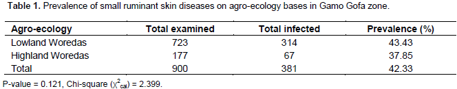
Animal was classified as positive to skin disease if it has at least one of the clinical abnormalities associated to skin disease or is infested with an external parasite. The overall prevalence of skin diseases in small ruminants was found to be 42.33% (381/900). On species bases, the prevalence was 52.22 and 38.66% in goats and sheep, respectively. Small ruminants from lowland agro-ecology revealed higher prevalence (43.43%) of skin diseases than the highland (37.85%) (Tables 1 and 2).
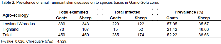
On species bases, statistically significant (p=0.026) variation in prevalence of skin disease was observed on goats 235 (52.22%) than sheep population 174 (38.66%). Except at Geze Gofa district, in which comparatively larger sheep populations manifested skin disease 52 (48.6%). In the rest two districts, higher prevalence was seen on goat population than sheep populations (Table 2).
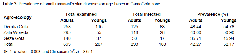
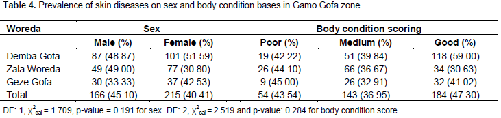
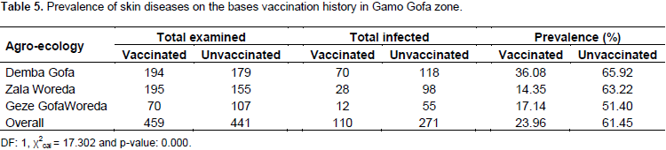
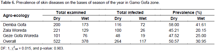
On age bases, there is statistically significant (p=0.003) difference on prevalence of skin diseases in young and adult small ruminants. Greater prevalence was recorded in young age groups as compared to adults (Table 3). Whereas, there were no statistically significant difference in the occurrence of skin diseases in small ruminants on sex bases (Table 4), body condition scores and season of the year (Table 6). On the bases of vaccination history, there was statistically significant difference (p=0.000) on occurrence of skin diseases in the study area. The study revealed higher prevalence of skin diseases, 61.45% in unvaccinated small ruminants as compared to vaccinated ones, 23.96 % (Table 5).
Prevalence of external parasites
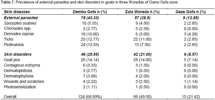
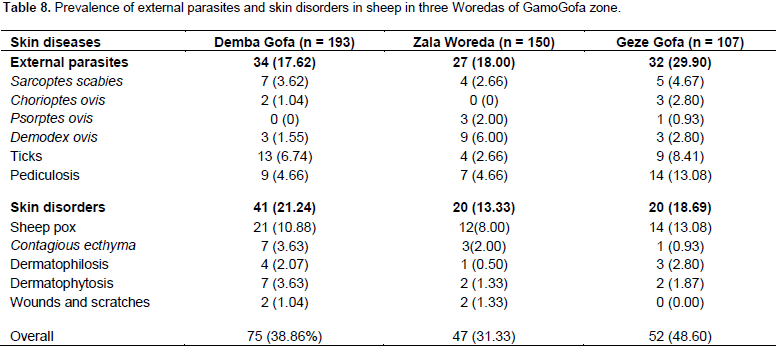
The study revealed significantly higher prevalence of external parasites (p<0.05) in goat population than sheep population. Furthermore, the study also revealed significantly higher prevalence (p<0.05) of external parasite infestation in males (30.38%) than in females (22.49%). When external parasite infestation was calculated, in goats: 78 (43.33%), 57 (28.50%) and 9 (12.85%) were infested at Demba Gofa, Zala and Geze Gofa Woredas, respectively. However, the prevalence of external parasites in goats was not showing statistically significant difference (P>0.05) among the three Woredas (Table 7). The current study also revealed the external parasite prevalence of 34 (17.62%), 27 (18.00%) and 32 (29.90%), respectively on sheep population from Demba Gofa, Zala and Geze Gofa Woredas. However, there was no statistically significant difference (P>0.05) in prevalence of external parasites of sheep among the three Woredas of Gamo Gofa zone (Table 8). In infested goats, the predominant external parasite identified was tick species (Ambyloma varigatum, Boophilus decoloratus and Rhipicephalus evertisi evertisi) accounted to be 23 (12.77%), 22 (11.00%) and 2 (2.85%), respectively at Demba Gofa, Zala and Geze Gofa Woredas. However the variation in prevalence of ticks was not statistically significant (P>0.05) with respect to agro-ecological zones, age, sex and body condition scoring. In goats, the other external parasite identified next to ticks was pediculosis (lice infestation) occurring with the prevalence of 13.33, 7.5 and 2.85% at Demba Gofa, Zala and Geze Gofa Woredas, respectively. The prevailing lice species identified were Linognathus species and Bovicola species from sucking and biting groups of lice (Table 7 and 8). As far as mange mite infestation in goat population was concerned, Sarcoptic mite was occurring at the prevalence of 5.55, 4.5 and 2.85% in Demba Gofa, Zala and Geze Gofa Woredas, respectively. Chorioptic mite (2.77, 2.50, and 0%), and Demodex caprae (10.00, 3.00, and 4.28%) were also recorded in the three respective Woredas above (Table 7 and 8). External parasites identified on the sheep was lice infestation, accounted to be 13.08, 4.66, and 4.66%, respectively at Geze Gofa, Zala and Demba Gofa Woredas. Similar species of lice as recovered in goats. Prevalence of pediculosis was not statistically significantly different (P > 0.05) among sheep of the three Woredas of Gamo Gofa Zone (Table 8). Ticks were the other dominant external parasites observed even though the prevalence was not showing statistically significant difference (P > 0.05) among small ruminants of the 3 Woredas of Gamo Gofa zone, Southern Ethiopia. Sarcoptic mite, Chorioptic mite, Psoroptic mite and Demodex ovis were also identified in sheep population, but their prevalence was at very low rate.
Prevalence of bacterial, fungal and viral infections
The predominant viral infection on small ruminants was caused by pox disease, prevalence of 14.44, 11.00, and 7.14% in goats and, 10.88, 10.66 and 9.35% in sheep at Demba Gofa, Zala and Geze Gofa Woredas, respectively. Occurrence of pox disease was not statistically significantly associated (P>0.05) with agro-ecology, species, sex and body condition scores, whereas, there was statistically significant variation (p<0.05) on age group base; higher in young as compared to adult (Table 8). The occurrence of pox disease in both sheep and goat was limited only to unvaccinated small ruminants.
DISCUSSION
The current study revealed the overall prevalence of 42.33% for small ruminant skin diseases. The record of significantly higher skin disease prevalence from goat species than sheep was suggested as due to variation in animal husbandry system. Traditionally, sheep and sheep products like skin are relatively expensive than goat and goat products in all three Woredas of the study areas and this finding is in agreement with the explanation underlined by Tekle et al. (2009) and Yacob (2013). So, the underlying reason for increased exposure of goats to skin disease was lack of care in terms of veterinary service, feeding, vaccination and housing. The finding of current study is in agreement with Rahmeto et al., (2011) who had reported an overall skin disease prevalence of 51.7% on sheep and 59.60% on goats at Tigri Regional State of Ethiopia and, a 48.1% overall skin disease prevalence reported by Yalew (2014) at Wolita-Sodo, Southern Ethiopia.
However, the current result is higher than that of previous works conducted by Dessie et al. (2010), on small ruminant mange mite; 1.98% on sheep and 5.85% on goats at three ecological zones of Wolita-Sodo, Southern Ethiopia. The variations could be attributed due to seasonal variation, variation in animal husbandry system and/or geographical location of the study areas. Relatively higher overall prevalence in current study (42.33%) could be due to very limited intervention for external parasite prevention and control in the area and overall poor veterinary facility in the area. The sharing of different flocks of animals to communal watering and grazing sites could have facilitated the establishment and spread of external parasite infestation and contagious skin infections in the area. This finding is also in agreement to Yacob et al. (2013). Statistically significant difference in prevalence (p<0.05) was observed from unvaccinated small ruminants than vaccinated groups. The finding suggests that immunizing the animals is an important strategy to prevent skin diseases especially those originated from bacterial and viral groups. Furthermore, the study revealed significantly higher overall small ruminant skin disease prevalence in young animals than adults, which could be because of their low acquired resistance compared to adults. Many previous works agrees with the finding of this research (Yacob et al., 2013; Fufa et al., 2012). The finding of higher prevalence of external parasite in goats than sheep in current report is in agreement with previous reports by Dessie et al. (2010), Kebede (2013), Yacob (2013) and Zenaw and Mekonnen (2012). The higher prevalence of external parasite in male small ruminants of both species could be due to the fact that anatomically the skins of male goats and sheep have heavy course grain nature that lacks tensile strength, while female skins have better strength in nature (Tekle, 2009), not favourable for external parasite infestation. The tick species identified by this study were Ambyloma varigatum, Rhipcephalus evertisi evertisi and Boophilus decoloratus in agreement to Yacob et al. (2008); reported the presence of these three tick genera in Ethiopia. The occurrence of small ruminants infested by ticks by current study is comparatively lower than the previous reports; 66.12% in goats and 80.7% in sheep by Fufa et al. (2012) around Bedelle district of Oromia regional state, Ethiopia. The current study revealed both burrowing (Sarcoptic mange, Demodectic mange) and non-burrowing mites (Chorioptic and Psoroptic mange) in both species of the study animals. These findings are in agreement to the reports of Dessie et al. (2010), Yacob et al. (2008) and Haffiz (2001), whereas, relatively larger prevalence of mange mites in sheep (69.3%) and goat (57.3%) was reported Kassa et al. (2013).
CONCLUSION AND RECOMMENDATIONS
Ethiopia has a huge small ruminant population, endowed with great potential of attractive global market for skin. However, the contribution of skin in the national export income and enhancing the earnings from the skin to its producers are disproportionately small as a result of various skin diseases. The causes of deteriorated quality of skin were external parasites infestation, bacterial, viral and fungal diseases, and the poor animal husbandry practices such as poor nutrition and improper slaughter and flying operations. Moreover, poor veterinary infrastructure, lack of awareness and absence of a designed strategy in prevention and control of skin diseases continued to be a problem of deterioration of skin quality.
1. Therefore, awareness creation among farmers about the impacts of skin diseases and improving livestock extension system was recommended.
2. Control of external parasites through combination rotational grazing, sound husbandry practices and application of acaricides should also be encouraged.
3. More importantly, vaccinating both sheep and goat population against pox diseases and bacterial pathogens across the study Woredas was also suggested.
4. Further study should be conducted in designing integrated skin diseases prevention and control regimen on the bases of species dynamicity and seasonal occurrence.
ACKNOWLEDGEMENTS
The authors would like to express heartfelt thanks and appreciation to Dr Gizat Almaw from Sebeta National Veterinary Research Institute and Yetinayet Aragaw from Addis Ababa University for their technical support. Deep appreciation also goes to staffs of Gofa Universal College, Department of Animal Health and the management of the college for allowing the use of their laboratory.
CONFLICT OF INTERESTS
The authors have not declared any conflict of interests.
REFERENCES
|
Abadi Y (2000). Current problems of leather industry: The opportunities and challenges of enhancing goat production in East Africa: Proceedings of Conference held at Debub University, Hawassa, Ethiopia. E(kika) de la Garza institute for Goat research. OK: Langston pp. 139-143. |
|
|
Assefa M, Tesfaye D, Taye M (2011). A study on the prevalence of sheep and goat skin defects in Bahir Dar tannery, Ethiopia. Onl. J. Anim. Feed Res. 2(4):384-387. |
|
|
Berhe A (2009). Assessment of hides and skin marketing in Tigri region. The case of Atsbi Wemberta Woreda, Eastern Tigri: Msc thesis: Addis Ababa University School of Graduate Studies College of Development Studies, pp. 1-37. |
|
|
Bersisa M, Jemberu A, Tamirat M (2013). Skin defects of small ruminants in Africa. Ethiop. Vet. J. 321-322. |
|
|
Dessie S, Hailu D, Dereje B (2010). Epidemiological study of small ruminant mange mites in three agro-ecological zones of Wolita, Southern Ethiopia. Ethiop. Vet. J. 14(1)31-38. |
|
|
Desta H, Girma A, Alemu Y (2001). Body condition scoring of sheep and goat. Technical Bulletin Number 8. Ethiopian Sheep and Goat Productivity Improvement Program (ESGPIP), Addis Ababa, Ethiopia, 2:109-134. |
|
|
Fufa A, Josen T, Alemayehu R (2012). Status of tick infestation in small ruminants of Bedelle district: Oromia region, Ethiopia. Glob. Vet. 8(5):459-462. |
|
|
Gatenby R (1999). Sheep major health in tropical production system. In: The Tropical Agriculturalist, Macmillan Education. London: UK pp. 7-8. |
|
|
Haffiz M (2001). Study on skin disease of small ruminants in central Ethiopia. DVM thesis: Faculty of Veterinary Medicine, Addis Ababa University, DebreZeit, Ethiopia, pp. 12-32. |
|
|
Kassa B, Asegid S (2013). Enhancing economic growth through control of livestock skin diseases in Ethiopia. 27th EVA Annual Conference Proceedings, Addis Ababa, Ethiopia. Ethiop. Vet. J. pp. 85-93. |
|
|
Kebede M (2013). Effects of small ruminant ecto-parasites in tanning industry in Ethiopia: O Revion. J. Anim. Sci. Adv. 3(9)424-430. |
|
|
Kumsa B, Beyecha K, Goleye M (2012). Ectoparasites of sheep in three -agro-ecological zones in central Oromia region, Ethiopia. J. Vet. Res. pp. 1-8. |
|
|
Mekonnen H, Nicholas M, Mwinyikione M (2013). Unlocking the potential of Ethiopian leather value chain: Livestock based extension role. COMESA Leather and Leather Products Proceedings (LLPI): 27th Ethiopian Veterinary Association Proceedings, Addis Ababa, Ethiopia. Ethiop. Vet. J. pp. 51-66. |
|
|
Rahmeto A, Dessie S, Mekalash T, Bekele M, (2011). Prevalence of small ruminant external parasites and associated risk factors in selected districts of Tigri region, Ethiopia. Glob. Vet. 7(5)433-437. |
|
|
Singla D (1995). A note on sub-clinical gastro-intestinal parasitism in sheep and goats in Ludhiana and Faridkot districts of Punjab. Indian Vet. Med. J. 19:61-62. |
|
|
Soulsby E (1982). Helminthes, arthropods and protozoa of Domesticated animals London: Bailliere Tindal. pp. 92-106, 136-166. |
|
|
Taylor M, Coop R, Wall R (2007). Veterinary Parasitology. 3rded . Oxford, UK: Blackwell Publishing, pp. 459-475. |
|
|
Tefera S (2012). Investigations on external parasites of small ruminants in selected sites of Amhara regional state and their impact on tanning industry. MSC thesis, Faculty of Veterinary Medicine, Addis Ababa University, Ethiopia, pp. 1-37. |
|
|
Tekle Z, Alemu Y, Merkel R (2009). Common defects of sheep and goat skins in Ethiopia and their causes: Technical Bulletin at Addis Ababa, Ethiopia, pp. 1-59. |
|
|
Tewodros F, Tsegedingle Y, Mersha C (2012). Bovine demodicosis: Threat to leather industry in Ethiopia. Asian J. Agric. Sci. 4(5)314-318. |
|
|
Thrusfield M (1995). Veterinary Epidemiology. 2 nd ed, Blackwell Science. |
|
|
Urquhart M, Armor J, Duncan L, Dunn A, Jennings F (1996). Veterinary Parasitology. 2nd ed. London: Blackwell Science, pp. 123-192. |
|
|
Yacob H (2013). Skin defects in small ruminants and their nature and economic importance: The case of Ethiopia. Glob. Vet. 11(5):552-559. |
|
|
Yacob H, Nesanet B, Dinka A (2008). Prevalence of major skin diseases in cattle, sheep and goats at Adama Veterinary Clinic, Oromia Regional State, Ethiopia. Rev. Med. Vet. 159:8-9. |
|
|
Yalew T (2014). Study on the prevalence of ecto-parasites in small ruminants in and around Wolita Sodo, Southern Ethiopia. Trop. Anim. Health Prod. 47:27-35. |
|
|
Zenaw Z, Mekonnen A (2012). Assessment of major factors that cause skin defects at Bahir Dar Tannery, Ethiopia. Adv. Biol. Res. 6(5)177-181. |
|
Copyright © 2024 Author(s) retain the copyright of this article.
This article is published under the terms of the Creative Commons Attribution License 4.0


