ABSTRACT
A cross sectional study was carried out from October 2010 to April 2011 in Gondar Elfora Abattoir to determine the prevalence of paramphistomosis in cattle (local, cross) breeds which were came from highland, mid highland and lowland areas. Three hundred eighty-four (384) cattle were included for routine ante-mortem and postmortem examination for the presence of paramphistomum. The parasite was examined grossly and under microscope to appreciate the morphology of adult paramphistomum. Out of 384 cattle examined, 199 (51.82%) were found to be positive for paramphistomosis. From 199 infected cattle fluke burden at organ level 125(62.81%) was in rumen, 40(20.1%) was in reticulum and 34(17.09%) was found mixed (rumen and reticulum). The existence of paraphistomum in respect to organ and origin, 56(44.80%) was in rumen and 20(50%) was found in reticulum predominantly in high and low land respectively. The highest infection of cattle with paramphistomum species was found during October to November. However, there is no statistical significance variation (p >0.05) between the prevalence of paramphistomum and that of origin, breed, and age groups of the animals. Integrated control approach using selected anthelmintic therapy and snail control to reduce the magnitude of the problem was suggested as a recommendation.
Key words: Elfora abattoir, cattle, Gondar, paramphistomosis, prevalence
Ethiopia has the largest livestock and draft animal population in the continent. There are approximately 44, 318, 877 cattle, 23, 619, 720 sheep, 23, 325, 113 of goats, 6 million equines, 2.3 million camels and 43 million poultry (CSA, 2011). Its livestock productivity, despite its huge population size, remains marginal due to prevalent diseases, malnutrition and management constraints. Parasitism represents a major obstacle to the development of sub-sector and bovine paramphistomosis is one of the most important parasitic diseases of cattle causing mortality and production losses in various parts of Ethiopia. Paramphistomosis is the priority disease in the highland as well as in lowland areas of Amhara regional state (CSA, 2010).
Paramphistomosis is distributed all around the world, but the highest prevalence has been reported in tropical and sub tropical regions, particularly in Africa, Asia, Australia, Eastern Europe and Russia. The epidemiology of Paramphistomum is determined by several factors governed by parasite-host-environment interactions. The major epidemiological variable influencing worm burdens of animals is the infection rate from pastures. It is also influenced by the climatic requirement for egg hatching, development and survival of the larvae in pasture (Ozdal et al., 2010).
The adult paramphistomum is found in the rumen and reticulum but the immature parasite is found in the duodenum. Adult paramphistomum are mainly parasitic in the fore stomachs of ruminants, although a few species occur in the intestine of ruminants, pigs and horses. Their shape is not typical of the trematodes, being conical rather than flat. All require a water snail as an intermediate host (Taylor et al., 2007).
Paramphistomosis causes a great economic loss in terms of decrease in milk and meat production, loss of weight treatment cost of diseased animals and additional labor required for handling such animals. Mortality rate in young animals is very high (Javed, 2008). It is caused by P. cervi, P. epiclitum, P. microbothriodes, (Chowdhry and Tada, 1994; Kassai, 1999; Rinaldi et al., 2005; Shanila and Hafeez, 2005; Sripalwit et al., 2007). This disease is accompanied by fatal diarroea, weakness, dehydration and deacresed milk yield, submaxillary edema and death thereby causing great economic loss to the livestock industry (Horak, 1971; Georgi et al., 1999; Mclaren et al., 2006; Merianos, 2007; Bianchin et al., 2007). Heavy infections with immature flukes in the upper small intestine can cause serious ill health and death (Panda, 1985; Urquhart et al., 2000).
Outbreaks of disease generally occur in the drier months of the year when the receding water uncovers herbage contaminated with encysted metacercariae in these areas. Dispersal of snails by flooding events and changes in farm-management practices may be responsible for the apparent emergence of the parasite (Foster et al., 2008). In spite of the aforementioned prevailing situation and the presence of a number of problems due to gastrointestinal parasites there is scarcity of well-documented information on the occurrence of Paramphistomum in ruminants in Ethiopia. The study was designed with the aims of determining the prevalence of paramphistomum in cattle slaughtered at Gondar Elfora Abattoir.
Study area description
The study was conducted in North Gondar, Northwestern part of Ethiopia. Gondar is located 727 km Northwestern of Addis Ababa in Amhara regional state. It is divided into three major agro-climatic zones: highland, mid-highland and lowland. The altitude ranges from 4620 m in the Semen Mountain in the North to 550 m in the West. The rainfall varies from 880 mm to 1772 mm, while the minimum and maximum temperatures are in the order of -10°C in the highland and 44.5°C in the West (low land). The area is also characterized by two seasons, the wet season from June to September, and the dry season from October to May. According to Gondar town agricultural office (2006), the livestock populations of Gondar registered were, 1,936,514 cattle, 524,083 sheep, 682,264 goats, 2,124,000 poultry, 223,124 donkeys, 12,473 mules, 36,828 horses and 606 camels (CSA, 2011).
Study population and sampling technique
The study animals were cattle (local, cross) breeds of different ages and body conditions brought from highland, mid highland and lowland areas to the abattoir for the purpose of meat production. Bovine breeds were categorized in to adult (3-7 yrs) and old (>7 yrs). Systematic random sampling technique was used to select the study units i.e. the animals were selected in a way that the first was taken randomly and the rest were selected in 5th round.
Study design and sample size determination
A cross sectional study was conducted to determine the prevalence of Paramphistomum infection in cattle from October 2010 to April 2011 in Gondar Elfora Abattoir. The desired sample size was determined by the formula given in (Thrusfield, 2007) with 95% of confidence interval and 5% desired precision with expected prevalence of 50%.
N = (1.96)2x pexp (1-pexp) / d2
Where N=number of sample size, pexp=expected prevalence, d2=Absolute precision.
Therefore, based on the aforementioned formula 384 were considered in the study.
METHODOLOGY
Ante mortem examination
Ante mortem inspection was carried out on the animals before slaughter to assess their general health status. During ante mortem examination, detail records about the species, breed, sex, age, origin and body condition of the animals was recorded. General physical examinations of animals were conducted.
Post mortem examination
During postmortem examination rumen and reticulum was systematically inspected for the presence or absence of adult paramphistomum and fluke burden using the routine meat inspection procedures which consist secondary examination, if evidence of paramphistomum were found they are recorded separately. The primary examination (visualization and palpation) is not showing positive or negative of paramphistomum where as the secondary examination involves further incisions of the rumen and reticulum to observe paramphistomum.
Identification of paramphistomum
For identification, the collected flukes were placed on Petri dish and observed through stereo microscope to appreciate the morphology. Final identification of Paramphistomum was done based on morphology of flukes; shape, posterior sucker (acetabulum), anterior sucker, terminal genitalium and tegumental papillae following the standard guidelines given by Urquhart et al. (1996).
Data analysis
All the data collected during the study from the abattoir were recorded in the format developed for these purpose and later on entered into Microsoft excel spreadsheet. Statistical analysis for categorical variables such as sex, age, origin and breed, was expressed in percentages by using Intercooled STATA 11.0 software.
Overall prevalence
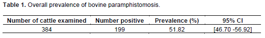
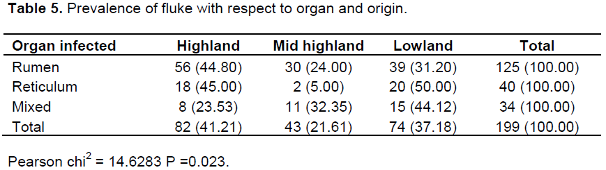
In this study, a cross-sectional investigation on the occurrence of bovine paramphistomosis was carried out between November 2010 and April 2011. A total number of 384 samples (cattle) were included to check the presence of paramphistomum in Gondar Elfora Abattoir. Out of 384 slaughtered cattle, 199 (51.82%) were found to harbor paramphistomum parasites (Table 1). Analysis of the result on the basis of origin and age was made. However, there is no statistically significant variation (P>0.05) in prevalence observed between origin and age of animals examined (Tables 2 to 5).
The study found that the overall prevalence of paramphistomosis in bovine was 51.82% (199/384). This finding is higher than the prevalence rate 20% found by Haridy et al. (2006) from Egypt, 16.6% Jithendran (2000) from India, 23.8% by Juyal et al. (2003) and 5.94% by Hafeez (2005) from India, 13.6% in Turkey by Sevimli et al. (2005), 17.1% by Phiri et al. (2006) from Zambia, 28% from Thialand by Morakot and Sakchai (2006). The difference may be due to difference in geographical regions and varied environmental conditions. The occurrence of paramphistomosis in an area is influenced by a multifactorial system that is composed of hosts, parasitic agents, transmission process and environmental effects (Radostits et al., 2000).
The current finding approaches that of Keyyu et al. (2006) which is 42.1% from Tanzania; Chingwena et al. (2002) which is 37.6% from Zimbabwe, and Phiri et al. (2006) which was 51.6% in Zambia. However, this finding is lower than that of Rolfe et al. (1991) reportedly 98%, and Lee and Lee (1971) reportedly 70%. The variation in the rate of prevalence may be attributed to environmental conditions, managemental conditions, parasites and use of antiparamphistome drug agents.
The presence of paramphistomosis and difference in their prevalence is influenced by local climatic conditions presence or absence of water reservoirs, lakes, rivers and availability of suitable intermidiate hosts. Maqbool et al. (2002, 2003), Narcis et al. (2004) and Diaz et al. (2007) reported that irrigation canals have a role in distribution of paramphistomosis eggs. An increased incidence of paramphistomosis in adult cattle has been reported in the present study. The finding agreed with the reports of Keyyu et al. (2006) who reported 75.2% prevalence in adults and 47.2% prevalence in young animals. These results differ with those of Juyal et al. (2003) 23.8%; and Shanila and Hafeez (2005) 5.94%. The relatively high frequencies could be associated with nutritional and climatic stress, such as altitude, rainfall, and temperature and livestock management system. As different herds of animals come in close contact at available communal watering and grazing sites (contact points) because of the feed scarcity, the establishment and spread of paramphistomosis were favored. Furthermore, adult animals were significantly more frequently affected than young regarding paramphistomosis because the young may not move to the grazing land (they stay around the house).
During the dry periods, breeding of the snails and development of the larval flukes slow down or stop completely and snails undergo a state of aestivation (Armour, 1975; FAO, 1994; Soulsby, 1982; Urquhart et al., 1986). Although a decreasing trend was observed along with the advancement of the dry season, relatively high prevalence rates were recorded throughout the study period. This may be attributed to infections acquired during previous peak snail activity season. In addition the existence of permanent suitable ecological conditions in areas like lake-borders, slowly flowing rivers and low lying marshy areas may contribute to persistent but low-grade infection during the dry season.
Two species of paramphistomum were identified during the study period; however, Paramphistomum clavula was the most prevalent (57.79%) species compared to Paraphistommum cervi (30.15 %) and mixed infection (12.06%). The highest prevalence rate was analyzed during October, when the wet-ecological conditions still prevailed. It has been described that the bionomic requirements for breeding of the planobid snails and development of the intermoluscan stages of the flukes often reach the optimum threshold during the wet months of the year. During the dry periods, breeding of the snails and development of the larval flukes slow down or stops completely and snails undergo a state of aestivation (Radostits et al., 2000).
CONCLUSION AND RECOMMENDATIONS
In general, paramphistomosis is one of the major obstacles for livestock development in Ethiopia by causing remarkable production losses at different parts of the country. This is due to the fact that the area of origin of the animals is suitable for the survival of the snail intermediate host and the parasite. Paramphistomum burdens varied seasonally and were dependent upon the number of infected host snails. Peak fluke burdens and clinical paramphistomosis occurred in late summer and early winter. Based on the aforementioned conclusion, integrated control approach using selected anthelmintic therapy and snail control should be implemented to reduce the magnitude of the problem. In addition, awareness of the producers about the disease should be raised to enable them actively participate in the control programs. Finally, further information on the epidemiology, ecology and biology of the intermediate host snail should be gathered to help in proposing and implementation of disease control programmes.
The authors have not declared any conflict of interest.
Authors would like to thank Gondar Elfora for their material and technical supports.
REFERENCES
|
Armour J (1975). The epidemiology and control of bovine paramphistomosis. J. Vet. Rec. 96:198-201.
Crossref
|
|
|
|
Bianchin C, Kichel T, Honer R (2007). The effect of the control of endo and ectoparasites on weight gains in cross breed cattle in the central region of Brazil. Trop. Anim. Health. Prod. 39(4):287-296.
Crossref
|
|
|
|
|
Central Statistical Authority (2010). Ethiopia Agricultural sample enumeration report on livestock and farm implement part IV. Addis Ababa, Ethiopia. pp. 29-136.
|
|
|
|
|
Central Statistics Authority (2011). Fedral Democratic Repopulic of Ethiopia. Agricultural sample enumeration statistical abstract.
|
|
|
|
|
Chowdhry N, Tada W (1994). Helminths of domesticated animals in India subcontinint. Helminthology pp. 73-120.
Crossref
|
|
|
|
|
Diaz P, Pedreira J, Arias M, Sanchez-Andrade R, Suarez JL, Arias MS, Francisco I, Fernandaz G, Diez-Banos P, Morrando P, Paz-Silva A (2007). Risk periods of infection by Calicophoron daubneyi (Digenea: paramphistomidae) in cattle from oceanic climate areas. Parasitol. Res. 101(2):339-342.
Crossref
|
|
|
|
|
Food Agricultural Organization of the United Nations (1994). Diseases of domestic animals caused by flukes: Epidemiology, diagnosis and control of Fasciola, Paramphistome, Dicrocoelium, Eurytrema and Schistosome infections of ruminants in developing countries. FAO/UN, Viale delle Terme di caracalla, Rome, Italy. P49.
|
|
|
|
|
Foster AP, Otter A, O'Sullivan T, Cranwell MP, Twomey DF, Millar MF, Taylor MA (2008). Rumen fluke (paramphistomosis) in British cattle. Vet. Record 162:528.
Crossref
|
|
|
|
|
Georgi JR, Georgi ME, Theodrides VJ (1999). Parasitology for veterinarians, 7th ed. W.B. saunkr company, Londan.
|
|
|
|
|
Gondar town Agricultural Office (2006).
|
|
|
|
|
Haridy FM, EL-sherbiny GT, Morsy TA (2006). Some parasitic flukes infecting farm animals in Al-santa center, gharbia govenorate, Egypt. J. Egypt Soc. Parasitol. 36(1):259-264.
|
|
|
|
|
Horak IG (1971). Paramphistomosis of domestic ruminants. Adv. Parasitol. 9:33-72.
Crossref
|
|
|
|
|
Javed UK (2008). Epidemiology, Economoc importance and therapy of paramphistomosis in cattle and buffaloes in Pakistan.
|
|
|
|
|
Jithendran KP (2000). Helminth parasites, a constraint in animal health management in Himachal Pradesh. Himalayan Ecol. Dev. 8(2).
|
|
|
|
|
Juyal PD, Kaur K, Hassan SS, Paramjit K (2003). Epidemiological status of paramphistomosis in domestic ruminants in Punjab. Parasites Dis. pp. 231-235.
|
|
|
|
|
Kassai T (1999). Veterinary Helminthlogy Butherworth, Heinemann, oxford.
|
|
|
|
|
Keyyu JD, Kassuku AA, Msalilwa LP, Monrad J, Kyvsgaard NC (2006). Cross sectional prevalence of helminth infections in cattle on traditional, small scale and large scale dairy farms in Iringa District, Tanzania. Vet. Res. Commun. 30(1):45-55.
Crossref
|
|
|
|
|
Lee WC, Lee KW (1971). Epizoological survey on infection rate of helminthes in Korean native cattle. Kisaengchunghak chapchi. 9(2):54-57.
Crossref
|
|
|
|
|
Maqbool A, Hayat CS, Tanveer A, Ahmad I (2003). Prevalence and ecology of dymnaea snails in Punjab. Iranian Vet. Res. Univ. Shiraz. 4(2):132.
|
|
|
|
|
Maqbool A, Hayat CS, Tanveer A, Hashmi HA (2002). Epidemiology of fasciolosis in buffaloes under defferent manegmental condition. Vet. Arhiv. 74(2):221-228.
|
|
|
|
|
Mclaren L, Duffield L, Kelton G (2006). The relationship between herd level disease incidence and a return over feed index in Ontario dairy herds. Can. Vet. J. 47(8):767-773.
|
|
|
|
|
Merianos (2007). Surveillance and response to disease emergence. Curr. Trop. Microbiol. Immunol. pp. 477-509.
Crossref
|
|
|
|
|
Morakot K, Sakchai W (2006). A preliminary survey of gastrointestinal of gastrointestinal and haemoparasites of beef cattle in the tropical livestock farming system in Nan province northern Thailand. J. Parasitol. Res. 99(3):306-308.
Crossref
|
|
|
|
|
Narcis BK, Simon B, Edridah MT, Francis K, Ambrose WO (2004). Epidemiology and geography of Schistosoma mansoni in Uganda: implications for planning control. Trop. Med. Int. Health 9(3):372.
Crossref
|
|
|
|
|
Ozdal N, Gul A, Deger S (2010). Prevalence of paramphistomum infection in Cattle and sheep in Vanprovince, Turkey. Helminthologia 47:20-24.
Crossref
|
|
|
|
|
Panda PG (1985). Outbreak of immature amphistomiasis in cattle in India. Indian J. Vet. Sci. 5:364-375.
|
|
|
|
|
Phiri AM, Phiri IK, Monrad J (2006). Prevalence of amphistomiasis and its association with fasciola gigantica infection in Zambian cattle from communal grazing areas. J. Helminthol. 80(1):65-68.
Crossref
|
|
|
|
|
Radostits OM, Gay CC, Blood DC, Hinchcliff IFF (2000). Veterinary medicine. 9th ed. W.B. saunkr company, Londan.
|
|
|
|
|
Rinaldi AG, Perugini F, Capuano D, Fenizia V, Musella V (2005). Characterization of the second internal transcribed spacer of ribosomal DNA of calioophoron daubneyi from virous host and locations in southern Italy. J. Vet. Par. 54:76-87.
|
|
|
|
|
Sevimli FK, Kose M, Kozan E, Dogan N (2005). Paramphistomosis and distomatosis in cattle in the Afyon province. Turkiye. Parasitol. Derg. 29(1):43-46.
|
|
|
|
|
Shanila PK, Hafeez M (2005). Prevalence of paramphistomosis in cattle in chittoor district of Andhra pardesh, India. J. Parasitic Dis. 29(1):01-08.
|
|
|
|
|
Soulsby EJL (1982). Helminthes, Arthropods and protozoa's of domestic Animals 7th ed. Bailliere Tindall, London.
|
|
|
|
|
Sripalwit P, Wongsawad C, Wongsawad P, and Anuntalabhochai S (2007). High annealing temperature-random amplified polymorphic DNA (HAT-RAPD) analysis of three paramphistome flukes from Thailand. Exp. Parasitol. 115(1):99-102.
Crossref
|
|
|
|
|
Taylor MA, Coop RL, Wall RL (2007). Veterinary parasitology 3rd ed. Blackwell publishing. P 52.
|
|
|
|
|
Thrusfield M (2007). Veterinary epidemiology. 3rd edition, Blackwell Publishing.
|
|
|
|
|
Urquhart GM, Armour J, Duncan JL, Dunn AM, Jennings FW (1996). Veterinary Parasitology, 2 edition, University of Oxford, Long man scientific and technical press, UK. pp. 100-110
|
|
|
|
|
Urquhart GM, Armour J, Duncan JL, Dunn AM, Jennings FW (2000). Veterinary parasitology. Longman scientific Technical U.K.
|
|