Full Length Research Paper
ABSTRACT
The diversity between the ruminants’ digits makes it necessary to study the anatomical features in all these animals. For this purpose, the left forelimb and hind limb of Brazilian male bovine breeds (Curraleira, n=15; Pantaneira, n=15; Nellore, n=15) and water buffaloes (n=12) were used. Dorsopalmar/plantar radiographies were obtained. Then they were photographed, digitalized and analyzing by computer program ImageJ software. The lengths of the condyles (LC), first (P1), second (P2), third phalanges (P3), and the overall length (OL) of the digits, on the forefeet (FF) and hind feet (HF) were determined. The LC, P1, P2, P3 and OL of the digits were similar in the Curraleira and Pantaneira breeds in the FF and HF. In Nellore, the lengths of all bone measurements were greater than the bone lengths of the Curraleira and Pantaneira. No differences for LC and P1 lengths between Nellore and Buffaloes were observed. The buffaloes showed the lengths of the P2, P3 and OL greater than the bone measurements of all bovine breeds. No statistical differences between lateral and medial digits, of the FF and HF, were seen in bovines and buffaloes. Nevertheless, it was interesting to observe that the lateral digits appeared some millimeters greater, in Curraleira (FF=2.4 mm, HF=0.8 mm), Pantaneira (FF=0.7 mm, HF=1.1 mm), Nellore (FF=0.7 mm, HF=1.6 mm) and buffaloes (FF=0.2 mm, HF=0.9 MM). The lateral forefoot condyles were longer than medial forefoot condyles in all animals. A higher correlation between the length of P3 and the body weight in the hind foot of buffaloes was seen. Canonical analysis demonstrated similarity between the bovine digits and proved that buffalo digits were longer than in all bovines. The lengths of the digital bones are different among Curraleira, Pantaneira and Nellore breeds, and Buffaloes.
Key words: Cattle, digit, ruminants, radiography, bone, Bovidae.
INTRODUCTION
Bovine breeds raised in Brazil can be classified into two groups, as Creole and Exotic (Egito et al., 2007). Creole breeds (Curraleira, Pantaneiro, Caracu, Crioulo Lageano and National Polled), also referred to as native, local or naturalized breeds, including those derived from the first cattle populations introduced by the European conquerors around 1500s (Mariante and Egito, 2002). The most prominent exotic breeds in Brazil are the Indian zebu breeds such as Nelore, Gyr and their hybrids, which have been introduced over the last 100 years (Egito et al., 2007).
Buffaloes of the Murrah, Mediterrâneo, Jafarabadi and Carabao breeds were introduced in Brazil in the beginning of the 20th century. Nowadays, the population is of 2.8 million in Brazil and increasing at a faster rate than cattle (Malhado et al., 2007).
These bovine breeds and the buffaloes have mainly been studied with the aim of determining the genetic diversity to establish conservation programs (Serrano et al., 2004; Ramos et al., 2006; Egito et al., 2007; Malhado et al., 2007; Salles et al., 2013; Malhado et al., 2013). Moreover, few studies have been done to describe the normal aspects of digits in Nellore (Gonçalves et al., 2014) or to report diseases that affect the limbs in cattle and buffaloes (Barbosa et al., 2014).
Taking into account the importance of these Bovideos for Brazil, this study investigates the digits of these animals to obtain relevant data using measurements taken using dimensions of the digits, their interrelation as well as establishing the correlation between these measures and body weight.
The aims are to compare the various digital bones of bovines (Nellore, Pantaneira, Curraleira) and water buffaloes (Murrah × Jafarabadi) using the radiographic anatomical measurements, in order to check the differences in the lengths of the metacarpal/metatarsal condyle, the three phalanges and the total lengths of the lateral and medial digits, in the thoracic and pelvic limbs.
MATERIALS AND METHODS
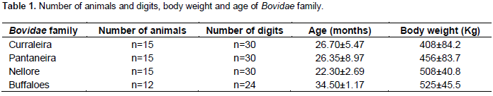
The left digits (forefeet and hind feet) of males in the Bovidae family, 45 bovines (Curraleira, Pantaneira and Nellore breeds) and 12 water buffaloes (cross breed Murrah × Jafarabadi), were used in this study (Table 1). The Research Ethics Committee of Federal University of Goiás has approved this study (Nº 090/2011).
The Nellore and Curraleira breeds came from state farms in Goiás. The Empresa Brasileira de Pesquisa Agropecuária (EMBRAPA) of Mato Grosso do Sul donated the Pantaneira breed. Moreover, the buffaloes came from state farms in Pará.
The animals were slaughtered following all the sanitary protocols indicated by the Federal Inspection in Goiás. The thoracic and pelvic limbs were disjointed on the carpal-metacarpal and tarsal-metatarsal joints. Only the left limbs were used in this experiment, while another study was carried out using the contralateral limbs.
Before the radiographic procedures the limbs were washed, dried, and examined to confirm the absence of digital diseases.
Dorsopalmar and dorsoplantar radiographies were obtained in a stationary X-ray device by Tur in the T-350 model (Röntgentechnik GmbH, Potsdam, Germany) with the capability for 600 mA. Exposure indices were defined considering the thickness of the member, varying from 60 kV to 70 kV, 25 to 30 mA and 0.2 s. The focus-film distance was maintained at 90 cm and the central beam was positioned perpendicular to the cassette at the level of the proximal interphalangeal joint between the two feet. The radiographies were developed in a Vision Line LX-2 (Lotus, Curitiba, Paraná, Brazil) automatic processor.
The radiographies were done and evaluated following the Bargai et al. (1989) and the Geissbühler et al. (2010) instructions. After, by using a digital camera (DSC-w130, Sony Brasil Ltda., São Paulo, São Paulo) the radiographies were photographed, digitalized and compressed in a Joint Photographic Experts Group (JPEG) format. Using the computer program ImageJ software (Version 1.36b for Mac OS X) the lengths of the bones were determined.
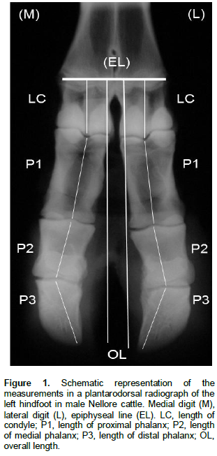
Figure 1 outlines the length of the condyles (LC), length of proximal phalanx (P1), length of medial phalanx (P2), length of distal phalanx (P3) and the overall length (OL) for the medial digit (M) and lateral digit (L) in the forefeet (FF) and hind feet (HF). The measurements were done according to Schwarzmann et al. (2007) and Muggli et al. (2011).
For all the measurements, the means and standard deviations were calculated. The mean lengths assessed in each group were compared by ANOVA followed by the Tukey test. A correlation between the body weight with the obtained measurements were evaluated using Pearson’s correlation. The Canonical analysis was used to express the similarities between the variables studied in each animal. All analyses were carried out by software R (R-Development Core Team, 2011) adopting p<0.05.
RESULTS
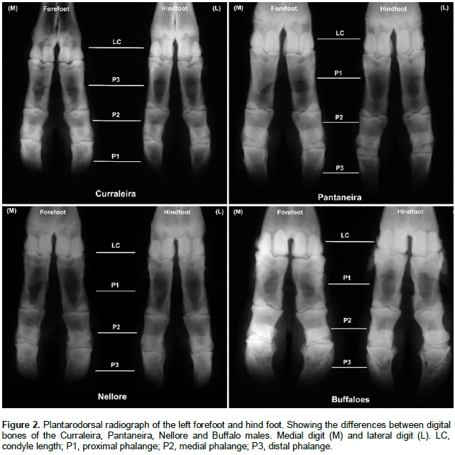
Examining the radiographies, the bovine bones silhouettes (condyles, first, second and third phalanges) appeared longer and thinner in Curraleira and Pantaneira than in Nellore. The bone silhouettes were bigger in Buffaloes (Figure 2). The lengths of the condyles, P1, P2, P3 and the overall length of the digits, in the forefoot and hind foot were similar (p>0.05) in the Curraleira and Pantaneira breeds (Table 2).
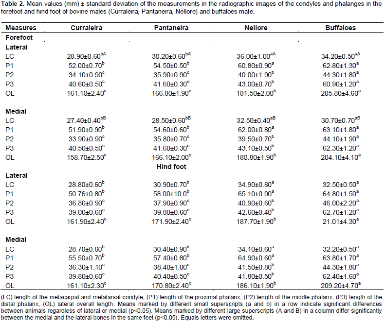
In Nellore, the length of all bones were greater than those of Curraleira and Pantaneira (p<0.05). No differences (p>0.05) were observed for LC and P1 lengths when the Nellore bone lengths were compared with bone buffaloes. The buffaloes showed greater lengths of the P2, P3 and OL than the bone measurements of all bovine breeds (p<0.05). Comparing the lateral and medial digits, of the FF and HF, no differences (p>0.05) were observed between all phalanges and OL for bovines and buffaloes. Nevertheless, it was interesting to observe that the OL of the lateral digits appeared some millimeters greater, but not significantly, in Curraleira (FF=2.4 mm, HF=0.8 mm), Pantaneira (FF=0.7 mm, HF=1.1 mm), Nellore (FF=0.7 mm, HF=1.6 mm) and buffaloes (FF=0.2 mm, HF=0.9 MM).
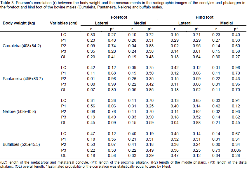
In the forelimb, the lateral condyles were longer than the medial condyles (Table 2) in Curraleira (p=0.001), Pantaneira (p=0.009), Nellore (p=0.005) and buffalo (p=0.004). No differences were observed in the condyles of the hind limb (p>0.05). The results of the statistical correlation showed higher correlation (r=0.73, p=0.006) between the length of P3 and the body weight in the hind foot of buffaloes (Table 3). Canonical analysis demonstrated similarity between bovine digits and,proved that buffalo digits were bigger than in all bovines.
DISCUSSION
The differences between the bone silhouettes in the radiographs of the bovines (Curraleira, Pantaneira, Nellore) and the buffaloes were confirmed by the length measurements of the condyles, P1, P2, P3 and OL in the forefoot and hind foot.
The bone lengths established in the present study were close to the lengths observed in different breeds of bovine, by direct measurements (Ocal et al., 2004; Nacambo et al., 2007; Radisic et al., 2012) and radiographic measurements (Schwarzmann et al., 2007; Muggli et al., 2011). As well as, in buffalo bones (Nourinezhad et al., 2012) and in wild animal radiographies (Keller et al., 2009).
The methods used for obtaining measurements on the radiographs had been established in previous studies (Schwarzmann et al., 2007; Nacambo et al., 2007; Keller et al., 2009; Muggly et al., 2011) and, in our study they were carried out thoughtfully to ensure accurate results. According to Nourinezhad et al. (2012) direct measurements have the advantage of direct visibility and better determination of measuring points, while in radiographic measurements, projection errors and poorer visibility of the surface of the bones may account for mistakes. The authors can assume that the measurements carried out in our study were accurate, since there was concordance with the results of the studies that used direct measurements.
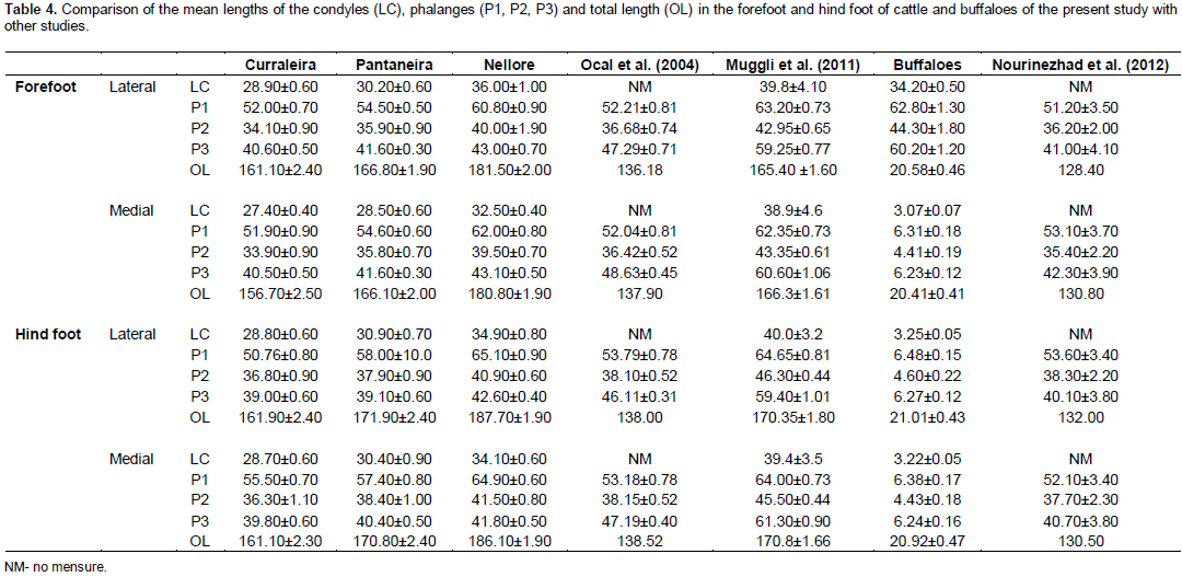
The mean lengths of the P1, P2, P3 and the OL (plus and minus the condyles) in our study were compared with the mean values of bone lengths in other studies (Table 4) of bovines and buffaloes. The Curraleira and Pantaneira measurements were similar to the measurements in Holstein male cattle (Ocal et al., 2004). The lengths of the Nellore bones were similar as those described by Muggli et al. (2011) in heifers and steers of different breeds. For buffaloes, the mean values in our study were distinctively greater than the values of the Khuzestan buffaloes (Nourinezhad et al., 2012).
There was no significant asymmetry between the lateral and medial bones, in the forefoot and hind foot, in both bovines and buffaloes. By direct measurements, Ocal et al. (2004) and Nourinezhad et al. (2012) reported no differences between the lengths of the phalanges and the total lengths of the three phalanges within the left and right thoracic and pelvic limbs. However, using radiographic measurements of Schwarzmann et al. (2007), Muggli et al. (2011) and Keller et al. (2009) described a lateral digit longer than medial.
Considering the differences between the bone lengths and the lateral and medial digits, it can be assumed that the digits bone length differences could be characteristics of artiodactyls (Keller et al., 2009), indicative of anatomical variations existent in individual cattle (Muggli et al., 2011) and/or, the differences among ruminants resulting from locomotion habits (Nourinezhad et al., 2012).
Even though the results have not pointed to a significant difference between the total lengths of the digits, the lateral digit were some millimeters longer than the medial in the forelimb (0.2 to 1.6 mm) and in the hind limb (0.8 to 2.4 mm). These results were similar with the findings observed by Muggli et al. (2011), the lateral digits were longer than the medial digits in the forelimb (1.8 mm) and in the hind limb (2.1 mm).
Nacambo et al. (2007), Keller et al. (2009) and Muggli et al. (2011) postulated that the length asymmetry between lateral and medial condyles were responsible for the difference in the length of the paired digits. Nourinezhad et al. (2012) hypothesized that such a length asymmetry of condyles bones exists in the water buffalo. In our study, the lateral metacarpal condyles were significantly longer than medial condyles in all animals of this study. Thus, our results support the assumptions reported in other studies.
A positive correlation between the body weight and the length of P3 was seen only in the buffalo hindfoot. Muggli et al. (2011) related no correlation between the body weight and measurements in the bone digits. Otherwise, Radišić et al. (2012) reported a higher degree of correlation between the body weight and hoof length in the front and hind limbs hooves of the Simmental bulls, ranged from 635 to 1370 kg.
It can be assumed that the greater P3 and, consequently, the wider capsule of the hooves in the buffaloes were the cause for the correlation that was observed in this study. According to Keller et al. (2009) the longer third phalanx correlated well with the longer dorsal wall length of the hooves in wild animals. Besides, Nacambo et al. (2007) hypothesized that the longer lateral condyles were responsible for the larger size of the lateral claws. In addition, Nuss et al. (2011) suggested that variations in shape and certain claw characteristics had been present in cattle individually, although the animal feet had been exposed to similar housing conditions.
The Canonical analysis confirmed the similarity between the Curraleira and Pantaneira bone lengths and that Nellore breeds showed bones with intermediate lengths compared to Pantaneira breeds and buffaloes. It also exposed that the digits of buffaloes were longer than digits of the bovine breeds.
The results showed that the measurements made in the radiographic image were consistent in identifying the anatomical differences among the Brazilian cattle and buffaloes. Therefore, data acquired in this present study might be useful as reference for researchers and for clinical practice, as well as to differentiate bone fragments in archaeological specimens.
CONCLUSION
The Curraleira and Pantaneira breeds have similar lengths of digital bones. The Nellore breed has intermediate lengths of the digital bones. Buffaloes have longer bones than the cattle bones
CONFLICT OF INTERESTS
The authors have not declared any conflict of interests.
REFERENCES
|
Barbosa JD, Lima DHS, Belo-Reis AS, Pinheiro CP, Sousa MGS, Silva JB, Salvarani FM, Oliveira CMC (2014) Degenerative joint disease in cattle and buffaloes in the Amazon region: A retrospective study. Pesq. Vet. Bras. 34:845-850. |
|
|
Bargai U, Pharr JW, Morgan JP (1989). Bovine Radiology. 1st ed: Iowa State University Press. |
|
|
Egito AA, Paiva SR, Albuquerque MSM, Mariante AS, Almeida LD, Castro SR, Grattapaglia D (2007). Microsatellite based genetic diversity and relationships among ten Creole and commercial cattle breeds raised in Brazil. BMC Genet. 8:83. |
|
|
Geissbühler U, Steiner A, Siegrist A, Mock L, Delley V, Stoffel M (2010). Bovine Radiology - Digital Diagnostic Atlas. Bern:University of Berne, Switzerland. (http://www.vetsuisse-bern.ch/bovine_radiology/Radioatlas.html#) |
|
|
Gonçalves PVR, Silva LAF, Silva LH, Costa APA, Bragato N, Cardoso JR, Kofler J, Borges NC (2014). Ultrasonography of the distal limbs in Nellore and Girolando calves 8 to 12 months of age. BMC Vet. Res. 10:102. |
|
|
Keller A, Clauss M, Muggli E, Nuss K (2009). Even- toed but uneven in length: the digits of artiodactyls. Zoology. 112: 270-278. |
|
|
Malhado CHM, Ramos AA, Souza CPL, Souza JC, Lamberson WR (2007). Genetic and phenotypic trends for growth traits of buffaloes in Brazil. Ital. J. Anim. Sci. 6(Suppl. 2):325-327. |
|
|
Malhado CHM, Malhado ACM, Ramos AA, Souza CPL, Souza JC, Pala A (2013). Genetic parameters for milk yield, lactation length and calving intervals of Murrah buffaloes from Brazil. R. Bras. Zootec. 42:565-569. |
|
|
Mariante AS, Egito AA (2002). Animal genetic resources in Brazil: Result of five centuries of natural selection. Theriogenology 57:223-235. |
|
|
Muggli E, Sauter-LOUIS C, Braun U, Nuss K (2011). Length asymmetry of bovine digits. Vet. J. 188:295-300. |
|
|
Nacambo S, Hässig M, Lischer C, Nuss K (2007). Difference in the length of the medial and lateral metacarpal and metatarsal condyles in calves and cows – a post-mortem study. Anat. Histol. Embryol. 36:408-412. |
|
|
Nourinezhad J, Mazaheri Y, Pourmahdi BM, Daneshi M (2012). Morphometric study on digital bones in native Khuzestan water buffaloes (Bubalus bubalis). Bulg. J. Vet. Med. 15:228-235. |
|
|
Nuss K, Sauter-louis C, Sigmund B (2011). Measurements of forelimb claw dimensions in cows using a standardised sole thickness: A post-mortem study. Vet. J. 190:84-89. |
|
|
Ocal MK, Sevil F, Parin U (2004). A quantitative study on the digital bones of cattle. Ann. Anat. 186:165-168. |
|
|
Radišić B, MatiÄić D, Vnuk D, Lipar M, Balić IM, Äitko B, Smolec O, Orak A, Capak H, Kos J (2012). Measurements of healthy and pathologically altered hooves, their interrelation and correlation with body mass in Simmental breeding bulls. Vet. Arch. 82:531-534. |
|
|
Ramos AA, Malhado CHM, Carneiro PLS, Gonçalves HS, Azevedo Dmmr (2006). Phenotypic and genetic characterization of the milk yield and calving interval in buffalo of the Murrah breed. Pesq. Agropec. Bras. 41:1261-1267. |
|
|
Salles PA, Barbosa VV, Sousa CM, Medeiros GR, Rocha LL, Weller M (2013). Breeding management and assessment of Curraleiro cattle in Northeastern Brazil. Anim. Genet. Resourc. 52:139-145. |
|
|
Schwarzmann B, Köstlin R, Nuss K (2007). Difference in the dimensions of the digital bones and claws in calves Größenunterschiede zwischen den lateralen und medialen Zehenknochen und Klauen von Kälbern. Tierarztl. Prax. 35(G):341-349. |
|
|
Serrano GM, Egito AA, Mcmanus C, Mariante AS (2004). Genetic diversity and population structure of Brazilian native bovine breeds. Pesq. Agropec. Bras. 39:543-549. |
|
Copyright © 2024 Author(s) retain the copyright of this article.
This article is published under the terms of the Creative Commons Attribution License 4.0


