African Journal of
Agricultural Research

  • Abbreviation: Afr. J. Agric. Res.
  • Language: English
  • ISSN: 1991-637X
  • DOI: 10.5897/AJAR
  • Start Year: 2006
  • Published Articles: 6849

Full Length Research Paper

Ecological diversity and economical importance of species from Aphanomyces genus

Bazyli Czeczuga*
  • Bazyli Czeczuga*
  • Department of General Biology, Medical University of Bialystok, Mickiewiecza 2c, 15-222 Bialystok, Poland.
  • Google Scholar
Ewa Czeczuga-Semeniuk
  • Ewa Czeczuga-Semeniuk
  • Department of General Biology, Medical University of Bialystok, Mickiewiecza 2c, 15-222 Bialystok, Poland.
  • Google Scholar
Adrianna Semeniuk- Grell
  • Adrianna Semeniuk- Grell
  • Department of General Biology, Medical University of Bialystok, Mickiewiecza 2c, 15-222 Bialystok, Poland.
  • Google Scholar
Bazyli Czeczuga
  • Bazyli Czeczuga
  • Department of General Biology, Medical University of Bialystok, Mickiewiecza 2c, 15-222 Bialystok, Poland.
  • Google Scholar
Ewa Czeczuga-Semeniuk
  • Ewa Czeczuga-Semeniuk
  • Department of General Biology, Medical University of Bialystok, Mickiewiecza 2c, 15-222 Bialystok, Poland.
  • Google Scholar
Adrianna Semeniuk- Grell
  • Adrianna Semeniuk- Grell
  • Department of General Biology, Medical University of Bialystok, Mickiewiecza 2c, 15-222 Bialystok, Poland.
  • Google Scholar


  •  Received: 19 March 2015
  •  Accepted: 09 October 2015
  •  Published: 26 November 2015

 ABSTRACT

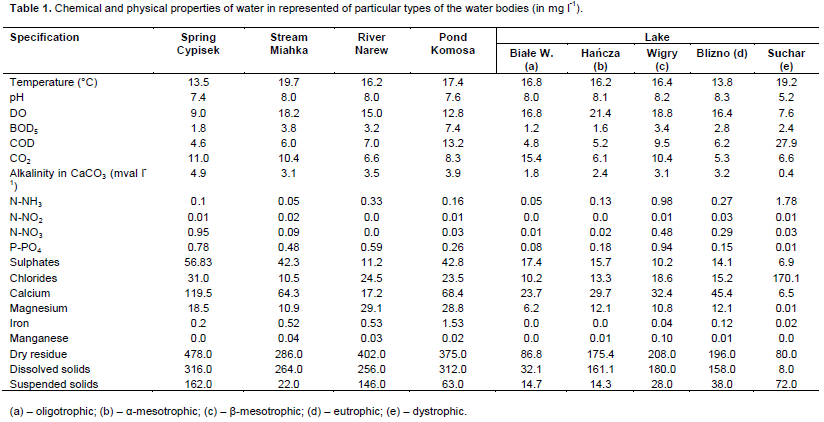
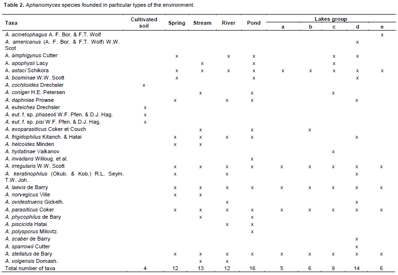
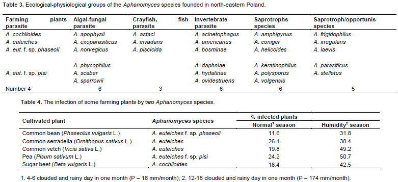
Species from Aphanomyces genus were investigated in 225 limnologically and trophically different water bodies (springs, rivers, ponds and lakes) as well as in humid soils of north- eastern Poland. Distribution of particular species and their role in diverse ecosystems has been investigated. Thirty taxa, including: 2 species of cultivated parasitic plants, 3 species of parasitic animals, 6 saprotrophic species and 5 species of saprotrophic/ opportunistic species were recorded. Some of Aphanomyces species occurred also as parasites of algae, straminipiles and invertebrates. Among parasitic species: A. cochlioides, A. euteiches (cultivated plant parasite), A. astaci (crayfish parasite), A. piscicida (fish parasite) playing economically important role were found. Amino acid, carbohydrate and urease assimilation tests were used.

Key words: Aphanomyces species, ecological diversity, hydrochemistry, parasite, economical importance.


 INTRODUCTION

The number of fish species bred in the control conditions increased in recent years according to Food and Agriculture Organization (FAO, 2012). On the other hand, death on a large scale of the particular fish populations may occur due to bacterial and mycotic infections (Bruno and Wood, 1988). Straminipila from the Saprolegniales order, especially Saprolegnia, Achlya and Aphanomyces species are responsible for eggs infection in the fish species. For example Hatai and Hoshiai (1992) dealt with saprolegniosis of Oncorhynchus kisutch demonstrating heavy losses caused by Saprolegnia parasitica in the breeding of this species, even up to 50%. Achlya flagellata and A. prolifera are known to have caused total damage to the incubated eggs of Tor tor Lac. in India (Sati and Khulbe, 1981).  Aphanomyces astaci  is  known to cause so-called “plague” of crayfish (Schikora, 1903) and A. laevis known in mass deaths of rainbow trout during reproduction on the Taiwan (Chien, 1981). Since the mid- 1980s epizootic ulcerative syndrome (EUS) have been described as a disease affecting wild and farmed freshwater and estuarine fish (Chinabut, 1998). It has been reported from Australia, North America, Asia and Africa (OIE, 2007). The agent associated with this disease is the straminipiles organism belonging to Aphanomyces genus. In 1997 Kitancharoen and Hatai (1997) described also A. frigidophilus as a parasite of the Japan charr eggs.Whereas, such species of the Aphanomyces genus as A. cochlioides and A. euteiches because of their agriculturally importance, are specialized to parasitize roots of sugar beet and Fabaceae  species,
 
respectively (Bangsund and Leistritz, 1993; Sauvage et al., 2007; Dieguez-Uribeondo et al., 2009).
 
On account of that, authors have decided to find out which of the already known straminipiles organisms belonging to Aphanomyces genus is responsible for the mycotic diseases in plants and animals and can grow in different ecosystems of the north-eastern Poland.


 MATERIALS AND METHODS

Description of Aphanomyces (de Bary, 1860) genus
 
Aphanomyces genus was described firstly in 1860 by de Bary (de Bary, 1860) and included initially following four species: A. laevis, A. phycophilus, A. scaber and A. stellatus. Other species from this genus have been described by Drechsler (1929) and Scott (1961). At present, according to Index Fungorum, Aphanomyces genus comprises 45 taxa and 40 species (David and Kirk, 1997), according to Ballesteros et al. (2006)- 30 species and according to Dieguez- Uribeondo et al. (2009)- 35-40 species. Dick (2001) classifies Aphanomyces together with Leptolegnia and Plectospyra to Leptolegniaceae family. Some species specialize in plant or animal parasitism, other ones are saprotrophic growing on decaying plant and animals debris (Scott, 1961; Fuller and Jaworski, 1987; Dick, 2001; Johnson et al., 2002; Dieguez-Uribeondo et al., 2009). There are also species which are primarily saprotrophic but in some instances become opportunistic pathogens (Royo et al., 2004; Patwardhan et al., 2005). Plant parasitic species include: A. cochlioides- pathogen of roots of sugar beet and P. euteiches which parasitize on Fabaceae species (Papavizas and Ayers, 1974; Levenfors and Fatehi, 2004). A. astaci is a parasite of a freshwater crayfish (Söderhäll and Cerenius, 1999). A. invadans and A. frigidophilus (A. piscicida) devastate both freshwater and estuarine fishes (Chnabut, 1998; Czeczuga et al., 2011a). A. laevis, A. stellatus and A. helicoides belong to opportunistic pathogens (Patwardhan et al., 2005; Dieguez-Uribeondo et al., 2009).
 
Occurrence environment of Aphanomyces species
 
Material was collected in north- eastern part of Poland within 1985 to 2012. Water samples were collected from 225 limnologically and trophically different water bodies-springs (31), rivers (45), ponds (21) and lakes (128). Oligotrophic, mesotrophic, eutrophic and dystrophic types of lakes were investigated. Water samples were collected at a depth of 10 (spring)- 30cm (other water bodies) and at a distance of 0.5m from the bank, at three sites. Nineteen water parameters were determined (Table 1, only as representatives for particular types of water bodies) according to generally accepted methods (Golterman and Clymo, 1969; APHA, 2005).
 
 
The soil species was collected from roots of common bean, common serradella, common vetch, pea and sugar been (Table 4) at farmed land of north-eastern Poland. The amino acid, carbohydrate and urease assimilation tests were performed according to Yuasa and Hatai (1996) and Kitancharoen and Hatai (1998).
 
Determination of Aphanomyces species
 
Thickness, height and branches of the hyphae were measured in Aphanomyces species. The diameter of zoosporangia, zoospores, cystospores, oospores, oogonia and antheridia were defined. Morphological structure of the oogonia, antheridia and their number were also studied. It was investigated, if  the  oogonia are  mono-, diclinous or androgynous. Some parasitic Aphanomyces species showed the repeated zoospore emergence (RZE) as adaptation to the parasitic mode of life (Cerenius and Söderhäll, 1985). For parasitic species (plant and animal) general principles of culture procedures were used (Seymour and Fuller, 1987; Roberts et al., 1993; Willoughby and Roberts, 1994; Paterson and Bridge, 1994; Watanabe, 2002). The parasitic species of the sugar beet and of some species of the legumes (soil species) were isolated according to Dyer et al. (2004) and Sauvage et al. (2007) methods. The isolates collected from the necrotic roots of the investigated plants were transferred to the corn meal agar (CMA, Difco, Detroit, MI) supplemented with rifampicin (38 mg/l). For each isolate, a single hyphal tip was transferred to a new CMA plate to insure only one genotype per isolate. The cultures were stored in sterile distilled water and were replaced in storage by culturing on CMA with rifampicin to repeat the process for storage (Parke and Grau, 1992).The taxa of Aphanomyces species were identified using the keys of Scott (1961), Batko (1975), Pystina (1994), Johnson et al. (2002) and Petrini and Petrini (2013).


 RESULTS

The water samples used for the analysis differed in nutrient content and other parameters (Table 1). The highest nitrogen, sulphates and chlorides content was found in Spring Cypisek, Akcent and Fosa, Komosa Pond. The lowest content of nitrogen, sulphates and chlorides was noted in Hancza Lake. 30 taxa from Aphanomyces genus were found in the water bodies and soil of north-eastern Poland (Table 2). Same species occurred in all types of the investigated water reservoirs. A. astaci, A. laevis, A. irregularis, A. parasiticus and A. stellatus belong to this group. Such species as A. apophysii, A. coniger, A. ovidostruens, A. polysporus, A. scaber and A. volgensis were found only on the plant debris, other species also on animals’ substratum (Table 3). A. cochlioides and A. euteiches are the parasit on cultivated plants (Table 4). 6 Aphanomyces species including: A. frigidophilus, A. invadans and A. piscicida and others were present on the fish eggs (Table 5). Investigated Aphanomyces species assimilated only three amino acids: alanine, glutamine and cysteine and from carbohydrates only glucose and starch, but they did not assimilate urease (Table 6).


 DISCUSSION

Thirty taxa Aphanomyces were found in the water bodies of north-eastern Poland (Table 2). Five species occurred in all types of investigated water bodies, two species- A. cochloides and A. euteiches occurred in humid soils. A. cochlioides is a parasite of roots of sugar beet, A. euteiches of pea and other legumes species roots. The infected roots of plants ranged from 31.8% (common bean) to 50.7% (pea) during humid years. Therefore, soil species of the Aphanomyces genus are economically important, especially for the food industry (pea, sugar beet) (Levenfors and Fatehi, 2004) and  for  some  plant species used as animal foodstuff for domestic animals (Papavizas and Ayers, 1974; Holub et al., 1991; Brantner and Windels, 2001).
 
 
Most Aphanomyces species were found in such eutrophic reservoirs of the stagnant water bodies as ponds (16) and eutrophic lakes (14 species), (Table 2). Only a few Aphanomyces species were found in water with a small content of the biogenic substances. There are oligotrophic, α-mesotrophic and dystrophic lakes (5-6 species). In the flowing waters (springs, streams, rivers) the authors found many (12-13) Aphanomyces species and such species as A. astaci, A. irregularis, A. laevis, A. parasiticus and A. stellatus were found in all types of the investigated water bodies. According to this fact they are called the eurytrophic species and have a wide range of the ecological tolerance. There are various organic compounds of carbon, free saccharides, free amino acids, numerous enzymes and other compounds (Hoagland et al., 1993) called extracellular products (being excreted by the phytoplankton and  makrophytes) which exert the influence on the mycotal species in water. They are a medium for different heterotrophic organisms including mycotal species. As it is known, in oligotrophic, α-mesotrophic and dystrophic lakes there is a little amount of biogenic compounds, the phytoplankton and the macrophytes excreting less extracellular products serving as nutrients for heterotrophic organisms.Such species as A. apophyscii, A. coniger, A. ovidostruens, A. polysporus,   A. scaber   and   A. volgensis    were
 
found only on the plant substratum, other species also on the animal substratum. The 9 Aphanomyces species present in fish, A. invadans and A. piscicida occurred only on fish eggs. A. euteiches is the parasite of Fabaceae family (Papavizas and Ayers, 1974). A. astaci, A. invadans and A. piscicida are animal parasites. A. astaci, which has been described by Schikora (1903), infects freshwater crayfish (Söderhäll and Cerenius, 1999). Both A. invadans and A. piscicida devastated natural and cultured stocks of freshwater and estuarine fish (Chinabut, 1998; Johnson et al., 2004). Some authors confirmed this both species as synonym (Dieguez-Uribeondo et al., 2009), others- as separate species (Phadee et al., 2004a, b). A. piscicida was described by Hatai (1980) and causes mycotic granulomatosis in fish (MG) (Egusa and Masuda, 1971), whereas A. invadans was described by Willoughby et al. (1995) and is an agent of EUS (Epizootic ulcerative syndrome). Kitancharoen and Hatai (1997) described new species of A. frigidophilus from eggs of Japan charr. In Europe, first A. frigidophilus was described on eggs of some species of coregonide fishes (Czeczuga et al., 2004). Two years later, Ballesteros et al. (2006) found this species in cuticule of dead crayfish Austropotamobius pallipes in Spain and Kiziewicz et al. (2013) observed its occurrence in water from the some springs of north- eastern Poland. Growth of A. frigidophilus was also stated on eggs of some species from Salmo genus (Czeczuga et al., 2004, 2011a), sturgeonids,  Chinook  salmon (Czeczuga et al., 2011b) and African catfish (Czeczuga et al., 2013).
 
As shown in Table 3, such species as: A. irregularis, A. laevis, A. parasiticus and A. stellatus were found on plant debris and on fish eggs. They all belong to opportunistic pathogens, which are sapro- and necrotrophic species similar to many species belonging to Achlya and Saprolegnia genera. Accourding to our study such saprotrophic species as A. amphiginus and A. helicoides were growing on decaging invertebrate animals and plant debris. According to Dieguez-Uribeondo et al. (2009). A. helicoides belongs to saprotrophic- opportunistic group. The most numerous pathogens from this group on fish eggs are: A. laevis (41 species) and A. stellatus (29). A. irregularis was found on eggs of 8 fish species, A. parasiticus- on 7. Mentioned species were also present on the eggs of lamprey (Czeczuga, 1997). According to Batko (1975) and Pystina (1994) A. irregularis is saprotrophic species and A. parasiticus-parasite on other fungal species, especially from Achlya genus. A. laevis both- eggs and adult individuals of many economically valuable fish species (Lartzeva, 1986; Dudka et al., 1989). A. bosminae and A. daphniae (occurring on fish eggs) were found in some of investigated water bodies. A. bosminae was firstly described by Scott (1961) and first investigated fungus in America. It is a parasite of freshwater crustacean from the Bosmina genus. The authors found it on the eggs of Leuciscus leuciscus in water    from   River    Suprasl   (Czeczuga     and Muszynska, 1999). A. daphniae is known to be a parasite of the plankton crustacean Daphnia hyalina (Leydig) (Prowse, 1954). In our study its growth was revealed on the eggs of Cobitis aurata (Filippi) in River Suprasl water (Czeczuga and Muszynska, 1997).
 
The occurrence of Aphanomyces species has been registered in water bodies of African continent by El- Sharouny and Badram (1985), Shaheen et al. (1999), El- Hissy et al. (2004) and Ali (2009). Aphanomyces species have been associated with skin lesions and mortality of some fish species. This phenomenon has been observed in two species of fishes from excavated earthen pond at the western shore of the Suez Canal, Egypt, during the winter of 1971 (Shaheen et al., 1999). According to Lilley et al. (2003) this invasive Aphanomyces fish infection has been reported by Shaheen et al. (1999) and has been caused by A. invadans species. It has also been observed on skin-muscles on aquarium species of Labeo bicolour Smith specimens and on the alevins of the Nile tilapia- Oerochromis niloticus L.
 
The assimilation of the amino acids, carbohydrate and urease by two investigated Aphanomyces species was very small. Specimens of A. frigidophilus from fish eggs have assimilated only 3 amino acids, 3 carbohydrates, whereas A. laevis assimilated only one amino acid and 2 carbohydrates. Whereas specimens of Saprolegnia, Achlya or Pythium assimilated 4 to 5 amino acids, 9 to 16 carbohydrates and urease (Kitancharoen and Hatai, 1998). The specimens of both species from Aphanomyces genus did not assimilate urease.


 CONCLUSIONS

All together, 30 taxa of Aphanomyces genus occurred in 225 water bodies and some farming land of north-eastern Poland. This straminipiles species was found in limnologically and trophically different springs, rivers, ponds and lakes. Two species were found in farming land. All 30 Aphanomyces taxa belonged to four groups: a) plant parasite species, b) animal parasites, c) saprotrophic/opportunistic species and d) saprotrophic species. Group a) consists of: A. cochlioides and A. euteiches, group b) A. astaci, A. invadans and A. piscicida. A. astaci are etiologically an agent of the crayfish plague. A. invadans and A. piscicida infect freshwater and estuarine fishes as well as devastation of natural and cultured stocks (epizootic ulcerative syndrome). Some species, for example: A. acinetophagus, A. exoparasiticus, A. hydatinae and A. sparrowii occurred on some algae, straminipiles and invertebrates species as their parasites. Group c) - A. frigidophilus, A. irregularis, A. laevis, A. parasiticus and A. stellatus belong to the group of saprotrophic/opportunistic. These species are growing on decaying plant and animal debris (saprotrophic) and on fish eggs (parasitic). Fourth group- d) includes saprotrophic species fragments which grow only on decaying plant and animal debrits. Representatives of this group are: A. amphiginus, A. coniger, A. helicoides, A. keratinophilus, A. polysporus, and A. volgensis. Pathogenic Aphanomyces species in soil and in water bodies play economically significant role.


 CONFLICT OF INTEREST

The authors have not declared any conflict of interests.


 REFERENCES

Ali EW (2009). Antifungal activity of sodium chloride on Saprolegnia diclina and Aphanomyces sp. Acta Mycol. 44:125-138.
Crossref


APHA (American Public Health Association) (2005). Standard methods for the examination of water and wastewater, APHA, Washington, DC, USA.


Ballesteros I, Martin MP, Dieguez-Uribeondo J (2006). First isolation of Aphanomyces frigidophilus (Saprolegniales) in Europe. Mycotaxon 95:335-340.


Bangsund DA, Leistritz FL (1993). Economic contribution of the sugarbeet industry to the economy of North Dakota and Minnesota. 1998 Sugarbeet Res. Ext. Rept. 29:160-179.


de Bary HA (1860). Einige neue Saprolegnieen. Jahrb. Wiss. Bot. 2:169-192.


Batko A (1975). Hydromycology – an overview. PWN, Warszawa.


Brantner JR, Windels CE (2001). Variability of spore production and aggressiveness of Aphanomyces cochlioides on sugarbeet 2000. Sugarbeet Res. Ext. Rept. 31:241-246.


Bruno DW, Wood BP (1999). Saprolegnia and other Oomycetes. In: Woo PTK & Bruno DW (Eds.), Fish Diseases and Disorders. Viral, Bacterial and Fungal Infections, vol. 3. CABI Publishing, Wallingford, Oxon, UK, pp. 599-659.


Cerenius L, Söderhäll K (1985). Repeated zoospore emergence as a possible adaptation to parasitism in Aphanomyces. Exper. Mycol. 9:259-263.
Crossref


Chien C–Y (1981). Observations on the growth and morphology of saprolegniaceous fungi isolated from rainbow trout (Salmo gairdneri). Fish Pathol. 15:241-247.
Crossref


Chinabut S (1998). Epizootic alcerative syndrome: information up to 1997. Fish Pathol. 33:321-326.
Crossref


Czeczuga B (1997). Aquatic fungi growing on lamprey eggs (Petromyzontidae). Bull. Lampetra 3:7-19.


Czeczuga B, Bartel R, Semeniuk A, Czeczuga–Semeniuk E, MuszyÅ„ska E, Godlewska A, Mazalska B, Grochowski A (2011a). Straminipilous organisms (Mycota) growing on the eggs of Atlantic salmon (Salmo salar L.) entering Polish rivers for spawning or reared in fresh water. Trends Comp. Biochem. Physiol. 15:73-81.


Czeczuga B, Czeczuga-Semeniuk E, Semeniuk A (2011b). Aquatic fungi developing on eggs of Chinook salmon Oncorhynchus tshawytscha and same of their biochemical characteristics. Trends Comp. Biochem. Physiol. 16:85-92.


Czeczuga B, Czeczuga-Semeniuk E, Semeniuk A, Semeniuk J (2013). Straminipiles (Oomycota) developing on the eggs of an African catfish, Clarias gariepinus Burchell in water bodies of Poland. Afr. J. Microbiol. Res. 7(20):2378-2384.


Czeczuga B, Kiziewicz B, Muszyńska E (2004). Presence of zoosporic fungus species on the eggs of whitefish from Lake Gołdapiwo, Mazury Region. Med. Weteryn. 60:379-383.


Czeczuga B, Muszyńska E (1997). Aquatic fungi growing on the eggs of Polish cobitid fish species. Acta Hydrobiol. 39:67-75.


Czeczuga B, Muszyńska E (1999). Aquatic fungi growing on the eggs of fishes representing 33 cyprinid taxa (Cyprinidae) in laboratory conditions. Acta Ichthyol. Piscat. 29:53-72.


David JC, Kirk PM (1997). Index of Fungi. Elsevier Publication 6:706.


Dick MW (2001). Straminipilous Fungi: Systematics of the Peronosporomycetes including accounts of the marine straminipilous protists the plasmodiophorids and similar organisms. Kluwer Academic Publishers, Dordrecht.
Crossref


Dieguez-Uribeondo J, Garcia MA, Cerenius L, Kozubikova E, Ballesteros I, Windels C, Weiland J, Kator H, Söderhall K, Martin MP (2009). Phylogenetic relationships among plant and animal parasites, and saprotrophs in Aphanomyces (Oomycetes). Fung. Gen. Biol. 46:365-376.
Crossref


Drechsler C (1929). The beet water mold and several related root parasites. J. Agric. Res. 38:335.


Dyer AT, Szabo LJ, Windels CE (2004). Characterization and spatial distribution of Aphanomyces in sugarbeet fields. J. Sugar Beet Res. 41:1-16.
Crossref


Egusa S, Masuda N (1971). A new fungal disease of Plecoglassus altivelis. Fish Pathol. 6:41-46.
Crossref


Dudka IA, Isaeva NM, Davydova ON (1989). Saprolegniaceae breeding fish mycoses. Mycol. Phytopatol. 23:488-498.


El-Hissy FT, Ali EH, Abdel-Raheem A (2004). Diversity of zoospore of fungi of recovered from the surface of water bodies in four Egyptian lakes. Ecohydrol. Hydrobiol. 4:77-84.


El-Sharouny HM, Badram RAM (1995). Experimental transmission and pathogenicity of some zoosporic fungi to Tilapia fish. Mycopathology 132:95-105.
Crossref


FAO (2012). Aquatic Sciences and Fisheries Information System (ASFSZ) Species List, Rome.


Fuller MS, Jaworski A (1987). Zoosporic Fungi in Teaching and Research. Southeastern Publishing, Athens.


Golterman HL, Clymo RS (1969). Methods for chemical analysis of fresh waters. IBP, Handbook No 8, Blackwell Scientific Publications, Oxford.


Hatai K (1980). Studies on pathogenic agents of saprolegniasis in fresh water fishes. Spec. Rep. Nagasaki Prefect. Inst. Fish 8:1-95.


Hatai K, Hoshiai G (1992). Mass mortality in cultured coho salmon (Oncorhynchus kisutch) due to Saprolegnia parasitica Coker. J. Wild Dis. 28:532-536.
Crossref


Hoagland KD, Rosowsky JR, Gretz MR, Roemet SC (1993). Diatom extracellular polymeric substances: function, fine structure, chemistry and physiology. J. Phycol. 29:537-566.
Crossref


Holub EB, Grau CR, Parke JL (1991). Evaluation of the forma specialis concept in Aphanomyces euteiches. Mycol. Res. 95:147-157.
Crossref


Johnson TW, Seymour RL, Padgett DE (2002). Biology and systematics of the Saprolegniaceae. 


Johnson RA, Zabrecky J, Kiryu Y, Shields JD (2004). Infection experiments with Aphanomyces invadans in four species of estuarine fish. J. Fish Dis. 27:287-295.
Crossref


Kitancharoen N, Hatai K (1997). Aphanomyces frigidophilus sp. nov. from eggs of Japan char, Salvelinus leucomaenis. Mycoscience 38:135-140.
Crossref


Kitancharoen N, Hatai K (1998). Some biochemical characteristics of fungi isolated from salmonid eggs. Mycoscience 39:249-255.
Crossref


Kiziewicz B, Dieguez-Uribeondo J, Martin MP (2013). Aphanomyces frigidophilus, fungus – like organisms isolated from water of springs in BiaÅ‚ystok, Poland. Afr. J. Biotechnol. 12(44):6310-6314.


Lartzeva LV (1986). Saprolegnia on the spawn of sturgeons and salmon. Hydrobiol. J. 22:103-107.


Levenfors JP, Fatehi J (2004). Molecular characterization of Aphanomyces species associated with legumes. Mycol. Res. 108:682-689.
Crossref


Lilley JH, Hart D, Panyavachira V, Kanchanak S, Chinabut S, Söderhäll K, Cerenius L (2003). Molecular characterization of the fish – pathogenic fungus Aphanomyces invadans. J. Fish Dis. 26:263-275.
Crossref


OIE (World Organisation for Animal Health) (2007). World animal health information database (WAHID) interface OIE, Paris. 


Papavizas GC, Ayers A (1974). Aphanomyces species and their root diseases in pea and sugar beet. US Dept. Agric. Technol. Bull. P 1485.


Parke JL, Grau CR (1992). Aphanomyces. In: Singleton LL, Mihail JD, Rush CM (Eds.) Methods for Research on Soilborne Phytopathogenic Fungi. APS Pr St. Paul, MN, pp. 27-30.


Paterson RRM, Bridge PD (1994). Biochemical Techniques for Filamentous Fungi. CAB International, UK.


Patwardhan A, Gandhe R, Ghule V, Mourya D (2005). Larvicidal activity of the fungus Aphanomyces (Oomycetes: Saprolegniales) agains Culex quinqefasciatus. J. Commun. Dis. 37:69-274.


Petrini LE, Petrini O (2013). Identyfying Moulds. A Practical Guide. J. Cramer, Stuttgart.


Phadee P, Kurata O, Hatai K (2004a). A PCR method for the detection of Aphanomyces piscicida. Fish Pathol. 39:5-32.
Crossref


Phadee P, Kurata O, Hatai K, Irono I, Aoki T (2004b). Detection and identification of the fish phatogenic Aphanomyces piscicida using polymerase chain reaction (PCR) with species – specific primers. J. Aquat. Anim. Health 16:220-230.
Crossref


Prowse GA (1954). Aphanomyces daphniae sp. nov., parasitic on Daphnia hyalina. Trans. Br. Mycol. Soc. 37:22-28.
Crossref


Pystina KA (1994). Ordines Saprolegniales, Leptomitales, Lagenidiales. Nauka, Sankt Petersburg.


Roberts RJ, Willoughby LG, Chinabut S (1993). Mycotic aspects of epizootic ulcerative syndrome (EUS) of Asian fishes. J. Fish Dis. 16:169-183.
Crossref


Royo F, Andersson G, Bangyeekhun E, Muzquiz JL, Söderhäll K, Cerenius L (2004). Physiological and genetic characterization of some new Aphanomyces strains isolated from freshwater crayfish. Vet. Microbiol. 104:103-112.
Crossref


Sati SC, Khulbe RD (1981). A new host record for the fungal genus Achlya. Curr. Sci. (India) 50(16):313.


Sauvage H, Moussarat A, Boist F, Tivioli B, Barray S, Laal K (2007). Development of a molecular method to detect and quantyfi Aphanomyces euteiches in soil. Ferms Microbiol. Lett. 273:64-69.
Crossref


Schikora F (1903). Über die Krebpest und ihren Erreger. Fischereiztg. (neudamm) 6:353-355.


Scott WW (1961). A monograph of the genus Aphanomyces. Wirginia Agric. Exp. Station Technol. Bull. 151:1-95.


Seymour RL, Fuller MS (1987). Colletion and isolation of water molds (Saprolegniaceae) from water and soil. In: Fuller M.S., Jaworski A. (Eds.). Zoosporic Fungi in Teaching and Research. Southeastern Publishing, Athens, pp. 125-127.


Shaheen AA, Elsayed E, Faisal M (1999). Isolation Aphanomyces sp(p) associated with skin lesions and mortalities in striped (Mugil cephalus) and the thin lip (Liza ramada) grey mullets. Bull. Eur. ass. Fish Pathol. 19:79-82.


Söderhäll K, Cerenius L (1999). The crayfish plaque fungus: history and recent advances. Fresh. Crayf. 12:11-35.


Watanabe T (2002). Pictorial Atlas of Soil and Seed Fungi: Morphologies of Cultured Fungi and Key Species. CRC Press Boca Raton, Florida.
Crossref


Willoughby LG, Roberts RJ (1994). Improved methodology for isolation of the Aphanomyces fungal pathogen of epizootic ulcerative (EUS) in Asian fishes. J. Fish Dis. 17:541-543.
Crossref


Willoughby LG, Roberts RJ, Chinabut S (1995). Aphanomyces invaderis sp. nov., the fungal pathogen of freshwater tropical fish affected by epizootic ulcerative syndrome. J. Fish Dis. 18:273-275.
Crossref


Yuasa K, Hatai K (1996). Some biochemical characteristics of the genera Saprolegnia, Achlya and Aphanomyces isolated from fishes with fungal infection. Mycoscience 37:477-479.
Crossref


          */?>