Full Length Research Paper

ABSTRACT
Aluminium is present in some manufactured medicines and foods. It is known that aluminium causes oxidative stress. Therefore, the present study was undertaken to determine the effectiveness of Haloxylon salicornicum extract in modulating aluminium chloride (AlCl3) induced oxidative stress in rats. Male rats (40 to 50 g) were divided into four groups of six animals each. The experimental protocol was based on the administration of AlCl3 (30 mg/kg/body weight) intraperitoneally (ip) every 5 days for 15 days. The groups treated with the plant receive daily dose of 0.05 g/kg/body weight. Increased level of aspartate aminotransferase (AST), alanine aminotransferase (ALT), lactate dehydrogenase (LDH), urea, and creatinine in serum indicated hepatic and renal dysfunction. The variation of catalase (CAT), superoxide dismutase (SOD), reduced glutathione (GSH), and lipid peroxidation (thiobarbituric acid reactive substances, TBARS) were assessed. These parameters indicated the extent of oxidative damage in liver and kidney, thus confirming the histology results in liver and kidney. It was found that the consumption of H. salicornicum extract protects the liver and kidney against aluminium chloride toxicity. The aim of the present study was to evaluate the protective effect of the H. salicornicum extract on the damages caused by administration of aluminium chloride (AlCl3) in young rats.
Key words: Aluminum chloride, Wistar rats, Haloxylon salicornicum, lipid peroxidation, superoxide dismutase (SOD), reduced glutathione, catalase.
INTRODUCTION
Aluminium is an abundant metallic element on the earth; it is used highly because of its excellent properties (Bai et al., 2012). Biological function of aluminium is not understood very well (Farina et al., 2002; Joshi et al., 2013). Aluminum presents 8% of all mineral components of earth. There is not a maximum level fixed for aluminium in food (AFSSAPS, 2011).
It can be used as a food additive and in materials in contact with foodstuffs (AFSSAPS, 2011), used as water clarifying agent (coagulation, flocculation, etc.). It is found in water intended for human consumption with a maximum concentration of 0.2 mg/L (AFSSAPS, 2011). This metal is also used in the preparation of some drugs (Ochmanski and Barabasz, 2000; Turkez et al., 2010). In mammals, aluminum is accumulated in kidney, liver, heart, blood, bones, and brain (Sanchez-Iglesias et al., 2007; Gonzalez et al., 2009; Al-Kahtani, 2010). The accumulation of aluminum in liver generates significant lesions (Nikolov et al., 2010; Shati and Alamri, 2010). Kidney prevents the accumulation of aluminium in different organs, this by urinary excretion (Shirley et al., 2004; Stoehr et al., 2006). However, kidney can be very vulnerable to the nephrotoxic effects of aluminium (Mahieu et al., 2003; Stoehr et al., 2006).
Aluminium can produce free radicals in the body. It induces a toxic effect due to its ability to transfer electrons (Taus et al., 2013). These free radicals resulting from aluminum can affect cell integrity, producing the peroxidation of the lipids in the intracellular membranes, and the cross-linking with the macromolecules in the membranes (Taus et al., 2013). Aluminium is known to affect the permeability of cellular membranes, of sub-cellular organelles, the structure and functions of proteins and the structure of nucleic acids (Taus et al., 2013).
Plants have always play a major role in treatment of human and animal diseases. Medical plants are a therapeutic resource much used in the traditional population of the world specifically for health care (Kadham, 2008).
Haloxylon salicornicum is a desert plant which belongs to the family of Chenopodiaceae which includes 100 genus and 1200 species (Ferheen et al., 2005). Most of the species of this family are weedy and grow in unfertile areas of soil (Ferheen et al., 2005). H. salicornicum is a diffuse shrub with a lot of branches, woody stem, 25 to 60 cm tall. It is widely distributed in Northern Africa and Asia, in both temperate and tropical regions (Ashraf et al., 2012). This plant contains, among other chemical constituent such as sodium (28.48%), potassium (9.49%), carbonate (61.06%), alkaloid, saponin and phenol (Al-Saeed, 2002). This plant was used effectively in folk medicine as diuretic (Al-Saeed, 2002), anti-ulcer (Shahana et al., 1990), hypoglycemic and anti-microbial (Abbas et al., 2006). Ahmad and Eram (2011) have shown that this plant has a hepatoprotective effect.
The aim of the present study was to evaluate the protective effect of the H. salicornicum extract on the damages caused by administration of aluminium chloride (AlCl3) in young rats.
MATERIALS AND METHODS
Plant
The plant was collected from May to April in El-bayadh region of Southern Algeria. Aerial parts of H. salicornicum were air-dried at room temperature for 30 min. The dried plants were collected and ground to fine powder.
Preparation of aqueous extract
The powdered aerial parts of H. salicornicum (10 g) were extracted with boiled water (100 ml) for 20 min. After this step, the decoction was filtered and was freeze dried. The yield of this extraction is 13.01%.
Animals
Wistar rats were used as an experimental model to evaluate the effect of aluminium on liver and kidney. Aluminium chloride (AlCl3) was administered intraperitoneally to rats at a dose of 30 mg/kg/body weight, every five days for 15 days. The lyophilized extract of H. salicornicum is dissolved in distilled water. The plant is administered daily to the rats using a feeding tube at a dose of 0.05 g/kg/body weight. Wistar rats (40 to 50 g) were randomly distributed in four groups containing 6 rats each: group (control): control rats were given nothing; group (Al): animals were given three injections of AlCl3 (30 mg/kg/body weight); group (Al+P): rats received three injections of AlCl3 (30 mg/kg/body weight) in addition to the plant (0.05 g/kg/body weight); group (P): animals were given only the plant (0.05 g/kg/body weight).
Rats were sacrificed after 5 days of the last dose and blood was collected in hemolysis tube. Liver and kidney were rinsed with saline solution (0.9% NaCl). For evaluation of oxidative status, liver and kidney were homogenized in suitable buffers: in 0.1 M phosphate buffer (pH 7.1) for superoxide dismutase (SOD), CAT, and reduced glutathione (GSH) in 1.15% KCl for thiobarbituric acid reactive substances (TBARS). Some portions of liver and kidney were fixed in formalin for histological study.
Biochemical parameters
Aspartate aminotransferase (AST), alanine aminotransferase (ALP), lactate dehydrogenase (LDH), creatinine, and urea were measured using Chronolab kits (Spain). Biochemical parameters were estimated in serum.
Catalase activity (CAT)
CAT was assayed colorimetrically at 620 nm, expressed as µmoles of H2O2 consumed/min/mg/protein (Sinha, 1972). The reaction mixture of 1.5 ml contained 1.0 ml of 0.01 M pH 7.0 phosphate buffer, 0.1 ml of homogenate, and 0.4 ml of 2 M H2O2. The reaction was stopped by the addition of 2.0 ml of dichromate-acetic acid reagent, 5% potassium dichromate, and glacial acetic acid was mixed in 1:3 ratios.
Lipid peroxidation (TBARS)
Lipid peroxidation was assessed by measuring substances which react with TBARS (Yagi, 1976). 125 μl of sample, 50 μl of TBA, and 125 μl of trichloroacetic acid-butylated hydroxytoluene (TCA-BHT) were mixed. After stirring, incubation was done at 80°C (10 min) and centrifugation was done (100 g for 10 min). The supernatant was removed. 200 μl of the supernatant were mixed with 40 μl of HCl (0.6 M) and 160 μl of Tris-TBA (26 mM Tris, 120 mM thiobarbituric acid). After stirring, and incubation at 80°C (10 min), the optical density was measured at 530 nm. The quantity of TBARS was calculated using an extinction coefficient of 156 mM-1 cm-1.
Reduced glutathione (GSH)
GSH was measured by the method of Ellman (1959). Samples were deproteinized with 5-5 'dithiobes (2-nitrobenzioc acid), and the optical density was measured at 412 nm.
Superoxide dismutase (SOD)
SOD was assessed using assay kit provided by Cayman (Chemical, USA).
Histological study
The sections were taken from liver and kidney and they were fixed in a formalin solution 1:10 to pH 7, dehydrated in acetone, clarified in xylene, and finally embedded in paraffin. Histological sections were cut at 3 µm using a microtome. The samples were stained with hematoxylin-eosin, and observed with an optical microscope.
Statistical analysis
The data were analyzed using Student's t-test. At p < 0.05, the results were considered significant. Values are expressed as the mean ± standard error.
RESULTS
Liver
The liver function is assessed by measuring of transaminases and LDH activities. AST, alanine aminotransferase (ALT), and LDH activities in the group (Al) were increased highly significantly (p˂0.001) as compared to the group (control) (Table 1). A significant decrease of these activities is recorded in the group (Al+P) as compared to group (Al). The comparison between groups (control) and (P) shows that there was no significant difference except for AST (Table 1). Activities decreased highly significantly for ALT and AST (p˂0.001), and significantly for LDH (p˂0.05) in group (Al+P) in relation to the group (Al) (Table 1).

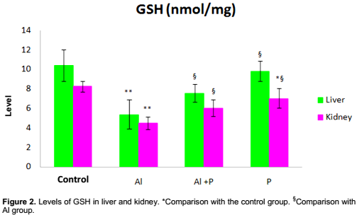
TBARS (Figure 1) and GSH (Figure 2) are higher in group (Al). The difference between groups (control) and (Al) is highly significant (p˂0.001) for TBARS and very significant (p˂0.01) for GSH. On the other hand, the com-parison between groups (Al) and (Al+P) is very significant (p˂0.01) for TBARS (Figure 1) and significant (p˂0.05) for GSH. In group (P), TBARS and GSH (Figure 2) decreased.

The CAT (Figure 3) and SOD (Figure 4) activities are decreased in group (Al). The difference between groups (control) and (Al) is highly significant (p˂0.001) for CAT and very significant (p˂0.01) for SOD. The comparison between groups (Al) and (Al+P) is very significant (p˂0.01) for CAT (Figure 3) and significant (p˂0.05) for SOD (Figure 4).


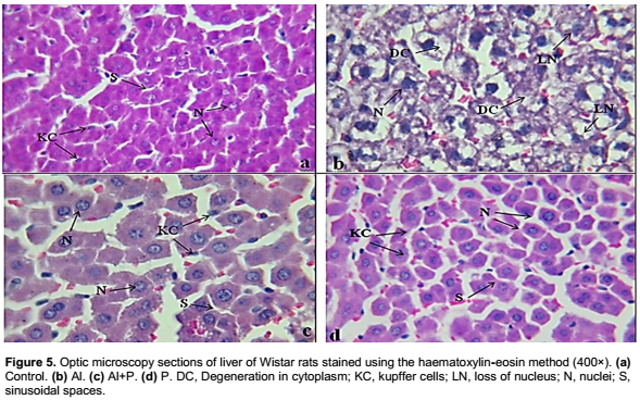
Figure 5a and d presents regular histological structure of the liver tissue of control rats with a granular cytoplasm, central core, and the open sinusoidal spaces of hepatocytes. Exposure to aluminium (Figure 5b) causes a loss of the arrangement of the hepatocytes with a degeneration of membrane, nucleus, and cytoplasm. Histological observations of liver of rats treated with H. salicornicum illustrate an improvement in some areas and a normal sinusoidal space (Figure 5c).
Kidney
Creatinine and urea were examined to verify the renal function. These parameters increased highly significantly (p˂0.001) in rats treated with aluminum (Al) relative to control) (Table 2). The consumption of plant leads a remarkable improvement (Table 2). The comparison between groups (Al) and (Al+P) shows that creatinine decreased very significantly (p˂0.01) and urea decreased highly significantly (p˂0.001).
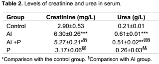
The difference between groups (control) and (P) is not significant; however, the comparison between groups (Al) and (P) parameters decreased very significantly (p˂0.01). The difference between groups (control) and (Al) is very significant (p˂0.01) for TBARS (Figure 1) and significant (p˂0.05) for GSH (Figure 2). The comparison between groups (Al) and (Al+P) is very significant (p˂0.01) for TBARS (Figure 1) and significant (p˂0.05) for GSH (Figure 2).
The comparison between groups (control) and (Al) is highly significant (p˂0.001) for CAT (Figure 3) and significant (p˂0.05) for SOD (Figure 4). The difference between groups (Al) and (Al+P) is very significant (p˂0.01) for CAT and significant (p˂0.05) for SOD. The consumption of plant only did not cause changes.
Figure 6a shows a normal histological structure of kidney cortex of control rats, Bowman capsules with an appropriate form, containing regular glomeruli and normal proximal and distal tubules with basal prominent nuclei (Figure 6a). Exposure to aluminium shows a kidney cortex with Bowman's capsule dilated, atrophic and destroyed glomerulus, tubular obstruction, and degeneration of tubular cells with pyknotic nuclei (Figure 6b). In group (Al+P), glomerulus adorned the greatest and not atrophied tubules are partially damaged. Despite these changes, the architecture of the renal cortex re-mained intact than that observed in the group treated with aluminum (Figure 6c). Kidney of the group treated with plant showed a normal histological structure (Figure 6d).

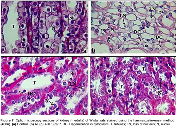
Kidney medulla of control rats shows that collecting tubules had a regular shape. The tubular cells are distinct and well defined (Figure 7a). Treatment with aluminium caused degeneration of tubular cells and loss of nuclei (Figure 7b). The group (Al+P) presents a degeneration of cytoplasm and tubular cells, however, the administration of the plant had induced a remarkable improvement in this tissue (Figure 7c). Kidney medulla of group treated with the plant shows a normal histological structure (Figure 7d).
DISCUSSION
The liver is a vital organ that is responsible for many of the processes that keep us alive. It drives a variety of metabolic substance and synthesizes a large number of enzyme (Bai et al., 2012). Hepatocytes are easily disintegrated by a variety of factors and harmful products (Bai et al., 2012). Exposure to high doses of aluminium may give an accumulation of this metal in the liver and cause alterations of the hepatic function (Nikolov et al., 2010). Degeneration, inflammation, and necrosis caused by hepatocyte damage can lead to an increase in the permeability of cell membranes. While AST and ALT are released into the blood through the cell membrane and their concentrations in the blood increases, ALT and AST are indicators of liver damage (Chinoy and Memon, 2001; El-Demerdash, 2004; Yeh et al., 2009; Shati and Alamri, 2010; Bai et al., 2012). In our study, the activities of AST and ALT in serum significantly increased in rats intoxicated with aluminium compared to controls. It was a sign of deterioration of liver function, these results were compatible with results of Zhuo et al. (2007), Ma et al. (2008), Yeh et al. (2009), Bai et al. (2012), Bhadauria (2012), Denen et al. (2015), and Kalaiselvi et al. (2015).
In group (Al+P), enzyme activities of AST and ALT in serum significantly decreased; therefore, the liver damage induced by aluminium can be improved by the plant.
LDH is an enzyme that is used to evaluate tissue damage of the affected organ (Anane and Creppy, 2001; Bhatti et al., 2014). Serum LDH is a biomarker of liver tissue lesions (Suzuki et al., 1995) and kidney (Karadeniz Cerit et al., 2013). Cell necrosis leads to increase LDH in tissue and serum (Türkez et al., 2011). LDH release in the blood is an indicator of cell death and disintegration of the cell membrane (Lindell et al., 1996). LDH is a marker of aluminium toxicity (Anan and Creppy, 2001). In this study, serum LDH was very high in rats treated with aluminium; it increased significantly in rats intoxicated with aluminium as compared to controls. The results of this study are consistent with results of Chinoy and Memon (2001), El-Demerdash (2004), and Türkez et al. (2011).
Exposure to aluminium causes changes in renal function (El-Demerdash, 2004; Åžener et al., 2007; Renugadevi and Prabu, 2010; Vijayaprakash et al., 2013). The assessment of harmful effects of aluminium is realized by measuring of biochemical parameters (Kowalczyk et al., 2004). Creatinine and urea are significant indicators of renal function (Al-Attar and Al-Taisan, 2010; Yakubu and Musa, 2012). According to the results of this study, the comparison between rats intoxicated with aluminium and control indicate a significant increase in both parameters. Yeh et al. (2009) reported that the plasma creatinine level is elevated in rats that received diets containing aluminium as compared to controls. According to Soudani et al. (2010), the increases of creatinine and urea concentrations indicate that the glomeruli and tubules are damaged.
Histology of organs is realized in order to verify the proper functioning of these organs. Microscopic observations in the control group (Figures 3a, 4a, and 5a) are similar to that of group (P) (Figures 3d, 4d, and 5d), respectively. These groups showed a normal structure of the liver and kidney, against group (Al) having abnor-malities in the liver and kidney (Figure 3b, 4b, and 5b). Treatment with H. salicornicum induced an improvement in histology of liver and kidney (Figure 3c, 4c, and 5c). Therefore, H. salicornicum was found to reshuffle the damage led by aluminium.
Bhadauria (2012) cited that aluminium exposure caused loss of cord arrangement of hepatocytes and sinusoidal spaces with degenerated plasma membranes and nuclei; for central vein filled with debris, severe necrosis was also seen. Buraimoh et al. (2012) mentioned that in their study, there were distorted sinusoids and congested central vein of the liver of the aluminium treated rats. Tehrani et al. (2013) reported that exposed animals showed cell damage in the liver. Al-Qayim and Mashi (2014) reported that light microscopic examination of kidney tissues sections revealed the negative and deleterious effects of aluminium chloride. Rats that received aluminium chloride showed infiltration of inflammatory cells and congestion of blood vessels. In spite of the material deposed in the lumen of tubules, there was enlargement of epithelial cells lining urinary tubules leading to occlusion of some urinary tubules and stenosis in others. Joshi et al. (2015) reported that aluminium induced kidney damage was illustrated not only by a significant alteration in serum metabolites, but also an altered histological feature in the kidney tissue reminiscent of some known diseases.
Accumulation of aluminum in organism can cause lesion directly or indirectly (Bai et al., 2012). It has been reported that aluminium exposure can increase lipid peroxidation rates (Anand et al., 2012). Lipid peroxidation is a chemical mechanism capable of disrupting the structure and the function of the biological membranes that occurs as a result of free radical attack on lipids (Yeh et al., 2009).
In this study, the rate of TBARS in liver and kidney of rats intoxicated is significantly increased as compared to the control rats. It was found out that the consumption of H. salicornicum induces a decrease in TBARS. From these results, it was shown that H. salicornicum has a potential protective effect against oxidative stress induced by aluminium. El-Demerdash (2004) reported that administration of 34 mg AlCl3/kg body weight induced free radicals and increased TBARS level in liver. Yeh et al. (2009) reported that the level of TBARS of the plasma and liver was increased with the increasing dose of aluminium. Mahieu et al. (2009) administered to male rats 0.57 mg aluminium per 100 g of body weight; they mentioned that their results indicated a significant increase of TBARS level of kidney in Al-treated rats when compared with the control animals. Joshi et al. (2013) administered 100 mg/kg of aluminium by gavage to young and adult rats; they reported a significant increase the lipid peroxidation in rats intoxicated versus control in liver and kidney.
The oxidant (reactive oxygen species [ROS] and nitrogen species [RNS]) and antioxidants are balanced in healthy individuals (Joshi et al., 2013). If the balance is disrupted due to over production of ROS, oxidative stress may occur, which influences oxidative damage to organs (Joshi et al., 2013). GSH is an important non-protein thiol present in the animal cells (Özkaya et al., 2010). It is a key component of the cellular defense cascade against injury caused by ROS (Hsu et al., 2000). The level of GSH in tissues serves as an indicator of oxidative stress (Hsu et al., 2000). Some antioxidant enzymes, such as SOD and CAT, play a principal role in antioxidation and elimination of ROS (Jihen et al., 2009). SOD catalyzes the dismutation of O2- to H2O2 which is decomposed by CAT. However, H2O2 may react with O2 to generate OHâ—¦, which is the initiator of the lipoperoxidation. The hydroxyl radical is highly toxic, because there is no specific enzyme system able to degrade it (Vamecq et al., 2004). According to our results, the comparison between groups (control) and (Al) demonstrate that the levels of GSH, SOD, and CAT decreased after exposure to aluminium. However, H. salicornicum consumption causes an increase in GSH levels, SOD, and CAT when compared with intoxicated rats. Based on the results of this study, it was concluded that H. salicornicum reshuffles oxidative stress caused by aluminium in liver and kidney.
These results are in good accordance with those obtained by Özkaya et al. (2010), Bai et al. (2012), and Joshi et al. (2013). Newairy et al. (2009) revealed that the level of TBARS was increased and the activities of GST, SOD, and CAT were decreased in liver, kidney, and brain of rat treated with 34 mg/kg body weight AlCl3 daily for 70 days. Yeh et al. (2009) mentioned that aluminium depleted GSH stores and reduced CAT activities.
CONCLUSION
Oxidative stress results from an imbalance between prooxidants and antioxidants. Biochemical indicators were measured and the redox status in liver and kidney was evaluated to check aluminium effect in both organs. From our results, exposure to aluminum release oxidative stress and a dysfunction in liver and kidney. Histological study confirmed the results. The administration of plant H. salicornicum induced improvement in all parameters studied. It was concluded that the plant reshuffle the harmful effects of aluminium.
CONFLICT OF INTERESTS
The authors have not declared any conflicts of interest.
REFERENCES
|
Abbas B, Al-Saeed M, Othman R (2006). Value of anti-microbial of Haloxylon salicornicum. J. Babylon Univ.14:3. |
|
|
AFSSAPS : Agence française de sécurité sanitaire des produits de santé (2011). Évaluation du risque lié à l'utilisation de l'aluminium dans les produits cosmétiques. Rapport d'expertise. |
|
|
Ahmad M, Eram S (2011). Hepatoprotective studies on Haloxylon Salicornicum: a plant from Cholistan desert. Pak. J. Pharm. Sci. 24 (3):377-382. |
|
|
Al-Attar AM, Al-Taisan WA (2010). Preventive effects of black seed (Nigella sativa) extract on Sprague Dawley rats exposed to diazinon. Aust. J. Basic Appl. Sci. 4:957-968. |
|
|
Al-Kahtani MA (2010). Renal damage mediated by oxidative stress in mice treated with aluminum chloride: Protective effect of Taurine. J. Biol. Sci. 10(7):584-595. |
|
|
Al-Qayim MAJ, Mashi S (2014). Renal effects of propolis and malic acid in Aluminum exposed male rats. App. Sci. Rep. 5(1):26-30. |
|
|
Al-Saeed AH (2002). Study the effect of some extract of Haloxylon sp. on blood glucose level in normal and hyperglycemic rabbits induced by alloxan. M.Sc. thesis, College Science, University of Basrah. |
|
|
Anand R, Kumari P, Kaushal A, Bal A, Wani WY, Sunkaria A, Dua R, Singh S, Bhalla A, Gill KD (2012). Effect of acute aluminum phosphide exposure on rats: a biochemical and histological correlation. Toxicol. Lett. 215(1):62-69. |
|
|
Anane R, Creppy EE (2001). Lipid peroxidation as pathway of aluminium cytotoxicity in human skin fibroblast cultures: prevention by superoxide dismutase+catalase and vitamins E and C. Hum. Exp. Toxicol. 20:477-481. |
|
|
Ashraf MA, Karamat M, Shahnaz K, Abdul W, Ismail Y (2012). Study of chemical and mineral constituents of Haloxylon salicornicum collected from Cholistan Desert, Bahawalpur, Pakistan. Wlfenia J. 19(10):306-327. |
|
|
Bai CS, Wang F, Zhao HS, Li YF (2012). Effects of Subchronic Aluminum Exposure on Liver Function in Rats. J. Northeast Agric. Univ. 19(2):62-65. |
|
|
Bhadauria M (2012). Combined treatment of HEDTA and propolis prevents aluminum induced toxicity in rats. Food Chem. Toxicol. 50:2487-2495. Bhatti GK, Sidhu IPS, Saini NK, Puar SK, Singh G, Bhatti JS (2014). Ameliorative role of melatonin against cypermethrin induced hepatotoxicity and impaired antioxidant defense system in Wistar. IOSR J. Environ. Sci. Toxicol. Food Technol. 8(1):39-48. |
|
|
Buraimoh AA, Ojo SA, Hambolu JO, Adebisi SS (2012). Effects of aluminium chloride exposure on the histology of the liver of adult wistar rats. IOSR J. Pharm. 2(3):525-533. |
|
|
Chinoy NJ, Memon MR (2001). Beneficial effects of some vitamins and calcium on fluoride and aluminium toxicity on gastrocnemius muscle and liver of male mice. Fluoride 34:21-33. |
|
|
Denen A, Samuel OO, Joseph ET, Egesie UG, Ejike DE (2015). Effects of ethanol extract of Garcinia Kola on biochemical markers of liver function of Wister rats. Int. J. Pharm. Sci. Invent. 4(5):5- 8. |
|
|
El-Demerdash FM (2004). Antioxidant effect of vitamin E and selenium on lipid peroxidation, enzymes activities and biochemical parameter in rats exposed to aluminium. J. Trace Elem. Med. Biol. 18:113-122. |
|
|
Ellman GL (1959). Tissue sulfhydryl groups. Arch. Biochem. Biophys. 82:70-77. |
|
|
Farina M, Lara FS, Brandao R, Jacques R, Rocha JBT (2002). Effects of aluminum sulfate on erythropoiesis in rats. Toxicol. Lett. 132:131-139. |
|
|
Ferheen S, Ahmed E, Afza N, Malik A, Shah MR, Nawaz SA, Choudhary MI (2005). Haloxylines A and B, Antifungal and Cholinesterase Inhibiting Piperidine Alkaloids from Haloxylon salicornicum. Chem. Pharm. Bull. 53(5):570-572. |
|
|
Gonzalez MA, Bernal CA, Mahieu S, Carrillo MC (2009). The interaction between the chronic exposure to Aluminium and liver regeneration on bile flow and organic anion transport in rats. Biol. Trace Elem. Res. 127:164-176. |
|
|
Hsu CH, Han BC, Liu MY, Yeh CY, Casida JE (2000). Phosphine-induced oxidative damage in rats: attenuation by melatonin. Free Radic. Biol. Med. 28(4):636-642. |
|
|
Jihen EH, Imed M, Fatima H, Abdelhamid K (2009). Protective effects of selenium (Se) and zinc (Zn) on cadmium (Cd) toxicity in the liver of the rat: effects on the oxidative stress. Ecotox. Environ. Saf. 72:1559-1564. |
|
|
Joshi D K, Tripathi S, Mishra S, Choudhary M, Rai R, Mahdi AA (2015). Docosahexenoic acid prevents aluminum induced metabonomic changes in rat urine: A Proton nuclear magnetic resonance study. Scholars. Acad. J. Biosci. 3(3):248-253. |
|
|
Joshi DK, Choudhary M, Tripathi S, Singh Negi MP, Mahdi AA (2013). Age dependent relative risk of aluminum toxicity: levels of metals and enzymic and non enzymic antioxidants status in liver, kidney and brain of aluminum treated young and old rats. Int. J. Biol. Pharm. Res. 4(3):176-185 |
|
|
Kadham MA (2008). Some pharmacological and toxicological investigations of Haloxylon salicornicum in rabbits. Coll. Vet. Med. 7(2):25-32. |
|
|
Kalaiselvi A, Aadhinath Reddy G, Ramalingam V (2015). Ameliorating Effect of Ginger Extract (Zingiber officinale Roscoe) on Liver Marker Enzymes, Lipid Profile in Aluminium chloride Induced Male Rats. Int. J. Pharm. Sci. Drug Res. 7(1):52-58. |
|
|
Karadeniz Cerit K, Karakoyun B, Yűksel M, Åzkan N, Çetinel Åž, DaÄŸli ET, YeÄŸen BÇ, TuÄŸtepe H (2013). The antifibrotic drug halofuginone reduces ischemia/reperfusion-induced oxidative renal damage in rats. J. Pediatr. Urol. 9:174-183. |
|
|
Kowalczyk E, Kopff A, Kędziora J, Błaszczyk J, Kopff M, Niedworok J, Fijałkowski P (2004). Effect of long-term aluminium chloride intoxication on selected biochemical parameters and oxidative-antioxidative balance in experimental animals. Pol. J. Environ. Stud. 13(1):41-43. |
|
|
Lindell SL, Hansen T, Rankin M, Danielewicz R, Belzer FO, Southard JH (1996). Donor nutritional status - A determinant of liver preservation injury. Transplantation 61:239-247. |
|
|
Ma YJ, Xu JG, Lin JY (2008). Effects of anetholtrithione on hepatic lipid peroxidation induced by aluminum in rats. Ind. Health Occup. Dis. 34(6):325-328. |
|
|
Mahieu S, Contini MDC. Gonzalez M, Miller NC (2009). Melatonin reduces oxidative damage induced by aluminum in rat kidney. Toxicol. Lett. 190:9-15. |
|
|
Mahieu SN, Gionotti M, Millen N, Elias MM (2003). Effect of chronic accumulation of aluminum in renal functions, cortical renal oxidative stress and cortical renal organic transport in rats. Arch. Toxicol. 77:605-612. |
|
|
Newairy AS, Salama AF, Hussien HM, Yousef MI (2009). Propolis alleviates aluminum-induced lipid peroxidaion and biochemical parameters in male rats. Food Chem. Toxicol. 47(6):1093-1098. |
|
|
Nikolov IG, Joki N, Vicca S, Patey N, Auchere D, Benchitrit J, Flinois JP, Ziol M, Beaune P, Drueke TB, Lacour B (2010). Tissue accumulation of lanthanum as compared to aluminum in rats with chronic renal failure-possible harmful effects after long-term exposure. Nephron Exp. Nephrol. 115:112-121. |
|
|
Ochmanski W, Barabasz W (2000). Aluminum-occurrence and toxicity for organisms. Przegl. Lek. 57:665-668. |
|
|
Özkaya A, Çelik S, Yüce A, Åžahin Z, Yilmaz Ö (2010). The Effects of ellagic acid on some biochemical parameters in the liver of rats against oxidative stress induced by aluminum. Kafkas Univ. Vet. Fak Derg 16(2):263-268. |
|
|
Renugadevi J, Prabu SM (2010). Quercetin protects against oxidative stress-related renal dysfunction by cadmium in rats. Exp. Toxicol. Pathol. 62:471-481. |
|
|
Sanchez-Iglesias S, Soto-Otero R, Iglesias-Gonzalez J, Barciela-Alonso MC, Bermejo-Barrera P, Mendez-Alvare E (2007). Analysis of brain regional distribution of aluminium in rats via oral and intraperitoneal administration. J. Trace Elem. Med. Biol. 21:31-34. |
|
|
Åžener G, Sehirli Ö, Tozan A, VelioÄŸlu-Övünç A, Gedik N, Omurtag GZ (2007). Ginkgo biloba extract protects against mercury (II)-induced oxidative tissue damage in rats. Food Chem. Toxicol. 45:543-550. |
|
|
Shahana MM, Mirhom YW, Generah AA, Aboutabi EA, Amer HA (1990). Study into wild Egyptian plants potential medicinal activity. Ninth communication: hypoglycemic activity of some selected plants in normal fasting and alloxanised rats. Arch. Exp. Vet. Med. 44(3):389-394. |
|
|
Shati AA, Alamri SA (2010). Role of saffron (Crocus sativus L.) and honey syrup on aluminum-induced hepatotoxicity, Saudi. Med. J. 31:1106-1113. |
|
|
Shirley DG, Walter MF, Walter SJ, Thewles A, Lote CJ (2004). Renal aluminum handling in rats: A micropuncture assessment. Clin. Sci. 107:159-165. |
|
|
Sinha AK (1972). Colorimetric assay of catalase. Anal. Biochem. 47:389-394. |
|
|
Soudani N, Sefi M, Amara IB, Boudawara T, Zeghal N (2010). Protective effects of selenium (Se) on chromium (VI) induced nephrotoxicity in adult rats. Ecotox. Environ. Saf. 73:671-678. |
|
|
Stoehr G, Leubbers K, Wilhelm M, Hoelzer J, Ohmann C (2006). Aluminum load in ICU patients during stress ulcer prophylaxis. Eur. J. Int. Med.17:561-566. |
|
|
Suzuki KT, Kano S, Misawa S, Aoki Y (1995). Copper-metabolism leading to and following acute hepatitis in lec rats. Toxicology 97:81-92. |
|
|
Taus N, Farraj M, Tănase S, Mironescu A, Boicu M, Necula V, Taus L (2013). Aluminum - a chemical neurotoxic agent. Bulletin of the Transilvania University of Braşov Series VI. Med. Sci. 2(55):1-8. |
|
|
Tehrani H, Halvaie Z, Shadnia S, Soltaninejad K, Abdollahi M (2013). Protective effects of N-acetylcysteine on aluminum phosphide-induced oxidative stress in acute human poisoning. Clin. Toxicol. (Phila). 51(1):23-28. |
|
|
Türkez H, GeyikoÄŸlu F, Çolak S (2011). The protective effect of boric acid on aluminum-induced hepatotoxicity and genotoxicity in rats. Turk. J. Biol. 35:293-301. |
|
|
Turkez H, Yousef MI, Geyikoglu F (2010). Propolis prevents aluminum-induced genetic and hepatic damages in rat liver. Food Chem. Toxicol. 48: 2741-2746. |
|
|
Vamecq J, Vallée L, Storme L, Gelé P, Bordet R (2004). Les acteurs immédiats du stress oxydant, La lettre du pharmacologue. 18:16-22. |
|
|
Vijayaprakash S, Langeswaran K, Kumar SG, Revathy R, Balasubramanian MP (2013). Nephro-protective significance of kaempferol on mercuric chloride induced toxicity in Wistar albino rats. Biomed. Aging Pathol. 3:119-124. |
|
|
Yagi K (1976). A simple fluorometric assay for lipoperoxide in blood plasma. Biochem. Med. 15:212-216. |
|
|
Yakubu MT, Musa IF (2012). Liver and Kidney Functional Indices of Pregnant Rats Following the Administration of the Crude Alkaloids from Senna alata (Linn. Roxb) Leaves. Iran. J. Toxicol. 6(16):615-625. |
|
|
Yeh YH, Lee YT, Hsieh HS, Hwang DF (2009). Effect of taurine on toxicity of aluminum in rats. Eur. J. Clin. Nutr. Metabol. 4:187-192. |
|
|
Zhuo JH, Liu P, Feng GC, Pingguo C (2007). Effect of aluminum chelating agent on liver zymogram and on essential elements of rats induced by aluminum. Chin. J. Public Health 23(3):316-317. |
|
Copyright © 2024 Author(s) retain the copyright of this article.
This article is published under the terms of the Creative Commons Attribution License 4.0


