African Journal of
Biotechnology

  • Abbreviation: Afr. J. Biotechnol.
  • Language: English
  • ISSN: 1684-5315
  • DOI: 10.5897/AJB
  • Start Year: 2002
  • Published Articles: 12488

Full Length Research Paper

ELISA, RT-PCR, semi-quantitative RT-PCR and sequencing methods for investigating an epidemic FMD virus serotype O outbreaks

Halla E. K. El Bahgy
  • Halla E. K. El Bahgy
  • Hygiene and Veterinary Care Department, Faculty of Veterinary Medicine, Benha University, 13736 Moshtohor, Toukh, Qalyubia, Egypt.
  • Google Scholar
Mahmoud M. A. Moustafa
  • Mahmoud M. A. Moustafa
  • Genetics and Genetic Engineering Department, Faculty of Agriculture, Benha University, 13736 Moshtohor, Toukh, Qalyubia, Egypt.
  • Google Scholar


  •  Received: 15 March 2018
  •  Accepted: 09 April 2018
  •  Published: 11 April 2018

 ABSTRACT

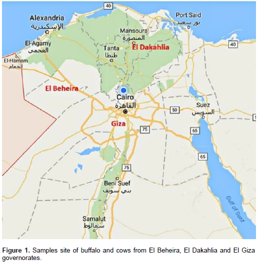
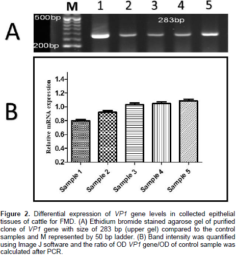
This study was conducted to investigate the seroprevalence of non-structure protein for foot and mouth disease virus (FMDV) and identify FMDV serotypes. A total of 3600 serum samples (1080 buffaloes and 2520 cattle) were collected randomly from different breeds, age, and sex at El Beheira, El Dakahlia, and El Giza. Nonstructure protein of FMDV was detected in 41 of 1080 (3.8%) Buffalo and 185 of 2520 (7.3%) cattle. The case fatality was 14.6 and 17.8% in the vaccinated buffalo and cattle, respectively. The prevalence of nonstructural protein FMDV serotyping O was 8.3, 7.5 and 2.9% in El Beheira, El Dakahlia, and El Giza, respectively. Moreover, the case fatality was 8% (El Beheira), 26.3% (El Dakahlia), and 20% (El Giza). The circulation of FMDV was prevalent during the winter season of the year. The frequency of positive case was significantly different between species, sex, and age, while it was non-significant with different breeds. The mean values of antibodies of a non-structural protein of FMDV were the highest in male cattle at 5 months to 1 year of age. Additionally, seven epithelial tissues were collected from tongue; buccal mucosa and teat of recently sudden dead animals. The obtained sequences by reverse transcription polymerase chain reaction (RT-PCR) were registered in GenBank under accession numbers MF980930.1, MF991123.1, MF991124.1, and MF991125.1. Phylogenetic analysis revealed that the obtained sequences belonged to deposited FMDV type O (KP121442.1) with similarity ratios of 98, 100, 99, and 99%, respectively. Also, the deduced amino acids of the obtained sequences are related to capsid protein of VP1 gene of FMDV.

Key words: Foot and mouth disease virus (FMDV), semi-quantitative reverse transcription polymerase chain reaction (RT-PCR), VP1 gene, vaccinated animals, phylogenetic analysis.


 INTRODUCTION

Foot and mouth disease (FMD) is the utmost spreadable disease of animals and has a great possibility for instigating serious economic damage in susceptible ruminant animals. The serotypes of FMD virus are O, A,  C, SAT 1, SAT 2, SAT 3, and Asia 1. Infection with one type does not give immunity against another type of virus. FMD is difficult to differentiate clinically with other vesicular diseases. The diagnosis of FMD is an emergency case (Cox and Barnett, 2009; Alam et al., 2015; Saduakassovaa et al., 2018).  The ruminant animals are sensitive to FMD (FAO, 1984). The wild infected animal species transfer FMD Virus to the domesticated animal species. Also, camels have been reported to be infected with FMD Virus (Larska et al., 2009). Foot and mouth disease virus belongs to family Picornaviridae and consists of seven different serotypes. Its RNA genome is about 8.5 kb that transcribes to 12 protein-coding genes and one viral genome protein (VPg) (Carroll et al., 1984; Forss et al., 1984; Li et al., 2007; Hwang et al., 2016; Abdulla et al., 2017). Serotypes A, SAT2, and O of FMDV were detected in Egypt since 1950 and caused outbreaks during, 1953, 1958 and 1960 and till 2006 in livestock (OIE, 2000; Aidaros, 2002). FMD serotype A outbreaks were noted in cows and buffalos in 2006 (OIE, 2006; Knowles et al., 2007). Moreover, the outbreaks of serotype O were registered in 2006, 2007, and 2008 for Egyptian animals (El-Buhayrah and Alexandria governorates) even though the animal received for routine vaccination by a local bivalent vaccine which comprise both O1 and A/Egypt/2006 (FAO, 2008). Egypt is an epidemic country of FMD that produces severe outbreaks almost every year and cause excessive economic damages (S1).
 
The native routine vaccination of the animals is not professional for controlling the dispersal of the disease due to the lack of cross-protection between the different viral serotypes and subtypes (Mason et al., 2003) and the viral mutations are at the high rate, especially in VP1 gene (Dopazo et al., 1988; Domingo et al., 1990; Samuel et al., 1999; Carrillo et al., 2005; Maryam et al., 2017; Islam et al., 2017). The control of FMD should be quick, more perfect and constant via developed diagnostic tools to assess the circulating serotypes for limiting its diffusion into vast terrestrial areas (Belak, 2007; El-Khabaz and Al–Hosary, 2016). The application of reverse transcription quantitative polymerase chain reaction (RT-qPCR) is highly sensitive and is considered as a valuable tool for the detection of viruses to reducing the risk of cross-contamination (Mackay et al., 2002; Mackay, 2004; Howson et al., 2017; Haidar et al., 2018). The present study aimed to investigate the seroprevalence of non-structural protein for foot and mouth disease virus (FMDV) and to identify its serotype in the collected samples. Also, it is performed to characterize and to determine circulating serotypes of FMDV during the risk time of year. On another hand, it was conducted to estimate the risk factor affecting the incidence of FMDV outbreaks. Also, FMDV outbreaks cause the big losses for farmers and Egyptian national income, so this study is an attempt for early diagnosis of FMDV and advise to policymakers to develop an effective hexa vaccine against FMDV serotypes.


 MATERIALS AND METHODS

Sampling
 
During the outbreaks of 2016 to 2017 in Egypt, a total of 3600 cattle and buffalo were investigated during the period between June 2016 and May 2017 in different farms located at El Beheira, El Dakahlia, and El Giza governorates (Figure 1). One hundred blood samples were collected from each governorate in a month. The susceptible animals composed of milking Friesian cows, buffalo and beef cow of different breeds, age, and sex.  In this study, a total of 3600 serum samples (1080 buffaloes and 2520 cattle) were collected from apparent and unapparent healthy animals of different breeds, age, and sex. Additionally, seven epithelial tissues were collected from the tongue; buccal mucosa and teat of recently sudden dead animals were post-mortems examined for the presence of tiger heart, swollen in a lymph node with hemorrhagic appearance and vasoconstriction of intestine leading to infection suspension with foot and mouth disease under national and international standard biosafety conditions and ethics. All samples were collected from vaccinated animals with local Tri-Aphthovac® MEVAC vaccine which composed of inactivated FMDV serotypes (An Iran 05, O Pan Asia 2, and SAT-2) with oil adjuvant manufactured by ME-VAC.  The blood samples were left in tightly closed tubes overnight at 4°C and then centrifuged at 3000 rpm for 10 min to separate the sera and non-sera fractions. The clear serum was obtained by using sterile tips and placed in clean Eppendorf tubes, labeled, and stored at -70°C in the laboratory for the detection of antibodies to non-structure proteins of FMDV in animal serum samples. All positive non-structural protein samples were tested by indirect sandwich enzyme-linked immunosorbent assay (ELISA) for detection of FMDV antigen and serotypes. Epithelial samples were collected in sterilized tubes that contained glycerol and phosphate-buffered saline (PBS), pH 7.2 to 7.6, (penicillin [1000 International Units (IU)], neomycin sulfate [100 IU], polymyxin B sulfate [50 IU], and Mycostatin [100 IU]). Samples stored at -80°C were used for isolation of the virus and identification for detection and confirmation using RT-PCR and sequencing methods.
 
Diagnostic tests
 
Serological diagnosis
 
Detection of non-structural protein of FMDV: Detection of IgG antibodies in animal's serum samples was carried out by FMDV 3ABC-Trapping ELISA to measure the antibodies of non-structure proteins of FMDV to differentiate between the antibodies against infection and antibodies against vaccination (Brocchi et al., 2006) produced by IZSLER, Biotechnology laboratory, via A. Bianchi, 9-25124 Brescia (Italy). These serological diagnoses were performed at the Animal Health Research Institute, Agriculture Research Center, Doki, Giza, Egypt.
 
FMD antigen typing detection ELISA Kits: All positive non-structural protein of FMDV samples were tested by indirect sandwich ELISA test for detection of FMD viral antigen and serotypes (Ferris and Dawson, 1988). The kits were produced at IZSLER Biotech Laboratory, Pirbright Institute, UK. These serological diagnoses were performed at the Animal Health Research Institute, Agriculture Research Center, Doki, Giza, Egypt.


Molecular detection of FMDV by RT-PCR: The viral RNA was extracted from all collected samples by QIAamp® Viral RNA Mini Kit (Qiagen, Hilden, Germany) following the mini spin protocol according to the manufacturer’s instructions and eluted in 50 μl of elution buffer. 1 μg of the obtained RNA was used as the template in a one-step RT-PCR (Ready-To-Go RT-PCR Beads; Amersham). The primers used were F:5/.CCTCCTTCAAYTTACGGTG.3/ (Parlak et al., 2002) and R: 5/.GACATGTCCTCCTGCATCTG.3/ (Bachanek-Bankowska et al., 2016). The thermal profile was used as follows: 42°C for 30 min; 94°C for 5 min; 35 cycles of 94°C for 60 s; 55°C for 60 s; and 72°C for 90 s; followed by a final extension at 72°C for 5 min. The amplified RT-PCR products were analyzed by electrophoresis in 1.5% agarose gel stained with Ethidium bromide using GeneRuler 50 bp DNA ladder (Cat. #: SM0373), then visualized under UV Transilluminator (BioRAD). The obtained RT-PCR products were purified by Qiaquick PCR purification kit (Qiagen) according to the manufacturer’s instructions. The RT-PCR products were eluted in nuclease-free water. The purified RT-PCR products were sent to Macrogen Company (South Korea) for sequencing service.
 
Semi-quantitative expression of VP1 gene in infected cows: Quantification of band intensities (OD) was measured using Image J software and a ratio OD candidate gene compared to OD of the control sample was calculated for each sample. The probability associated with one-way ANOVA. Bioinformatics analysis. The obtained partial sequence for Vp1 gene of FMDV was about 283 bp as shown in Figure 2. NEBcutter V2.0 software was used to create restriction map and to identify GC and AT ratios of the obtained sequence (Vincze et al., 2003; http://nc2.neb.com/NEBcutter2/). Jalview software was used to show SNPs that resulted from the alignment of the obtained sequences and the nearest sequences in NCBI database (http://www.jalview.org/). The obtained sequences were registered at GenBank under accession numbers MF980930.1, MF991123.1, MF991124.1 and MF991125.1 of Foot-and-mouth disease virus-type O isolates HM1, HM2, HM3, and HM4, respectively (http://www.ncbi.nlm.nih.gov). Construction of the phylogenetic trees was done by using Clustal Omega and MEGA7 software. Moreover, the amino acid sequences of the current sequences were obtained by using ExPASy translate tool (http://web.expasy.org/translate/). Also, the restriction maps of the current sequences were obtained through NEBcutter V2.0 tool (http://nc2.neb.com/NEBcutter2/) to display the possible endonuclease sites within the obtained sequences. GC and AT contents ratios were calculated by NEBcutter V2.0 tool of the obtained sequences. 


Statistical analysis
 
The statistical analysis of deltamethrin DATA was done by one-way ANOVA and Independent T-tests using SPSS software version 20. The results were considered significantly different at P<0.05.


 RESULTS

The highest incidence and case fatality of FMDV were 7.3 and 17.8%, respectively in the vaccinated cattle serum samples (Table 1). The prevalence of non-structural protein serotyping O of FMDV was 8.3, 7.5 and 2.9% with case fatality 8, 26.3 and 20% in El Behera, El Dakahlyia, and El Giza governorates, respectively (Table 2). The incidence of FMDV was the most prevalent in the period between January and April (winter season) with the highest mortality rate in January in all governorates during a period of the study (Figure 3). The frequency of positive case was significantly different between species, sex, and age; on the other hand, there was no significant difference with the breed. The mean values of antibodies of non-structural protein of FMDV were the highest in male cattle at 5 months to 1 year (Table 3). The obtained current sequences MF980930.1, MF991123.1, MF991124.1, and MF991125.1 were closed to foot-and-mouth disease virus-type o (KP121442.1) with high similarity ratios so they were located in the same clade (Figure 4). The single nucleotide polymorphism (SNP) showed little variation among the obtained current sequences (MF980930.1, MF991123.1, MF991124.1, and MF991125.1) and the nearest deposited sequence (KP121442.1) in DNA DATA base (Figure 5).


Moreover, the aligned protein sequences showed that the expected amino acids sequences, the current study, were related to polyprotein of foot-and-mouth disease virus-type O. The number of SNPs for the obtained amino acid sequences varied among the obtained sequences and the nearest ones registered in DNA database (Figure 6). This indicated that the obtained current sequences MF980930.1, MF991123.1, MF991124.1 and MF991125.1 related to deposited foot-and-mouth disease virus-type o (KP121442.1) in GenBank. Also, the collective phylogeny tree reported that the current sequences (MF980930.1, MF991123.1, MF991124.1 and MF991125.1) located in the same clade in the phylogeny tree. This revealed that the obtained sequences were more related to each other as shown in Figure 4. On the other hand, the obtained amino acids sequences converted from the current sequences showed that they belonged to VP1 gene which encoded to protein coat of foot-and-mouth disease virus-type o (Figure 6). The restriction map image showed many possible endonuclease sites of the obtained sequences and GC and AT ratios.The expected endonuclease sites can be used as DNA markers for detecting foot and mouth disease virus in the future. The restriction maps revealed that endonuclease site (CTGAAG) of the Acu1 enzyme was common in the four obtained sequences. Furthermore, this common site sequence as single nucleotide polymorphism (SNP) with a PCR-RFLP application as specific SNP of foot and mouth disease virus type 0 can be exploited in the future survey studies. Semi-quantitative expression of VP1 gene in infected cows showed little differences in the incidence of mRNA transcript levels among different dead cows; where the highest expression of VP1 gene was shown in sample 5 as shown in Figure 2. The results of differential expression of VP1 gene revealed that the FMDV was widespread in epithelial tissues of dead cows.


 DISCUSSION

In Egypt, FMD has taken an enzootic form and many outbreaks had occurred since 1950 till now. FMDV type O was the most prevalent until serotype A appeared in 2006 (Moussa et al., 1984; Daoud et al., 1988; Farag et al., 2005; El-Khabaz and Al-Hosary, 2016); then during April and May 2012, six outbreaks of FMD type SAT 2 were reported in Egyptian governorates (Abd El-Moety et al., 2013; El-Khabaz and Al-Hosary, 2016). Up to date, several FMD outbreaks are still stroking the livestock in Egypt despite routine massive vaccination. The vaccination of livestock in Egypt with the triple vaccine appears not to be enough to control six FMDV serotypes. The vaccine should be included in all known serotypes to avoid any outbreak of FMDV. Then the big losses of farmers and national income can be reduced. The serotype O of FMDV, in this study, was detected in El Behera, El Dakahlyia and El Giza governorates. IT was the most predominant in all regions from January to April 2017. FMDV mostly occured during winter as the virus is sensitive to high temperature. Tomasula et al. (2007) and SareyyüpoÄŸlu and Burgu (2017) reported that at high temperature, there might be the destruction of virus receptors, which ultimately declined its infectivity.
 
The obtained results showed that the cattle were more sensitive than buffaloes to FMDV due to the fact that immunity of buffaloes is more than cattle. Also, the susceptibility of buffaloes to FMDV is less than cattle. This finding agrees with Ahmed et al. (2012) and Valdazo et al. (2012) who reported that the clinical picture of FMD in the affected animals was characterized by severe clinical signs in cattle, buffalos, small ruminants and young animals. These results agreed with Alexandrov et al. (2013) who reported that both mouth and foot lesions can occur in water buffalo, but the clinical signs are reported to be milder than in cattle and lesions may heal more rapidly, as its resistance is higher than that of cattle. The obtained results clarified that 1 to 3 years old age is the least susceptibility to infection as may be attributed to the fact that they have a complete mature immune system and have previous exposure to FMD infection or vaccination. FMDV infected animals up to 5 months to 1 year of age. This may be attributed to colostral antibodies which protect animals to 3 to 6 months of age which in turn decrease the rate of FMDV infection among calves of less than 6 months of age (Alam et al., 2016). However, most deaths were among animals less than 1 year ago.
 
This could be attributed to that naïve calves which develop more prominent clinical FMD signs and lesions than older animals including cardiac complications which are considered the main cause of high mortalities among younger animals (Geering and Lubroth, 2002). This also highlights the importance of proper vaccination of the dams to protect their young calves. The prevalence of FMD disease was found significantly higher in male than female indicating that the male are more susceptible to FMDV than the female. The obtained results agreed with the previous findings of other authors (Alam et al., 2016; Mannan et al., 2009; Sarker et al., 2011; Rahman et al., 2015; Tomasula et al., 2007).  The obtained DNA data showed that the isolated FMDV serotype was type O. The phylogeny tree of the current sequences (MF980930.1, MF991123.1, MF991124.1, and MF991125.1) showed that they located with foot-and-mouth disease virus-type O (KP121442.1) in the same clade. Moreover, the differential expression of VP1 gene confirmed that the cause of the death of cows was due to the spread of FMDV in the epithelial tissues.


 CONCLUSION

Egypt is an epidemic country for FMD expressing many outbreaks of the disease almost every year producing excessive economic damages. The native routine vaccination of the animals is not professional for controlling the dispersal of the disease due to the lack of cross-protection between the different viral serotypes and subtypes and the high rate of viral mutations, especially in VP1 gene; so the control of FMD requires quick, more perfect and constant well-developed diagnostic tools to assess the circulating serotypes for limiting its diffusion into vast terrestrial areas and will aid in the proper vaccine choice and consequently reduce disease damages. Moreover, ELISA, RT-PCR, semi-quantitative RT-PCR and sequencing methods are very informative for a screening of FMD virus and prediction of more effective vaccines.


 CONFLICT OF INTERESTS

The authors have not declared any conflict of interests.


 REFERENCES

Abd El-Moety MS, Abd El-Aty MM, Fakry HM, Daoud HM, Ibrahim EE, Gamal El-Din WM, Rizk SA, Abu El-Naga H, Mohamed AA, Abd El-Krim AS, Farouk EM (2013). Isolation and molecular characterization of foot and mouth disease SAT2 virus during outbreak 2012 in Egypt. J. Vet. Adv. 3(2):60-68.
Crossref


Abdulla NM, Mohran KA, Haroun M, Ausama AA, Shalaby MA (2017). Identification of Foot and Mouth Disease Virus Strains Originating from Multispecies Susceptible Animals. J. Vet. Sci. Med. Diagn. 6:1.
Crossref


Ahmed HA, Salem SA, Habashi AR, Arafa AA, Aggour MG, Salem GH, Gaber AS, Selem O, Abdelkader SH, Knowles NJ, Madi M, Valdazo-Gonzalez B, Wadsworth J, Hutchings GH, Mioulet V, Hammond JM, King DP (2012). Emergence of foot and mouth disease virus SAT2 in Egypt during 2012. Transbound. Emerg. Dis. 59(6):476-481.
Crossref


Aidaros HA (2002). Regional status and approaches to control and eradication of foot and mouth disease in the Middle East and North Africa. Rev. Sci. Technol. 21:451-458.
Crossref


Alam MA, Amin MR, Paul TK, Rizon MK. (2016). Clinically detection of food and mouth disease at Kapasia upazila under Gazipur district in Bangladesh. J. Bangladesh Agric. Univ. 14(2):185-190.
Crossref


Alam MA, Rahman M, Hossen ML, Ahmed S, Parvej MS, Khan MFR, Rahman MB (2015). Reverse transcription polymerase chain reaction (RT-PCR) based detection and serotyping of FMD Virus from field samples of Gazipur, Bangladesh, and adaptation of the virus in BHK-21 cell. J. Adv. Vet. Anim. Res. 2(3):291-295.
Crossref


Alexandrov T, Stefanov D, Kamenov P, Miteva A, Khomenko S, Sumption K, Meyer Gerbaulet H, Depner K (2013). Surveillance of foot and mouth disease (FMD) in susceptible wildlife and domestic ungulates in southeast of Bulgaria following a FMD case in wild boar. Vet. Microbiol. 166(1-2):84-90.
Crossref


Bachanek-Bankowska K, Merob HR, Wadswortha J, Miouleta V, Salluc R, Belshamd GJ, Kasangab CJ, Knowlesa NJ, Kinga DP (2016). Development and evaluation of tailored specific real-time RT-PCR assays for detection of foot-and-mouth disease virus serotypes circulating in East Africa. J. Virol. Methods 237:114-120.
Crossref


Belak S (2007). Molecular diagnosis of viral diseases present trends and future aspects: A view from the OIE collaborating centre for the application of polymerase chain reaction methods for diagnosis of viral diseases in veterinary medicine. Vaccine 25:5444-5452.
Crossref


Brocchi E, Bergmann IE, Dekker A, Paton DJ, Sammin DJ, Greiner M, Grazioli S, De Simone F, Yadin H, Haas B, Bultu N, Malirat V, Neitzert E, Goris N, Parida S, Sorensen K, De Clercq K (2006). Comparative evaluation of six ELISAs for the detection of antibodies to the non-structural proteins of foot-and-mouth disease virus. Vaccine 24:6966-79.
Crossref


Carrillo C, Tulman ER, Delhon G, Lu Z, Carreno A, Vagnozzi A, Kutish GF, Rock DL. (2005). Comparative genomics of foot-and-mouth disease virus. J. Virol. 79:6487-6504.
Crossref


Carroll AR, Rowlands DJ, Clarke BE (1984). The complete nucleotide sequence of the RNA coding for the primary translation product of foot and mouth disease virus. Nucl. Acids Res. 12:2461-2472.
Crossref


Cox SJ, Barnett PV (2009). Experimental evaluation of foot-and-mouth disease vaccines for emergency use in ruminants and pigs: a review. Vet. Res. 40:13-43.
Crossref


Daoud A, Omar A, El-Bakry M, Metwally N, El-Mekkawi M, El-Kilany S (1988). Strains of foot and mouth disease virus recovered from 1987 outbreak in Egypt. J. Egypt. Vet. Med. Ass. 48:63-71.


Domingo E, Mateu MG, Martínez MA, Dopazo J, Moya A, Sobrino F (1990). Genetic Variability and Antigenic Diversity of Foot-and-Mouth Disease Virus. In. Kurstak E, Marusyk RG, Murphy FA, Van Regenmortel MHV (eds) Virus Variability, Epidemiology and Control. Applied Virology Research, vol 2. Springer, Boston, MA. pp. 233-266.
Crossref


Dopazo J, Sobrino F, Palma E, Domingo EL, Moya A (1988). Gene encoding capsid protein VP1 of Foot-and-mouth disease virus: Aquasispecies model of molecular evolution. Proc. Natl. Acad. Sci. USA 85:6811-6815.
Crossref


El-Khabaz KAS, Al-Hosary AAT (2016). Detection and identification of Foot and Mouth disease virus serotypes in Assiut governorate, Egypt. J. Adv. Vet. Anim. Res. 4(1):32-38.


Farag MA, Aggour MA, Daoud AM (2005). ELISA as a rapid method for detecting the correlation between the field isolates of Foot and Mouth Disease and the current used vaccine strain in Egypt. Vet. Med. J. Giza 53(4):949-955.


Ferris N, Dawson M (1988). Routine application of enzyme-linked immune sorbent assay in comparison with complement fixation for diagnosis of food-and-mouth and swine vesicular disease. Vet. Microbiol. 16:201-209.
Crossref


Food and Agricultural Organization of the United Nations (FAO) (1984). Emerging Diseases of Livestock. The Diseases and their Diagnosis Geering WA ed FAO Rome Italy 1:43-51.


Food and Agricultural Organization of the United Nations (FAO) (2008). Foot and mouth disease report Food and Agriculture Organization of the United Nations Italy.


Forss S, Strebel K, Beck E, Schaller H (1984). Nucleotide sequence and genome organization of foot-and-mouth disease virus. Nucleic Acids Res. 12:6587-6601.
Crossref


Geering WA, Lubroth J (2002). Preparation of foot-and-mouth disease contingency plans. Food Agriculture Org. pp. 39-57.


Haidar F, Waheed U, Haque SE, Arshad M (2018). Molecular Detection of Foot and Mouth Disease Virus (FMDV) from 2017 Outbreaks in Punjab by RT-PCR and RT-LAMP Assays. J. Vet. Sci. Technol. 9(1):1-4.
Crossref


Howson ELA, Armson B, Lyons NA, Chepkwony E, Kasanga CJ, Kandusi S, Ndusilo N, Yamazaki W, Gizaw D, Cleaveland S, Lembo T, Rauh R, Nelson WM, Wood BA, Mioulet V, King DP, Fowler VL (2017). Direct detection and characterization of foot-and-mouth disease virus in East Africa using a field-ready real-time PCR platform. Transbound. Emerg. Dis. 65:221-231.
Crossref


Hwang JH, Shin YK, Park SY, Kim J, Kim SM, Kim B, Park JH, Lee JS, Lee KN (2016). Robust real-time reverse transcription-PCR for detection of foot-and mouth disease virus neutralizing carryover contamination. J. Clin. Microbiol. 54:216-219.
Crossref


International Office of Epizootics (OIE) (2000). Foot and mouth disease Chapter 211 in Manual of Standards for Diagnostics Tests and Vaccines, 5th Edn Foot and Mouth Disease in Egypt Report OIE International Office of Epizootics http://www.oie.int.


International Office of Epizootics (OIE) (2006). Foot and mouth disease Chapter 211 in Manual of Standards for Diagnostics Tests and Vaccines, 4th Edn OIE International Office of Epizootics http://www.oie.int.


Islam MS, Habib MA, Saha PC, Das PM, Khan M (2017). Distribution of foot and mouth disease virus serotypes in cattle of Bangladesh. SAARC J. Agric. 15(1):33-42.
Crossref


Knowles NJ, Wadsworth J, Reid SM, Swabey KG, El-Kholy AA, El-Rahman AA, Soliman HM, Ebert K, Ferris NP, Hutchings GH, Statham RJ, King DP, Paton D (2007). Foot and mouth disease virus serotype A in Egypt. Emerg. Infect. Dis. 13(10):1593-1596.
Crossref


Larska M, Wernery U, Kinne J, Schuster R, Alexandersen G, Alexandersen S (2008). Differences in the susceptibility of dromedary and Bactrian camels to foot-and-mouth disease virus. Epidemiol. Infect. 8:1-6.


Li D, Shang YJ, Liu ZX, Liu XT, Cai XP (2007). Comparisons of the complete genomes of two Chinese isolates of a recent foot-and-mouth disease type Asia 1 virus. Arch. Virol. 152:1699-1708.
Crossref


Mackay IM (2004). Real-time PCR in the microbiology laboratory. Clin. Microbiol. Infect. 10:190-212.
Crossref


Mackay IM, Arden KE, Nitsche A (2002). Real-time PCR in virology. Nucleic Acids Res. 30:1290-1305.
Crossref


Mannan MA, Siddique MP, Uddin MZ, Parvez MM (2009). Prevalence of foot and mouth disease (FMD) in cattle at Meghnaupazila in Comilla in Bangladesh. J. Bangladesh Agric. Univ. 7(2):317-319.


Maryam S, Rasheed T, Latif A, Zahra R, Bin Zahur A, Ahsan A, Afzal M, Farooq U (2017). One-step real-time loop-mediated isothermal amplification (RT-LAMP): evaluation and its application for the detection of foot-and-mouth-disease virus and its serotypes. Turk. J. Vet. Anim. Sci. 41:435-443.
Crossref


Mason PW, Grubman MJ, Baxt B (2003). Molecular basis of pathogenesis of FMDV. Virus Res. 91:9-32.
Crossref


Moussa AAM, Daoud A, Hussein K, Hassan NA, Fahmy F, Azab A, El- Shehawy L (1984). Prevalence of FMD in Egypt. Agric. Res. Rev. 62(5B):55-63.


Parlak Ü, AktaÅŸ S, Özyörük F (2002). Genetic characterisation of type O and A viruses isolated from Turkey between 2000 and 2002 Appendix 11 FMD Institute PK 714 Ulus Ankara Turkey.


Rahman MM, Jalil MA, Hossain KMM, Alam KJ, Salam R, Reza MA (2015). Occurrence of foot and mouth disease in cattle in Magura district of Bangladesh. Int. J. Nat. Soc. Sci. 2(3):1-4.


Saduakassovaa MA, Sultanova AA, Kutumbetova LB, Wadsworth J, Wood BA, Knowles NJ, King DP, Bachanek-Bankowska K (2018). Development and evaluation of a novel real-time RT-PCR to detect foot-and mouth disease viruses from the emerging A/ASIA/G-VII lineage. J. Virol. Methods 252:37-41.
Crossref


Samuel AR, Knowles NJ, Mackay DKJ (1999). Genetic analysis of type O viruses responsible for epidemics of foot-and-mouth disease in North Africa. Epidemiol. Infect. 122:529-538.
Crossref


SareyyüpoÄŸlu B, Burgu I (2017). Development of multiplex RT-PCR for detection and differentiation of foot-and-mouth disease virus O and A serotypes in Turkey. Turk. J. Vet. Anim. Sci. 41:764-769.
Crossref


Sarker S, Talukder S, Haque MH, Islam MH, Gupta SD (2011). Epidemiological study on foot-and-mouth disease in cattle: Prevalence and risk factors assessment in Rajshahi Bangladesh Wayamba. J. Anim. Sci. 3:71-73.


Tomasula PM, Kozempel MF, Konstance RP, Gregg D, Boettcher S, Baxt B, Rodriguez LL (2007). Thermal inactivation of foot and mouth disease virus in milk using high-temperature short-time pasteurization. J. Dairy Sci. 90:3202-3211.
Crossref


Valdazo-Gonzalez B, Knowles NJ, Hammond J, King DP (2012). Genome sequences of SAT-2 foot and mouth disease viruses from Egypt and Palestinian Autonomous Territories (Gaza strip). J. Virol. 86:8901-8902.
Crossref


Vincze T, Posfai J, Roberts RJ. (2003). NEBcutter: a program to cleave DNA with restriction enzymes. Nucleic Acids Res. 31:3688-3691.
Crossref


          */?>