ABSTRACT
Ageratum conyzoides Linn is a medicinal plant used for diverse ethnomedicinal applications including anti-ulcer treatment. Usually, protection of gastric mucosa from injury or ulceration is dependent on the efficacy of intrinsic or induced protective factors against erosive effects of aggressive factors. In this study, our aim was to ascertain the gastroprotective activity of methanolic leaf extracts of A. conyzoides L. and assess the associated roles of gastric mucous cells and p53 protein. This study involved 25 adult male Wistar rats divided into five groups (A-E). Groups A and E were used as normal and test controls while B-D were administered with extracts at 100, 300 and 500 mg/kg body weight for 28 days. Gastric mucosal injury was induced via pyloric ligation method. Gastric tissues were processed, stained with periodic acid-Schiff and immunostained for p53 protein (using monoclonal antibody). Stained sections were quantified using image-J software, data obtained were statistically analyzed. The results showed significant increase (p<0.05) in mucous cell population but no significant increase in p53 protein expression in gastric tissues of treated animals. This implied that increase in mucous cell count and down-regulation of p53 protein in gastric tissues play key role in gastroprotective activity of methanolic extracts of A. conyzoides L.
Key words: Ageratum conyzoides, mucous cell, p53 protein, gastroprotection, rats.
Ageratum conyzoides Linn (Asteraceae) is a medicinal, tropical plant commonly found in West Africa and parts of Asia and South America. It is an annual herbaceous plant not usually eaten by humans largely because of the characteristics bad odour, likened to the smell of a male goat but has an age-long record of diverse traditional medicinal uses in several countries (Ming, 1999; Okunade, 2002; Shekhar and Anju, 2012). Historically, it is popularly known for its usage in treatment of burns, wounds, infectious diseases, arthritis and fever (Kamboj and Saluja, 2008). The study by Elisabetsky and Wannmacher (1993) and Ming (1999) reported the application of A. conyzoides L. extracts as anti-inflammatory agent, analgesic and for treatment of malaria and yellow fever. The study by Dung and Loi, (1991) and Sharma and Sharma (1995) described the application of whole plant of A. conyzoides L. as antiallergic agent and also for treatment of gynaecological diseases. Further studies by Perumal-Samy et al., (1999) and Ayyanar and Ignacimuthu, (2005) reported the application of A. conyzoides L. leaf extracts as coagulant and in treatment of diarrhea. The study by Sofowora (1993) reported the application of A. conyzoides L. leaf extracts to treat conjunctivitis; otitis and head ache in some parts of Cameroon. In Madagascar, Novy (1997) described the application of juice extracted from A. conyzoides L. leaves as a coagulant and for treatment of diarrhoea. Another study by Gurib-Fakim et al., (1993) in Mauritius, reported the medicinal application of A. conyzoides L. leaves to cure diarrhoea, skin infection and as diuretic in urinary diseases. In some parts of Kenya, leaves and roots of A. conyzoides L. are used for treatment of stomach ache while the seeds are used for treatment of epilepsy in Tanzania (Geissler et al., 2002; Moshi et al., 2005). The study by Mahmood et al., (2005) reported the anti-ulcer effects of leaf extracts of A. conyzoides L. in ethanol-induced gastric ulceration in rats. However, with several reported and documented pharmacological uses of A. conyzoides L., there remains a conspicuous dearth of explanation of the mechanisms of its numerous pharmacological applications. This study was therefore carried out to ascertain gastroprotective activity of methanolic leaf extracts of A. conyzoides L. and to determine the role of gastric mucous cells and tumor suppressor (p53) protein during the activity in male Wistar rats treated with the extracts prior to induction of gastric mucosal injury through pyloric-ligation method.
Plant material
Fresh whole plant of A. conyzoides L. was identified at the Herbarium of Department of Plant Biology and Biotechnology, University of Benin, Nigeria and documented with a voucher number 344.
Method of extraction
The leaves of plant were detached, air-dried and pulverised into powdered form using mechanical grinder. 1000 g of the powdered leaves was dissolved in 5 L of methanol for 72 h. Thereafter, the preparation was filtered and the filtrate evaporated to dryness by using rotary evaporator. The residue obtained was cooled at room temperature, weighed and used as the methanolic extracts for this study.
Animal used
This study involved 25 male Wistar rats weighing between 180-200 g and divided into five groups (A -E) comprising 5 animals per group (that is, n = 5).
Experimental design
Group A animals were given distilled water (5 mls/kg body weight). This group represented normal control that was not treated and not induced by pyloric ligation.
Group B animals were given 100 mg/kg methanolic extracts of A. conyzoides L.
Group C animals were given 300 mg/kg methanolic extracts of A. conyzoides L.
Group D animals were given 500 mg/kg methanolic extracts of A. conyzoides L.
Group E animals were given distilled water (5 mls/kg body weight). This group represented test control that was not treated but induced by pyloric ligation.
Period and mode of study
The treatment period of this study was 28 consecutive days and all treatments were done orally using a flexible orogastric gavage.
Induction of gastric mucosal injury using pyloric ligation method
The animals were fasted for 24 h in separate cages but allow free access to water. Animals were anesthetized by intraperitoneal injection of Ketamine/Xylazine (50 mg/kg at 1:1). A small midline incision was made on the abdomen of animals to access pyloric part of the stomach. The pyloric end of stomach was gently pulled up, ligated and gently returned into the abdominal cavity and the abdomen was closed. After an observatory period of 5 h, the animals were sacrificed and stomach tissue harvested and prepared for tissue processing (Shay et al., 1945).
Tissue processing
The tissues were fixed in 10% neutral buffered formalin, dehydrated using ascending grades of alcohol (two changes each of 70 and 90%, respectively, and absolute alcohol for 30 min each), cleared in xylene for 30 min and embedded in molten paraffin and allowed to cool to form tissue blocks.
Sectioning
Blocks of tissue were cut into sections at 3 and 5 µ thickness by using rotary microtome and mounted on microscope slides.
Staining:
The 3 and 5 µ tissue sections were used for the histochemical (Periodic acid–Schiff) staining and immunohistochemical (Horseradich-peroxidise-3, 3-Diaminobenzidine) staining respectively.
Periodic acid–Schiff (PAS) staining
Sections were deparaffinised in xylene 15 min, hydrated from absolute alcohol to distilled water, immersed in 0.5% periodic acid solution at room temperature for 5 min, rinsed in several changes of distilled water, immersed in Schiff’s reagent at room temperature for 15 min, washed in running tap water, counterstained in Mayer’s Haematoxylin solution for 3 min, rinsed in running tap water, dehydrated in alcohol, cleared in xylene and mounted with DPX.
Horseradich-peroxidise-3, 3-Diaminobenzidine (HRP-DAB) immunostaining for p53 protein (using monoclonal antibody)
Sections brought to water, antigen retrieval performed by using citric acid solution (pH 6.0) in a microwave at power 100 Watts for 15 min, equilibrated by gently displacing hot citric acid with running tap water for 3 min, endogenous peroxidases blocked using peroxidase block for 15 min, sections washed in phosphate buffer saline (PBS) mixed with Tween 20 for 3 min, protein blocked with Nevocastra protein block for 15 min, sections washed with PBS for 3 min, sections incubated in primary antibody (1 in 100 dilution ratio) for 45 min, washed with PBS for 3 min, secondary antibody added and allowed for 15 min, sections washed with PBS for 3 min, polymer added and allowed for 15 min, sections washed twice with PBS and treated with 3, 3-Diaminobenzidine (DAB) substrate for 5 min each (DAB was prepared in 1/100 dilution ratio with the DAB substrate), washed with water and counterstained with Haematoxylin for 2 min, washed in water, dehydrated in alcohol, cleared in xylene and mounted with DPX.
Photomicrography
Photomicrographs were generated from stained microscope slides using 10 MP digital camera for microscope.
Analysis of photomicrographs
All photomicrographs were analyzed using the image-J software to quantify mucous cell population in PAS sections and distribution of p53 protein in HRP-DAB sections. All values obtained were recorded.
Statistical analysis
The recorded data were analyzed using IBM-SPSS (version 20) and presented as mean ± SEM. Relevant statistical values were derived using T-test and one-way analysis of variance (ANOVA). (P<0.05 was considered as statistically significance level).
Histochemical results
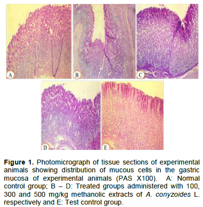
The PAS-stained tissue sections showed the distribution of mucous cell population in the gastric mucosa of experimental animals in normal control group A, treated groups B-D and test control group E (Figures 1 and 2).
Immunohistochemical results
The immunostained gastric tissue sections showed the distribution of p53 protein in the gastric mucosa of experimental animals in normal control group A, treated groups B-D and test control group E (Figure 3).
The protection of gastric mucosa integrity is usually a function of equilibrium between protective and aggressive factors or their stimulators and inhibitors. Among the primary protective factors of gastric mucosa is the mucus produced by mucous cells (Johnson, 2003). From the histochemical result of this study, there was observable prominence of mucous cell population in gastric tissues of treated groups B-D (Figure 1) and there was corresponding significant increase (P<0.05) in the mucous cell count in gastric tissues of animals in all treated groups compared to the control groups (Figure 2). According to Table 1, the values of mucous cell count for treated groups B (9.88 ± 0.40), C (9.63 ± 0.86), and D (9.75 ± 0.80) were significantly (P<0.05) higher compared to the control groups A (5.75 ± 0.31) and E (4.55 ± 0.40). The increased gastric mucous cell population implied increased secretion of viscous and alkaline mucus which constitutes a vital part of primary gastric mucosal protective factors. The mucus forms a protective layer of gel on gastric mucosal surface which protects it against the digestive action of pepsin and erosive effect of acidic gastric juice (Sembulingam and Sembulingam, 2010). There are two types of mucus secreted by mucous cells of gastric mucosa. These include soluble mucus secreted by neck mucous cells which help to neutralize gastric contents and insoluble mucus secreted by surface mucous cells after exposure to chemical or physical irritants (Rhoades and Tanner, 2004; Sembulingam and Sembulingam, 2010). The study by Olaibi et al., (2014) identified one mechanism of gastric mucosal injury to involve disruption of mucus production by gastric mucous cells. The result of their study also showed that preservation of these mucus secreting cells in stomach tissue is vital in gastric mucosal protection. Another study by Ige et al., (2016) reported that increase in mucus secretion (by gastric mucous cells) is one mechanism of protecting gastric mucosa from effects of aggressive factors gastric mucosal injury. In this study, the gastroprotective activity of methanolic extracts of Ageratum conyzoides L. may result from significant increase in mucous cell population.
Moreover, factors that shift the equilibrium between protective and aggressive factors positively towards cell survival or negatively toward cell death will either maintain or disrupt the gastric mucosal integrity respectively. One factor that projects cell toward cell death is the tumor suppressor (p53) protein. The immunohistochemical result of this study revealed a staining intensity which range from moderate immuno-staining in normal control group A and treated group B to mild or concentric immunostaining in treated groups C and D but intense or widespread staining in test control group E (Figure 3). Consequently, the distribution of p53 protein was not markedly increased in all treated groups compared to the normal control group A. However, there was a spike in the p53 protein expression in test control group E due to exposure to the aggressive factor (acidic gastric juice) (Figure 4). From Table 2, the values of p53 distribution obtained among the treated groups B (16.67 ± 1.20), C (14.00 ± 2.08) and D (14.67 ± 0.88) were not significantly different from the value in normal control group (16.33 ± 0.88) but was significantly increased in test control group E (32.00 ± 3.03). The p53 is a nuclear protein commonly described as guardian of the genome due to its vital regulatory role during different events of cell cycle which include DNA damage repair, cellular division and cell death (Kern et al., 1991; Lane, 1992; Volgelstein and Kinzler, 1992). It is a tumor suppressor protein which plays an important role in cell proliferation and apoptosis (Alshenawy and Alshafey, 2012). In a stable or unstressed tissue, p53 protein exhibits low level of expression and its short half-life is due to continuous ubiquitination via an inhibitory interaction with MDM-2 and degradation by 26S proteasome (Brooks and Gu, 2004; Yang et al., 2004; Bond et al., 2006). As a guardian of the genome, the p53 is expressed as an important inducer of apoptosis especially in condition of irreparable genomic alterations, over-expression of oncogenes or other cellular stresses which may be caused by exposure of cells to cytotoxic agents (Brooks and Gu, 2004; Yang et al., 2004). The study by Teh et al., (2002) reported the critical role that p53 protein plays during initiation of apoptosis such that the mutation of p53 gene or inactivation of wild-type p53 protein cause unregulated gastric epithelial plasticity. The study by Arab et al., (2015) opined that gastroprotective mechanisms involve down-regulation of apoptotic triggers such as p53 protein expression so as to shift the balance of gastric mucosal cells toward survival. From this study, the exposure to methanolic extracts of A. conyzoides L. have resulted into gastroprotective activity in the experimental animals also through suppression of pro-apoptotic signals generated by p53 protein which may follow exposure to gastric mucosal aggressive factors.

The methanolic extracts of A. conyzoides L. exhibited potent gastroprotective activity against exposure to aggressive factors of gastric mucosal injury at all the dosage levels in this study. The identified mechanisms of gastroprotective activity of methanolic extracts A. conyzoides L. include increase in mucous cell population leading increase in protective mucus secretion and down-regulation of p53 protein expression leading to suppression of pro-apoptotic signals that follows exposure to the aggressive factors.
The methanolic extracts of A. conyzoides L. is a potent gastroprotective agent. However, further studies are recommended to identify other mechanisms of its gastroprotective activity and veracity of its potency against other aggressive factors.
The authors have not declared any conflict of interests.
Authors appreciate the efforts of the following during the tissue processing and staining: Mrs Okoro, Histopathology Department, University of Benin Teaching Hospital, Benin city, Nigeria and Mr Jonathan, Histopathology Department, National Hospital, Abuja, Nigeria.
REFERENCES
|
Alshenawy HA, Alshafey AM (2012). Does eradication of Helicobacter pylori decreases the expression of p53 and c-Myc oncogenes in the human gastric mucosa? Current Topics in Gastritis.
Crossref
|
|
|
|
Arab HH, Salama SA, Omar HA, Arafa EA, Maghrabi IA (2015). Diosmin protects against ethanol-induced gastric injury in rats: Novel Anti-ulcer actions. PLoS One 10(3):e0122417.
Crossref
|
|
|
|
|
Ayyanar M, Ignacimuthu S (2005). Traditional knowledge of Kani tribals in Kouthalai of Tirunelveni Hills,Tamil Nadu, India. Journal of Ethnopharmacology 102(2):246-255.
Crossref
|
|
|
|
|
Bond GL, Hirshfield KM, Kirchhoff T, Alexe G, Bond EE, Robins H, Bartel F, Taubert H, Wuerl P, Hait W, Toppmeyer D, Offit K, Levine AJ (2006). Mdm2 snp309 accelerates tumor formation in a gender-specific and hormone-dependent manner. Cancer Research 66(10):5104-5110.
Crossref
|
|
|
|
|
Brooks CL, Gu W (2004). Dynamics in the p53-mdm2 ubiquitination pathway. Cell Cycle 3(7):895-899.
Crossref
|
|
|
|
|
Dung NX, Loi DT (1991). Selection of traditional medicine for study. Journal of Ethnopharmacology 32(1-3):57-70.
Crossref
|
|
|
|
|
Elisabetsky E, Wannmacher L (1993). The status of ethnopharmacology in Brazil. Journal of Ethnopharmacology 38(2-3):137-143.
Crossref
|
|
|
|
|
Geissler PW, Harris SA, Prince RJ, Olsen A, Odhiambo RA, Oketch-Rabah H, Madiega PA, Anderson A, Molgaard P (2002). Medicinal plants used by Lou mothers and children in Boudo district, Kenya. Journal of Ethnopharmacology 83(1-2):39-54.
Crossref
|
|
|
|
|
Gurib-Fakim AI, Gueho J, Sewraj-Bissoondayal M, Dulloo E (1993). Medical ethnobotany of some weeds of Mauritius and Rodrigues. Journal of Ethnopharmacology 39(3):175-185.
Crossref
|
|
|
|
|
Ige AO, Adewoye EO, Okwundu NC, Alade OE. and Onuobia PC (2016). Oral magnesium reduces gastric mucosa susceptibility to injury in experimental diabetes mellitus. Pathophysiology 23(2):87-93.
Crossref
|
|
|
|
|
Johnson LR (2003). Essential Medical Physiology. Third Edition. Elsevier Academic Press, Carlifonia, USA pp. 502-512.
|
|
|
|
|
Kamboj A, Saluja AK (2008). Ageratum conyzoides L. - a review of its phytochemical and pharmacological profile. International Journal of Green Pharmacy 2(2):59-68.
Crossref
|
|
|
|
|
Kern SE, Kinzler KW, Bruskin A, Jarosz D, Friedman P, Prives C, Vogelstein B (1991). Identification of p53 as a sequence-specific DNA-binding protein. Science 252(5013):1708-1711.
Crossref
|
|
|
|
|
Lane DP (1992). P-53, Guardian of the genome. Nature 358:15-16.
Crossref
|
|
|
|
|
Mahmood AA, Sidik K, Salmah I, Suzainur KAR, Philips K (2005). Antiulcerogenic activity of Ageratum conyzoides leaf extract against ethanol-induced gastric ulcer in rats as animal model. International Journal of Molecular Medicine and Advance Sciences 1(4):402-405.
|
|
|
|
|
Ming LC (1999). Ageratum conyzoides: a tropical source of medicinal and agricultural products. Perspective on new crops and new uses. J. Janick (ed), ASHS Press, Alexandria, Virginia, USA pp. 469-473.
|
|
|
|
|
Moshi MJ, Kagashe GAB, Mbwambo ZH (2005). Plants used to treat epilepsy by Tanzanian traditional healers. Journal of Ethnopharmacology 97(2):327-36.
Crossref
|
|
|
|
|
Novy JW (1997). Medicinal plants of the Eastern region of Madagascar. Journal of Ethnopharmacology 55(2):119-126.
Crossref
|
|
|
|
|
Okunade AL (2002). Review: Ageratum conyzoides L. (Asteracaea). Fitoterapia 73(1):1-16.
Crossref
|
|
|
|
|
Olaibi OK, Ijomone OM, Olawuni IJ, Adewole SO, Akinsomisoye SO (2014). Mucus secreting activity and nitric oxide concentrations of ethanol-induced pylorus and duodenum of rats pretreated with Moringa oleifera. Journal of Experimental and Integrative Medicine 4(2):123-130.
Crossref
|
|
|
|
|
Perumal-Samy R, Ignacimuthu S, Patric-Raja D (1999). Preliminary screening of ethnomedicinal plants from India. Journal of Ethnopharmacology 66(2):235-240.
Crossref
|
|
|
|
|
Rhoades RA, Tanner GA (2004). Medical Physiology. Second Edition. Lippincott Williams and Wilkins pp. 484-488.
|
|
|
|
|
Sembulingam K, Sembulingam P (2010). Essentials of Medical Physiology. Fifth Edition. Jaypee Brothers Medical Publishers Ltd, St Louis, USA pp. 218-226.
Crossref
|
|
|
|
|
Sharma PD, Sharma OMP (1995). Natural products chemistry and biological proportions of the Ageratum plant. Toxicological and. Environmental Chemistry 50(1):213-232.
Crossref
|
|
|
|
|
Shay JP, Komorov SA, Fels SS, Meranze D, Grunstein M, Simpler H (1945). A simple method for the uniform production of gastric ulceration in the rat. Gastroenterology 5:43-61.
|
|
|
|
|
Shekhar TC, Anju G (2012). A comprehensive review on Ageratum conyzoides Linn (Goat weed). International Journal of Pharmaceutical and Phytopharmacological Research 1(6):391-395.
|
|
|
|
|
Sofowora EA (1993). Medicinal plants and traditional medicine in Africa. Spectrum Books Limited. Ibadan, Nigeria pp. 142-147.
|
|
|
|
|
Teh M, Tan KB, Seet BL, Yeoh KG (2002).Study of p53 immunostaining in the gastric epithelium of cagA-positive and cagA-negative Helicobacter pylori gastritis. Cancer 95(3):499-505.
Crossref
|
|
|
|
|
Volgelstein B, Kinzler KW (1992). P-53 function and dysfunction. Cell 70(4):523-526.
Crossref
|
|
|
|
|
Yang Y, Li CC, Weissman AM (2004). Regulating the p53 system through ubiquitination. Oncogene 23(11):2096-2106.
Crossref
|
|