Full Length Research Paper
ABSTRACT
Phytomedicine coupled with nanotechnology has shown possibilities in treating different ailments including cancer. In the current study, silver nanoparticles (AgNPs) were biogenically synthesized using methanolic extracts of Azadirachta indica (neem) barks and tested for cytotoxic and anti-proliferative activities on normal cells (Vero E6) and human prostate cancer cells (DU-145), respectively by resazurin assay. Characterization of the AgNPs using UV-Vis, Fourier transmission infrared spectroscopy (FTIR) and Zetasizer Nano were performed. The UV-Vis spectroscopy showed surface plasmon resonance of 428 nm indicating the successful formation of silver nanoparticles. The FTIR spectra indicated the presence of ketones, carboxylic acid, amines, aldehydes, secondary alcohols, alkenes, and carbohydrates. Zetasizer revealed the average particle size and zeta potential of 58.8nm and -33Mv respectively for the biogenic AgNPs. IC50 values were found to be 41.78±0.82, 8.02±0.18 and 6.37±0.34 µg/ml for crude extract, biogenic AgNPs and control drug (doxorubicin), respectively. While there was no significant difference (p>0.05) in anti-proliferative activities between biogenic AgNPs and control drug (doxorubicin), results showed a significant difference (p<0.05) in their cytotoxic activities. Selectivity index of the AgNPs was recorded to be 2.1 while that of control drug was 1.4; showing the potential of AgNPs to select cancerous cells over normal cells. We deduce that, the biogenic AgNPs has good anti-proliferative activities against human prostate cancer cells (DU 145) in a selective manner.
Key words: Silver nanoparticles, Azadiracta indica, biogenic synthesis, cancer, prostate cancer.
INTRODUCTION
Cancer burden in developing countries has increased to 80% and estimated to increase to 85% by 2030 in sub-Saharan Africa (Morhason-Bello et al., 2013). However, the region receives only 5% of the global resources dedicated to cancer (Morhason-Bello et al., 2013)prompting for an urgent need for novel therapeutics to combat the situation. Currently, cancer is managed through different ways such as surgery, chemotherapy, radiation therapy and immunotherapy (Suh et al., 2020). Regarding chemotherapy, there are several issues reported such as toxicity, resistance of cancer cells, high cost and poor delivery systems (Senapati et al., 2018)and therefore there is a need for effective and less toxic drugs for cancer. To date, there is a global resurgence in interest and use of plant-based therapies due to the fact that plant derived products have shown promise in treating different cancer types such as colorectal, breast, prostate, stomach, esophageal and liver cancer among others (Roy and Bharadvaja, 2017).
Despite of this pike in the use of plant derived materials for cancer treatment, there are issues reported on limited bioavailability of active phytochemicals to the target cells (Siddiqui and Sanna, 2016). Such, the limitation entails poor stability due to gastric and colonic acidity, poor solubility of the ingredient, poor metabolism by the effect of gut microflora, poor absorption across the intestinal wall, poor active efflux mechanism and first-pass metabolic effects hampering the success of these products through pre- and clinical trials (Siddiqui and Sanna, 2016). In that view, developed novel drug delivery system and carriers for herbal d rugs should preferably attain some prerequisites including proper delivering of the drug at a rate oriented by the needs of the body, over a given period of treatment and ultimately present the active ingredients of the drug to the site of action (Martínez-Ballesta et al., 2018). Several tactics have been explored to increase drug solubility, sustainability, bioavailability and gastrointestinal permeability including nanotechnology that has tremendously gained attention in the development of new pharmaceutical carrier and delivery systems. Encapsulation of plant phytochemicals into a biodegradable and biocompatible nanoparticle remains a solution to this problem. Nanomaterials can overcome limitations associated with conventional crude extract delivery, increase the action of plant extracts, reduce the required dose and side effects, and eventually improve the activity (Gudise et al., 2021).
Among other plants, Azadadiracta indica extracts have been encapsulated in various metallic nanoparticles (NPs) such as silver and gold (Hareesh et al., 2016)among others. The silver nanoparticles (AgNPs) is one of the most studied and utilized nanoparticles in drug formulation due to its potent broad spectrum of activities (for instance inhibitory activity towards nearly 650 microbes), extremely large surface area, strong permeability and little drug resistance (Ivanova et al., 2018).
Silver nanoparticles biogenically synthesized using methanolic bark extracts of A. indica has shown antiproliferative activity against CT26 colorectal cancer cell lines (Sokei, 2018). In the study, results showed that the nanoformulation had significantly higher (p>0.05) activity (IC50 15.13 µg/ml) compared to the methanolic extract (IC50 87.46 µg/ml) with a higher selectivity index (2.03) compared to the methanolic crude extract (1.08) and that of doxorubicin (1.56). Furthermore, the NPs increased in vivo tumor growth inhibitory activities in Swiss albino rats at an inhibitory capacity of 71.96% at 30 mg/kg body weight compared to the crude extracts (Sokei, 2018). Given good activities of the NPs on colorectal cancer, it is of great interest therefore to explore their potential in treating other adenornocarcinoma (cancer developing from secretory cells). In this study, we studied cytotocity and antiproliferative activities of the silver nanoparticles biosynthesized using methanolic barks extracts of A. indica on normal cells (Vero E6) and human prostate cancer cell lines (DU 145), respectively.
MATERIALS AND METHODS
Collection of plant
About 7 kg of barks of the neem plant (A. indica) was harvested from a farm sustainably (by cutting branches and later pealing them) at farm in Juja town, Kiambu County, Kenya Republic 10 5’ S, 370 10’ E, 1520 m alt. in September, 2020. After proper authentication by a botanist, the materials were harvested and a voucher specimen deposited at Jomo Kenyatta University of Agriculture and Technology (JKUAT) botany herbarium and assigned voucher specimen number SK-001.
Methanolic extraction
The method as described by Sokei (2018)was adopted. Briefly, the barks of A. indica were washed with distilled water, air-dried and crushed into coarse powder. The powdered bark (100 g) was soaked in 400 ml, 70% methanol. The mixture was allowed to stand for 72 h with vigorous shaking using a shaker (GYROMAXTM 727, Amerex Instruments, Inc.). After incubation, the methanolic extract was then filtered and the filtrate was concentrated under pressure at 60°C in a rotary evaporator (LabTech®, Daihan Labtech Co. Ltd.) to remove methanol. The resulting extract broth was kept at 4°C for downstream use. In case of crude extract preparation, the methanolic extract was dried in the water bath for 96 h at 60°C to obtain the dry powder of the crude extracts.
Biogenic synthesis of AgNPs using A. indica methanolic barks extracts
The AgNPs were formulated using the extract broth with 1 mM of silver nitrate (AgNO3) solution following the protocol followed by Nghilokwa et al. (2020). Primary characterization was done using UV spectrophotometer at wavelength between 300 and 800 nm and later washed three times with deionized water for complete purification and later frozen at -80°C for 48 h before drying in lyophilizer (freeze dryer, FDL-10N-50-BA, MRC, Laboratory equipment manufacturer, UK) and stored at 4°C.
Classification and characterization of A. indica silver nanoparticles
The synthesized nanoparticles were subjected to UV-Vis Spectroscopy (JENWAY, Bibby Scientific Ltd, UK) and Fourier-transform infrared spectroscopy (Perkin-Elmer FTIR spectrophotometer Norwalk, CT, USA) as in Sokei (2018). Also, Zetasizer Nano (Malvern Instruments Ltd., United Kingdom) was used to uncover the size and zeta potential.
Cell lines
DU 145 (human prostate cancer) and Vero E6 (normal) cells obtained from ATCC (Manassas, VA, USA) were used. As inSokei (2018), the cells were grown in MEM medium supplemented with 10% Fetal Bovine Serum (FBS), 1% L-Glutamine and 1% antibiotic (Penicillin/Streptomycin) in a T75 culture flask and incubated at 37°C and 5% CO2 to attain confluence of 80%.
In vitro anti-proliferative activity of A. indica extract and A. indica AgNPs on human prostate cancer cell line (DU145)
DU145 prostate cancer cells were prepared for anti-proliferative assay in conditions previously reported by Sokei (2018)and later by Gavamukulya et al. (2021). In this case, 20 µl of resazurin dye (0.15 mg/ml) was used and optical density (OD) was read using the MULTISKAN GO plate reader (Thermo Scientific) at 562 nm and a reference wavelength of 690 nm. From the readings, IC50s were established.
In vitro cytotoxic activity of A. indica extract and A. indica AgNPs on Vero E6 (Normal) cell line
Exponentially growing Vero E6 cells were washed and prepared for cytotoxicity assay in conditions previously reported by Sokei (2018). Subsequently, 20 µl of resazurin dye (0.15 mg/ml) was added and optical density (OD) read using the MULTISKAN GO plate reader (Thermo Scientific) at 562 nm. From the readings, CC50s were established.
Tumor selectivity index (TSI)
Tumor selectivity index (TSI) indicates the ability of the extract/drug to exert selective toxicity to cancer cells while sparing the normal ones. It was calculated by the following equation (Indrayanto et al., 2020):
TSI = mean CC50 against normal cells/mean IC50 against tumor cells,
Where CC50 is the concentration of extract/product that exerted cytotoxic effect to 50% of the normal cells (Vero E6) while IC50 stands for the concentration of the products that inhibited the growth of cancer cells by 50%.
Statistical analysis
Data from UV-Vis spectrophotometry and FTIR were handled with origin software for plotting graphs while absorbance data from Resazurin assay were handled in GraphPad Prism 9.2.0. First, the drug concentration and absorbance readings were log transformed and normalized, respectively. Secondly, nonlinear regression was used to compute the IC50 and CC50 for each treatment. Thirdly, differences in groups were compared using One-way ANOVA followed by post hoc analysis using Tukey’s test. The IC50 and CC50 values were expressed as mean ± standard deviation (sd) and a significance level was set at p ≤0.05.
Ethical approval
Approval for experimentations was granted by the University of Nairobi Animal Ethics Committee at department of veterinary physiology and anatomy and issued in a letter with reference number: FVM BAUEC/2021/308.
RESULTS AND DISCUSSION
Biogenic synthesis and characterization of silver nanoparticles
UV-Vis spectroscopy
The UV-Vis spectra illustrated in Figure 1, depicted absorption peaks for the particle sizes at different time intervals. Color change from rusty orange at time 0 to dark brown at 72 h and peak formation at about 380 to 480 nm confirmed bio-reduction of silver ions by the extract and the successful nanoformulation. In this case, the highest peak corresponds to 428 nm surface plasmon resonance (SPR). The similar results were reported earlier on silver nanoparticles synthesized by leaf extract of Amorphophallus paeoniifolius (Gomathi et al., 2019). This optical properties is due to collective electron oscillations induced by electromagnetic radiation which depends on the shape, geometrical arrangement and degree of interaction between particles (Chen and Jensen, 2016). The correlation between the SPR and geometrical properties therefore qualifies the SPR to be used as fast and simple morphological characterization tool for particles.
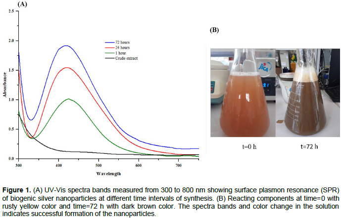
The SPR obtained in this study therefore indicates the synthesis of spherical molecules. When the amount of the particles was small, the absorption band was broad with a low peak intensity while as the amount of the particles increased with time, the absorption spectrum became narrower with an increase in the intensity of the absorption band (Jain and Mehata, 2017).
Fourier-transform infrared spectroscopy (FTIR) analysis
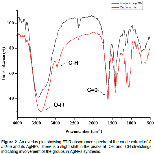
FTIR was done in order to study different functional groups present in the products and more importantly those involved in reduction and capping processes during synthesis of the NPs. Results showed the presence of different functional groups such as the polyenes, ketones, phenols, alcohol, alkane, alkene, amines, carbohydrates, amino acids and ethers, as reported by other authors (Nghilokwa et al., 2020; Sokei, 2018). Biomolecules such as phenolics, flavonoids, sesquiterpenes, and terpenoids found in the crude extract is implicated in the conversion of the ionic form of silver (Ag+) to the metallic nanoform (Ag0).
In the results presented in Figure 2, the wider spectral peak between 3369.5 and 2959 cm-1 are assigned to –OH and –CH stretching, respectively as it was also earlier reported (Velmurugan et al., 2015). Other peaks at 1633 cm-1 for C=O showing ketones and carboxylic acid, 1396.4 cm-1 for N-CH3 of amines and aldehydes, 1211.2 cm-1 for –CHOH indicating secondary alcohols and 1110.9 cm-1 for In-CH3 indicating alkenes. Also, 1072.2 cm-1 for C-N stretching indicates aliphatic amines and C-O stretching for aliphatic aryl, unsaturated diacylperoxides and carbohydrates, 702 cm-1 for phenols and 617.2 cm-1 for vinyl hydrocarbons. Shifts in the peaks where O-H (for phenols) and C-H (for alkane) functional groups resides, is an indication of involvements of such groups in bio-reduction and capping of ionic silver to its metallic nanoform. Other studies indicated that the reducing phytochemicals in the neem (A. indica) leaf consisted mainly of terpenoids, nimbin and quercetin which served as capping and stabilizing agents in addition to reduction (Sironmani, 2016). This concludes that, plants and particularly neem contains phytochemicals that can synthesize metallic nanoparticles in an ecofriendly, less toxic and cost effective manner.
Particle size determination and zeta potential measurements
Particle size determination of the biogenic AgNPs were performed by zeta sizer (Malvern, Zetasizer Nano, ZSP) and presented as percentage intensity. Laser diffraction revealed that particles obtained are polydisperse mixture with the size ranging from 24.4 to 141 nm (Figure 3). The average diameter of the particles was found to be 58.8 nm.
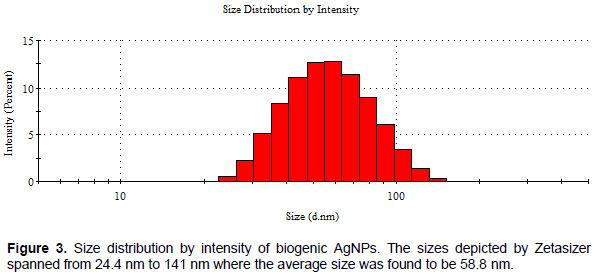
The size of the nanoscale materials provides significant control over physical and chemical properties including their interaction with biomolecules and cell organelles (Tavakol et al., 2017). Nanoparticles with smaller size conferring higher surface area can easily penetrate into the cells and mitochondria. However, NPs with higher surface area can play an important damaging role in cell through interaction with proteins due to more exposed atoms and active sites on the surface of small particles than the larger ones (Tavakol et al., 2017). In general, the size of NPs affects their toxicity levels whereby the smaller sized NPs becoming more toxic that large sized ones exhibiting the highest activity in cell morphological changes, cell membrane damage, ROS generation, cell cycle arrest and cell apoptosis among others (Zhang et al., 2016). Silver nanoparticles of average size above 50 nm show to have less toxicity compared to those of smaller size below 50 nm (Zhang et al., 2016)and that entails that the biogenic AgNPs used in this study of average size 58.8 nm can be less toxic.
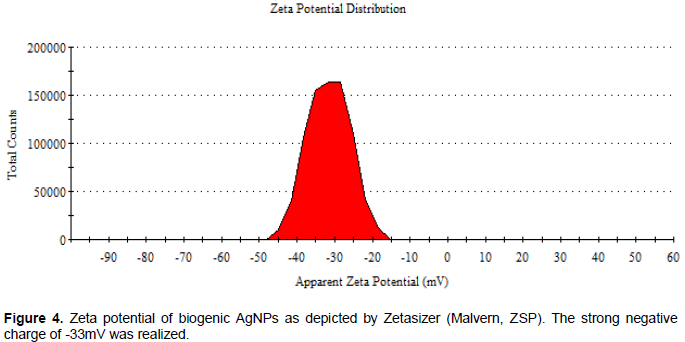
On the other hand, zeta potential measurement of the AgNPs was determined in water as solvent and found to be -33 mV (Figure 4) confirming the repulsion among the particles and thus increase in stability of the formulation. Zeta potential being the electrical charge on the surface of materials surrounded in the medium, is another important factor affecting different cell responses as compared to particle size (Tavakol et al., 2017). Overall, with higher zeta potential, the stability and assimilation of the materials to the body is guaranteed since the adherence and clumping is minimized (Santana et al., 2019). While nanoparticles with a zeta potential between -10 and +10 mV are considered approximately neutral, the nanoparticles with zeta potentials of greater than +30 mV or less than -30 mV are considered strongly cationic and strongly anionic, respectively (Santana et al., 2019). Results from this report concurs with silver nanoparticles (AgNPs) bio-stabilized using aqueous callus extract of Gymnema sylvestre and showed to have a zeta potential of -36.1 mV (Netala et al., 2016).
Resazurin assays
Table 1 shows results for anti-proliferative activities and cytotoxicity of crude extracts of A. indica and its silver nanoparticles. Antiproliferative effects are as shown in Figure 5 with dose-response curve for cell viability on DU154 prostate cancer cells in which the increase in drug concentration decreases the cell viability. The study on human prostate cancer DU145 cells showed that doxorubicin had a 6-fold higher activity (6.37 µg/ml) than the A. indica extract (IC50 41.78 µg/ml). Also, from this study, biogenic AgNPs showed higher activity (8.02 µg/ml) compared to their crude counterpart (IC50 41.78 µg/ml), a 5-fold increase with no significant difference to doxorubicin (p>0.05). This concurs with results by Sokei (2018)where AgNPs increased activity on CT26 colorectal cancer cell lines at ~6-folds compared to bark methanolic crude extracts. Different studies show varying activities of A. indica crude extracts for instance, Kashif et al. (2018)reported on the activity of methanolic oil extract of A indica (157.15 μg/mL) against DU-145 human prostate cancer cells after 24 h exposure. The reason behind these discrepancies could be attributed to differences in cell types, extracts and assays used.
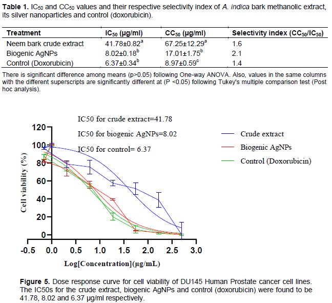
Generally, the findings suggest that nanoformulating the A. indica extract significantly improved its anti-proliferative activity. Many studies reported the enhancement of activities given to crude extracts of plants through nanoformulation including antidiabetic and antihyperlipidemic effects (Gudise et al., 2021), antitumor activities against colorectal cancer (Sokei, 2018)among others. This is the reason behind an extensive proposal to combine herbal medicine with nanoscience since nanomaterials can overcome limitations associated with conventional crude extract delivery leading to improvement of the activity (Gudise et al., 2021).
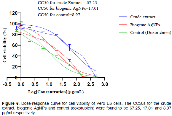
On the other hand, resazurin assay revealed the control drug (doxorubicin) to be cytotoxic to Vero E6 cells (8.97 µg/ml) with significant difference (p<0.05) from the biogenic AgNPs (17.01 µg/ml). On the other hand, the crude extract showed less cytotoxicity (67.25 µg/ml) with a significant difference (p<0.05) to both control and biogenic AgNPs. Figure 6 presents the cytotoxicity results with dose-response curve for cell viability on Vero E6 cells in which the increase in drug concentration also decreased the cell viability. Cytotoxicity being the measure of the ability of a test substance to kill normal cells is used as one of the screening tests to observe the cell growth, reproduction and morphological effects by medical devices. In this study, A. indica extract was nearly 7-times less cytotoxic than the doxorubicin but 3 times less toxic compared to biogenic AgNPs, results correspond with Sokei (2018).
Also, the results are consistent with findings where A. indica was reported to contain phytoconstituents that can combat oxidative damage, enhance the immunity, reduce inflammation, and interfere with the growth of cancer cells thereby making it non-toxic to normal cells (Sironmani, 2016). However, doxorubicin was 2-times more cytotoxic than the AgNPs which agrees with the findings by Buttacavoli et al. (2018), where the AgNPs were found to be 2.8 fold less cytotoxic than doxorubicin. In this study, the nanoparticles showed a 4-fold increase in cytotoxicity compared to that of crude extract, implying that nanoformulating plant phytochemicals increase cytotoxicity. Explanation to this is backed up by other reports which implicate both the AgNPs and Ag+ released by AgNPs involvement in the mechanism of cytotoxicity in different forms. First, AgNPs perhaps provide a perfect surface outside the mitochondria for the univalent reduction of oxygen to superoxide from electron through the electron transport chain (Buttacavoli et al., 2018). Secondly, Ag+ may interfere with the functioning of proteins and DNA as it binds to them (Abdelsalam et al., 2018). Thirdly, oxidative stress coupled with generation of reactive oxygen species (ROS) occurring as an early event, leads to NP-induced toxicity (Mao et al., 2016).
Nevertheless, from the results in Table 1, it was revealed that biogenic AgNPs had the highest selectivity index (SI) of 2.1, while the control (doxorubicin) had the lowest selectivity index of 1.4, and thus the higher toxicity of the later to non-target cells. Selectivity index (SI) can be defined as the ratio of the toxic concentration of a sample against its effective bioactive concentration, thus the ideal drug should have a relatively high toxic concentration but with a very low active concentration. A selectivity index greater than 2 is appropriate for a good anticancer drug candidate (Koch et al., 2005). From the current study, A. indica extract and doxorubicin had poor selectivity (SI < 2). As the SI demonstrates the differential activity of a drug, the greater the SI value, the more selective it is. Therefore, SI value less than 2 indicates general toxicity of the pure compound (Koch et al., 2005). Results from the ongoing report synchronizes with other findings where biogenic AgNPs acted more selectively on human lung cancer cells and caused less toxicity on normal cells (S.I = 2.2) (Gurunathan et al., 2015). Also AgNPs biosynthesized by Annona muricata fruits acted selectively against cervical and prostate cancer cells sparing normal cells with a selectivity index above 7 (Gavamukulya et al., 2021)and neem synthesized AgNPs induced cytotoxicity selectively in human gastric cancer cells indicating induction of apoptosis (Sironmani, 2016). The improved selectivity can be attributed to the nature of the size and surface modifications of the nanoparticles (Sokei, 2018). Since the biogenic AgNPs can kill cancer cells more selectively, they can therefore be an effective approach for controlling and ultimately eradicate cancer. Consequently, they can serve as drug delivery strategies, allowing the use of lower doses of drugs to reduce cytotoxic effects and therefore increase therapeutic efficacy. The biogenic AgNPs synthesized and tested in this report can act as a good candidate for cancer management due to its good selectivity index, size and stability. Therefore, preclinical trials can be set using experimental animals to test its efficacy and safety in vivo.
CONCLUSION
The present study confirms that phytochemicals present in bark methanolic extracts of A. indica can synthesize AgNPs in an ecofriendly manner. Also, our data suggest that the biogenic AgNPs synthesized hereby has good anti-proliferative activities against DU145 prostate cancer in vitro. The results further provide promising evidence that the biogenic AgNPs can selectively kill cancer cells while sparing normal cells with a selectivity index above 2 which is indicative of a potential anticancer candidate. Therefore, we explore the relevance of these findings for further safety and efficacy assessment in vivo against human prostate cancer or any other adenocarcinoma.
CONFLICT OF INTERESTS
The authors have not declared any conflict of interests.
ACKNOWLEDGEMENTS
The authors appreciate the financial support from the African Union through the Pan African University, Institute for Basic Sciences and Technology.
REFERENCES
|
Abdelsalam NR, Abdel-Megeed A, Ali HM, Salem MZM, Al-Hayali MFA, Elshikh MS (2018). Genotoxicity effects of silver nanoparticles on wheat (Triticum aestivum L.) root tip cells. Ecotoxicology and Environmental Safety 155:76-85. |
|
|
Buttacavoli M, Albanese NN, Di Cara G, Alduina R, Faleri C, Gallo M, Pizzolanti G, Gallo G, Feo S, Baldi F (2018). Anticancer activity of biogenerated silver nanoparticles: an integrated proteomic investigation. Oncotarget 9(11):9685. |
|
|
Chen X, Jensen L (2016). Understanding the shape effect on the plasmonic response of small ligand coated nanoparticles. Journal of Optics 18(7): 074009. |
|
|
Gavamukulya Y, Maina EN, El-Shemy HA, Meroka AM, Kangogo GK, Magoma G, Wamunyokoli F (2021). Annona muricata silver nanoparticles exhibit strong anticancer activities against cervical and prostate adenocarcinomas through regulation of CASP9 and the CXCL1/CXCR2 genes axis. Tumor Biology 43(1):37-55. |
|
|
Gomathi M, Prakasam A, Rajkumar PV (2019). Green synthesis, characterization and antibacterial activity of silver nanoparticles using Amorphophallus paeoniifolius leaf extract. Journal of Cluster Science 30(4):995-1001. |
|
|
Gudise V, Chowdhury B, Manjappa AS (2021). Antidiabetic and antihyperlipidemic effects of Argyreia pierreana and Matelea denticulata: Higher activity of the micellar nanoformulation over the crude extract. Journal of Traditional and Complementary Medicine 11(3):259-267. |
|
|
Gurunathan S, Park JH, Han JW, Kim JH (2015). Comparative assessment of the apoptotic potential of silver nanoparticles synthesized by Bacillus tequilensis and Calocybe indica in MDA-MB-231 human breast cancer cells: targeting p53 for anticancer therapy. International Journal of Nanomedicine 10:4203. |
|
|
Hareesh K, Williams JF, Dhole NA, Kodam KM, Bhoraskar VN, Dhole SD (2016). Bio-green synthesis of Ag-GO, Au-GO and Ag-Au-GO nanocomposites using Azadirachta indica: its application in SERS and cell viability. Materials Research Express 3(7):075010. |
|
|
Indrayanto G, Putra GS, Suhud F (2020). Validation of in-vitro bioassay methods: Application in herbal drug research. Profiles of Drug Substances, Excipients and Related Methodology 46:273-307. |
|
|
Ivanova N, Gugleva V, Dobreva M, Pehlivanov I, Stefanov S, Andonova V (2018). Silver nanoparticles as multi-functional drug delivery systems. In Nanomedicines. IntechOpen. |
|
|
Jain S, Mehata MS (2017). Medicinal plant leaf extract and pure flavonoid mediated green synthesis of silver nanoparticles and their enhanced antibacterial property. Scientific Reports 7(1):1-13. |
|
|
Kashif M, Kim D, Kim G (2018). In vitro antiproliferative and apoptosis inducing effect of a methanolic extract of Azadirachta indica oil on selected cancerous and noncancerous cell lines. Asian Pacific Journal of Tropical Medicine 11(10):555. |
|
|
Koch A, Tamez P, Pezzuto J, Soejarto D (2005). Evaluation of plants used for antimalarial treatment by the Maasai of Kenya. Journal of Ethnopharmacology 101(1-3):95-99. |
|
|
Mao BH, Tsai JC, Chen CW, Yan SJ, Wang YJ (2016). Mechanisms of silver nanoparticle-induced toxicity and important role of autophagy. Nanotoxicology 10(8):1021-1040. |
|
|
Martínez-Ballesta Mc, Gil-Izquierdo Á, García-Viguera C, Domínguez-Perles R (2018). Nanoparticles and controlled delivery for bioactive compounds: Outlining challenges for new "smart-foods" for health. Foods 7(5):72. |
|
|
Morhason-Bello IO, Odedina F, Rebbeck TR, Harford J, Dangou JM, Denny L, Adewole IF (2013). Challenges and opportunities in cancer control in Africa: a perspective from the African Organisation for Research and Training in Cancer. Lancet Oncology 14(4):e142-e151. |
|
|
Netala VR, Kotakadi VS, Domdi L, Gaddam SA, Bobbu P, Venkata SK, Ghosh SB, Tartte V (2016). Biogenic silver nanoparticles: efficient and effective antifungal agents. Applied Nanoscience 6(4):475-484. |
|
|
Nghilokwa E, Sokei J, Mwitari P, Maina N (2020). Sub-acute and chronic toxicity of silver nanoparticles synthesized by Azadirachta indica extract. African Journal of Biotechnology 19(6):320-331. |
|
|
Roy A, Bharadvaja N (2017). Medicinal plants in the management of cancer: a review. International Journal of Complementary and Alternative Medicine 9(2):291. |
|
|
Santana JS, de Carvalho Costa ÉK, Rodrigues PR, Correia PRC, Cruz RS, Druzian JI (2019). Morphological, barrier, and mechanical properties of cassava starch films reinforced with cellulose and starch nanoparticles. Journal of Applied Polymer Science 136(4):47001. |
|
|
Senapati S, Mahanta AK, Kumar S, Maiti P (2018). Controlled drug delivery vehicles for cancer treatment and their performance. Signal Transduction and Targeted Therapy 3(1):1-19. |
|
|
Siddiqui IA, Sanna V (2016). Impact of nanotechnology on the delivery of natural products for cancer prevention and therapy. Molecular Nutrition and Food Research 60(6):1330-1341. |
|
|
Sironmani TA (2016). Therapeutic potential of neem synthesized silver nanoparticles on human gastric cancer cells in vitro. World Journal of Nano Science and Engineering 6(2):90-110. |
|
|
Sokei J (2018). Azadirachta Indica Bark Extract Stabilized Silver Nanoparticles: Antiproliferative Activity, Acute Toxicity Study and Antitumour Activity. Pan African University Institute for Basic Sciences Technology and Innovation (PAUSTI). |
|
|
Suh JH, Kotecha R, Chao ST, Ahluwalia MS, Sahgal A, Chang EL (2020). Current approaches to the management of brain metastases. Nature Reviews Clinical Oncology 17(5):279-299. |
|
|
Tavakol S, Hoveizi E, Kharrazi S, Tavakol B, Karimi S, Rezayat Sorkhabadi SM (2017). Organelles and chromatin fragmentation of human umbilical vein endothelial cell influence by the effects of zeta potential and size of silver nanoparticles in different manners. Artificial cells, Nanomedicine and Biotechnology 45(4):817-823. |
|
|
Velmurugan P, Cho M, Lim SS, Seo SK, Myung H, Bang KS, Sivakumar S, Cho KM, Oh BT (2015). Phytosynthesis of silver nanoparticles by Prunus yedoensis leaf extract and their antimicrobial activity. Materials Letters 138:272-275. |
|
|
Zhang XF, Shen W, Gurunathan S (2016). Silver nanoparticle-mediated cellular responses in various cell lines: an in vitro model. International Journal of Molecular Sciences 17(10):1603. |
|
Copyright © 2024 Author(s) retain the copyright of this article.
This article is published under the terms of the Creative Commons Attribution License 4.0


