Full Length Research Paper

ABSTRACT
Enterobacteriaceae are the main etiological agents of environmental bovine mastitis and are often resistant to antimicrobials, especially β-lactam class due to the production of β-lactamases. These can be classified into several groups according to the molecular structure and substrate activity. The present study evaluated the profile of pheno-genotypic resistance to β-lactams, with emphasis on the functional detection of β-lactamases. The isolates were obtained from milk samples from farms located in the interior region of the state of Rio de Janeiro. 47.6% (20/42) of the bacterial isolates producing enzymes classified as Group 1 (AmpC type) were detected, and in disk approximation test, 30% were classified as the inducible type and 70% as not inducible. To the best of the authors’ knowledge, this is the first report on AmpC β-lactamase production in enterobacteria involved in mastitis etiology in Brazil.
Key words: Raw milk, environmental mastitis, dairy cattle, antimicrobial resistance.
INTRODUCTION
Mastitis is an inflammation of mammary gland and udder tissue and it is described as the most common and costly disease of dairy cattle and presents clinically or subclinically (Langoni et al., 2009; Brasil, 2012). This inflamemation is caused by stress, physical injury, metabolism and infection (Freitas et al., 2005; Tozzetti et al., 2008).
Enterobacteria are opportunistic pathogens often infecting the udder skin and teat during milking and via contact of the animal with contaminated environments. They are the most important agents in the etiology of environmental mastitis (Moreira et al., 2008).
Causative agents are very often resistant to several antimicrobials and also have developed resistance due to intensive selective pressure caused by use of other antimicrobials (Srinivasan et al., 2007; Langoni et al., 2009; Brasil, 2012).
The most common mechanism of resistance for β-lactam antimicrobials in clinically important Gram-negative bacteria is hydrolysis of these antimicrobial agents by β-lactamases (Bush and Jacoby, 2010). The broad-spectrum β-lactamases (ESBL) are enzymes that hydrolysis the penicilins and cephalosporins but are susceptible when β-lactam is associated with inhibitor but others cephalosporinases are resistant to β-lactamases inhibitors and can hydrolyze carbapenem as AmpC and carbapenemases (Dalmarco, 2006; Jacoby, 2009; Thomson, 2010). The molecular and functional classifycation of the β-lactamases was proposed by Ambler (1980) and Bush and Jacoby (2010), respectively. AmpC β-lactamases are only produced by Gram negative bacteria and belong to the functional group 1 by Bush and Group C classification by Ambler (Aguilar, 2009).
These enzymes can be detected by phenotypic antimicrobial markers and confirmed by specific tests. The research is done by searching for bla genes, which may be present in the bacterial chromosome or plasmid. Bla gene encode enzyme production and its expression can be induced by the presence of β-lactam or can be continuously activated (Livermore, 1995).
The group of β-lactamases is increasing due to the discovery of new enzymes (ANVISA, 2013; Deshpande et al., 2013). Bacteria producing β-lactamases already widely studied in clinical microbiology can be retrieved from cow's milk, beef, pork and chicken, along with animal faeces (Li et al., 2007; Locatelli et al., 2009; Bush and Jacoby, 2010; Geser et al., 2012; Silva and Lincopan, 2012; Dahmen et al., 2013). β-Lactamases-producing Enterobacteriaceae in raw milk can pose a risk to public health in countries even developed ones, where raw cheese is commonly consumed (Dahmen et al., 2013). ESBL and transferable AmpC-producing Escherichia coli and Salmonella was isolated by broiler meat in Europe and may be considered a public health problem (Egervärn et al., 2014).
To date, there is lack of information on the distribution of β-lactamases in animal isolates in South America.
The impact of healthy animals as a potential reservoir for genes coding β-lactamases should be monitored to prevent the spread of resistance to β-lactam antibiotics and to choose suitable prophylactic and therapeutic measures. The present study evaluated specifically production of AmpC β-lactamases in enterobacteria isolated from dairy herds in the interior region of the state of Rio de Janeiro, Brazil.
MATERIALS AND METHODS
Milk samples, isolation and identification of Enterobacteriaceae
The milk samples were collected from nine dairy farms in interior region of Rio de Janeiro. All animals were evaluated by clinical examination and no animal were considered clinical mastitis. These animals were submitted to the California Mastitis Test (CMT) to detect subclinical mastitis and 20% of the positive cows were included in the study. A total of 381 milk samples were collected according to the method described by Sampaio (1998). The characteristics enterobacteria colonies on blood agar (Columbia Blood Agar Base – HiMedia – with 5% defibrinated sheep blood) were picked in selective medium and all Gram negative glucose-fermenting, oxidase-negative and catalase-positive strains were identified by phenotypic laboratory tests in accordance with Koneman et al. (2010). These identification of enterobacteria were confirmed by matrix-assisted laser desorption ionization time-of-flight mass spectrometry (MALDI-TOF MS) (MALDI-TOF LT Microflex Bruker; MALDI Biotyper 2.0 program, Bruker) (Welham et al., 1998; Pribil and Fenselau, 2005; Suarez et al., 2014) and all isolates identified in the laboratory as E. coli were confirmed by phenotypic tests by the Oswaldo Cruz Foundation - FIOCRUZ (report number: 384/12).
Antimicrobials
To detect the AmpC β-lactamase producing β-lactams, the following were used: amoxicillin with clavulanic acid (AMC) (30 µg), cefoxitin (CFO) (30 µg), ceftazidime (CAZ) (30 µg), ceftriaxone (CRO) (30 µg), ertapenem (ERT) (10 µg), imipenem (IPM) (10 µg) (Sensidisc DME).
Susceptibility and confirmatory testing
The inoculum for these tests was obtained from overnight BHI broth (Merck) cultures and adjusted to achieve turbidity equivalent to a 0.5 McFarland standard and the standard strains used as control were E. coli ATCC 25922 and Klebsiella pneumoniae ATCC 700603 (CLSI, 2012).
Susceptibility was determined by agar diffusion techniques by using Muller-Hinton agar plates (HiMedia) and by following the current recommendations of the National Committee for Clinical Laboratory Standards (CLSI, 2008; 2012). Isolates that showed resistant to cefotaxime, ceftazidime and clavulanate associated with a β-lactam, were suspected of producing AmpC type beta-lactamases.
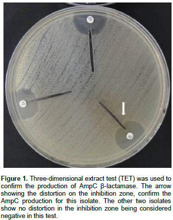
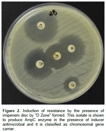
These isolates were submitted to the three-dimensional extract test (Shahid et al., 2004, 2010). The characteristic of AmpC expression was revealed by disk approximation for classification as inducible or not (Martínez-Rojas, 2009).
Modified Hodge test to confirm carbapenemase-producing isolates
An overnight culture of indicator organism E. coli ATCC 25922 was adjusted to a turbidity 0.5 of Mc Farland scale and these were used to swab inoculate the surface of the Muller-Hinton agar plates. After drying the surface, test organisms were heavily streaked from the center to the periphery of the plate using an inoculating loop and a 10 µg ertapenem disk was placed at the center, and incubated for 18 h. The test is interpreted as positive by the presence of distortion of the inhibition zone (CLSI, 2012).
Genotypic resistance evaluation
The DNA extraction was performed according to the method of Chapman et al. (2001) for the PCR technique, using the blaampC was amplified with primers 5’ CCC CGC TTA TAG AGC AAC AA and 5’ TCA ATG GTC GAC TTC ACA CC to produce a 634 pb product (Sobia et al., 2011).
A typical 20 μl PCR reaction mixture for every primer set consisted of 10X Buffer (10 mM Tris-HCl, pH 9.0, 50 mM KCl and 0.1% Triton X-100), 1.25 mM MgCl2, 5 pmol of each primer (Invitrogen), 0.2 mM dNTP (Thermo Scientific), and 2 U Taq polymerase (Fermentas) and 2 μl of DNA extract. The cycling conditions were: initial denaturation at 95°C for 15 min; 35 cycles of 94°C for 60 s; 58°C for 2 min; 72°C for 3 min; and a final elongation at 72°C for 10 min.
The PCR products were analyzed by electrophoresis on gel with 1.5% agarose and revealed with diluted Sybr Green (Invitrogen). The gel was observed under UV gel documentation (L-PIX EX) using 100 pb DNA ladder (Fermentas) to confirm the product molecular weight.
RESULTS
Milk samples, isolation and identification of Enterobacteriaceae
A total of 381 milk samples were collected from 339 subclinical mastitic cows and 356 bacteria samples were isolated, with the presence of 88.2% Gram-positive (Staphylococcus sp.) and 11.8% Gram-negative bacteria (n=42/356). In 78% of cases (33/42) was isolated the enterobacteria in combination with other bacterial groups, like Staphylococcus spp., Streptococcus spp., Listeria spp. and Corynebacterium spp., indicating a mixed infection.
The most enterobacteria isolated were Proteus mirabilis (45.3%) and E. coli (40.5%), the other enterobacteria isolated were Citrobacter freundii (4.8%), Serratia marcescens (4.8%), Serratia rubidae (2.4%) and Enterobacter aerogenes (2.4%). The MALDI-TOF confirmed 92.86% of phenotypical identification. All E. coli were confirmed by FIOCRUZ.
Susceptibility and confirmatory test for AmpC-β-lactamase production
The antimicrobial susceptibility showed 35.7% resistance to CRO, 38.1% resistance to CAZ, 47.6% resistance to CFO and 83.3% resistance to AMC. The β-lactamase study showed that Group 1 β-lactamase (AmpC type) was predominant, being detected in 47.6% of the species, these isolates also submitted to TET (Figure 1) confirmed the production of the enzyme AmpC in 25%. Resistance to ceftazidime divided Group 1 into two subgroups: 1 (21.4%) and 1e (26.2%). These isolates were tested by disc approximation with imipenem and revealed 30% as the inducible type (S. marcescens, S. rubidae, E. coli, P. mirabilis and E. aerogenes) and 70% as not inducible type (Figure 2).
Modified Hodge test to confirm carbapenemase-producing isolates
Three isolates belonging to Group 1 were suspected to be carbapenemase producers due to their resistance to imipenem. These isolates were tested phenotypically by disk diffusion with ertapenem and by the modified Hodge test (THM) and none of the isolates were found to express phenotypic carbapenemase production in both tests.
Genotypic resistance evaluation
The gene blaampC were detected in 23.8% of the isolates. Only 15% (3/20) of the isolates of the group 1 (1 and 1e) had the blaampC gene.
DISCUSSION
Enterobacteria were not the main cause of mastitis agents in these farms despite being present in samples. Thus, it was observed that low environmental hygiene contributed to these microorganisms which were present in the udders of these animals and affected by subclinical mastitis. In a study conducted by Santos (2006), enterobacteria were individually isolated in only 2.44% (2/82) and mixed infections with Staphylococcus spp. were more frequent (12/18), especially for the enterobacteria in association with coagulase-negative staphylococci.
The MALDI-TOF is widely used to identify microbial species and enzymes produced by these organisms (Pribil and Fenselau, 2005; Seng et al., 2010; Wang et al., 2013). For bacterial identification, some differences were observed when comparing MALDI identification with conventional identifications. Risch et al. (2010) observed 86.8% correct species identification by MALDI-TOF and Veen et al. (2010) observed the correct identification in 97.7% of Enterobacteriaceae.
E. coli was not the predominant species as expected. E. coli are considered one of the main agents of bovine mastitis and infection is characterized by environmental origin. Infections are related to opportunistic behavior of the agent, which infects animals mainly through contact of faeces with the teats. Poor sanitary conditions of farms available contribute to E. coli infections (Moreira et al., 2008).
The involvement of P. mirabilis in the etiology of bovine mastitis is underestimated and few reports in the literature point to its importance (Hogan and Smith, 2003). Proteus spp. commonly contaminates drop hoses used to wash udders before milking. These species were found in higher frequency in farms where drop hoses were used to wash the udder before milking.
These bacteria are present in the environment and are susceptible to a wide genetic exchange. Antimicrobials have often been detected in hospital effluents, municipal water supply treated wastewater, surface water and in some cases in groundwater (Meireles, 2008). According to Srinivasan et al. (2007), E. coli isolated from cows with mastitis were multiresistant and even carried several resistance genes.
The β-lactamase were classified based on resistance to cephalosporins, with cefoxitin the score for enzyme production type AmpC. The AmpC-type enzymes have activity on oxyimino-cephalosporin and on penicillins and monobactams. Some of these enzymes are weak hydrolyzing imipenem when expressed in large quantities and are weakly inhibited by clavulanic acid (Bush et al., 1995; Kao et al., 2010). This group belonging to molecular class C is mainly encoded for chromosomes in many enterobacteria and other microorganisms (Bush and Jacoby, 2010).
For phenotypical confirmation, the TET was not able to detect the most enzymes, but this may be linked to other resistance mechanisms (Shahid et al., 2004; Sobia et al., 2011). Villar et al. (1997) described bacterial isolates belonging to the Enterobacteriaceae that had absence or modification of porins. Recently, Hernández et al. (2010) studied the relationship between changes in porins expression and decreased sensitivity to β-lactams. These authors found isolated deficient in porins less sensitive to β-lactam regardless of β-lactamase production, and those isolated producers of β-lactamases were also resistant to carbapenems. Thus, this explanation is relevant as phenotypic resistance with low percentage of isolates confirmed production of the enzyme.
Many species including C. freundii, E. cloacae and S. marcescens, are natural producers of AmpC and its expression is low but inducible, unlike the Proteus spp., which normally have plasmid genes (Martínez-Rojas, 2009; Bush and Jacoby, 2010).
The imipenem resistance in three bacteria may be due to a variant of the AmpC enzyme which was recently described with hydrolytic activity against this antimicrobial (Bush and Jacoby, 2010; Kao et al., 2010).
Seven isolates not classified in Group 1 presented gene blaampC but probably did not express these traits phenotypically. The low detection of gene in Group 1 isolates can be explained by presence of other AmpC families genes or another mechanism resistance that express this resistance. The dissemination of genes conferring resistance to β-lactams can be broad in interior region of Rio de Janeiro, confirming the ease of genetic transmission, either by contamination of the environment or by human-animal contact.
The distinction between a β-lactamase AmpC chromo-somal and plasmid by the gene is not always easy, but is of interest in terms of epidemiological surveillance, it has important consequences because of the easy spread of these plasmids. So in the future, other genes to detect AmpC enzymes will be studied, along with determination of gene origin by bacterial transformation.
CONFLICT OF INTERESTS
The authors declare no potential conflict of interest with respect to the research, authorship, and/or publication of his article.
REFERENCES
|
Aguilar MAP (2009). Molecular characterization of carbapenem resistance in Enterobacteriaceae isolated in Brazilian hospitals. Dissertation. São Paulo University. |
|
|
Ambler RP (1980). The structure of β-lactamases. Phil. Trans. R. Soc. B 289: 321-331. |
|
|
ANVISA (2013). National Health Surveillance Agency. Technical note Nº 01/2013. Prevention measures and Infection Control for multiresistant Enterobacteriaceae. <http://portal.anvisa.gov.br/wps/wcm/connect/ea4d4c004f4ec3b98925d9d785749fbd/Microsoft+Word++NOTA+T%C3%89CNICA+ENTEROBACTERIAS+17+04+2013(1).pdf?MOD=AJPERES> |
|
|
Brasil (2012). Bovine Masitis: control and prevention. In: Technical Report. UFLA, Lavras, MG. |
|
|
Bush K, Jacoby GA (2010). Updated functional classification of β-lactamases. Antimicrob Agents Chemother 54 (3): 969-976. DOI: 10.1128/AAC.01009-09 PMid: 19995920. |
|
|
Bush K, Jacoby GA, Medeiros AA (1995). A funcional classification scheme for β-lactamases and its correlation with molecular structure. Antimicrob Agents Chemother 39 (6):1211-1233. |
|
|
Chapman PA, Ellinn M, Ashton R, Shafique W (2001). Comparison of culture, PCR and immunoassays for detecting Escherichia coli O157 following enrichment culture and immunomagnetic separation performed on naturally contaminated raw meat products. Intl. J. Food Microbiol. 68:11-20. |
|
|
CLSI (2008). Performance Standards for Antimicrobial Disk and Dilution Susceptibility Tests for Bacteria Isolated from Animals. Clinical and Laboratory Standards Institute, Wayne, PA. |
|
|
CLSI (2012). Performance Standards for Antimicrobial Susceptibility Testing. Clinical and Laboratory Standards Institute,Wayne, PA. |
|
|
Dahmen S, Métayer V, Gay E, Madec JY, Haenni M (2013). Characterization of extended-spectrum β-lactamase (ESBL)-carrying plasmids and clones of Enterobacteriaceae causing cattle mastitis in France. Vet. Microbiol. 162:793-799. |
|
|
Dalmarco EM, Blatt SL, Córdova CMM (2006). Laboratory Identification of extended spectrum β-lactamases – A review. RBAC 38: 171-177. |
|
|
Deshpande LM, Davies TA, Blandino G, Nicoletti G, Jones RN, Castanheira M (2013). IMP-33, a New IMP Variant Detected in Pseudomonas aeruginosa from Sicily. Antimicrobial Agents and Chemotherapy 57: 6401-6403. |
|
|
Egervärn M, Börjesson S, Byfors S, Finn M, Kaipe C, Englund S, Lindblad M (2014). Escherichia coli with extended-spectrum β-lactamases or transferable AmpC β-lactamases and Salmonella on meat imported into Sweden. Intl. J. Food Microbiol., 171: 8-14. |
|
|
Freitas MFL, Pinheiro Jr JW, Stamford TLM, Rabelo SSA, Silva DR, Silveira FVM, Santos FGB, Sena MJ, Mota RA (2005). Antimicrobial susceptibility in vitro profile of Staphylococcus positive coagulase isolated from dairy cows with mastitis in the Arid Zone of Pernambuco. Arquivos do Instituto Biológico, 72(2):171-177, 2005. |
|
|
Geser N, Stephan R, Hächler H (2012). Occurrence and characteristics of extended spectrum β-lactamase (ESBL) producing Enterobacteriaceae in food producing animals, minced meat and raw milk. BMC Vet. Res. 8:1-9. DOI: 10.1186/1746-6148-8-21 PMid: 22397509. |
|
|
Hernández JR, Conejo MC, Pascual A (2010). Comparative activity of ertapenem against Klebsiella pneumoniae producing extended-spectrum β-lactamases or plasmid AmpC β-lactamase: inoculum effect and role of loss porins. Enfermedades Infecciosas y Microbiología Clínic 28(1):27-30. |
|
|
Hogan J, Smith KL (2003). Coliform mastitis. Vet. Res. 34(5): 507–519. |
|
|
Jacoby GA (2009). AmpC β-lactamases. Clinical Microbiology Reviews 22(1): 161–182 |
|
|
Kao CC, Liu MF, Lin CF, Huang YC, Liu PY, Chang CW, Shi ZY (2010). Antimicrobial susceptibility and multiplex PCR screening of AmpC |
|
|
genes from isolates of Enterobacter cloacae, Citrobacter freundii, and Serratia marcescens. J. Microbiol. Imunol. Infect. 43 (3):180-187. |
|
|
Koneman EW, Allen SD, Janda WM, Schreckenbergere PC, Winn WC (2010). Microbiologic Diagnostic. MEDS, Rio de Janeiro, RJ. |
|
|
Langoni H, Laurino F, Faccioli PY, Silva AV, Menozzi BD (2009). Microbiologic Cultive and the Sensibility in Pathogenic Isolates from Bovine Mastites. Vet. Zootec. 16:708-715. |
|
|
Li XZ, Mehrotra M, Ghimire S, Adewoye L (2007). Review: β-lactam resistance and β-lactamases in bacteria of animal origin. Vet. Microbiol. 121(3-4):197-214. |
|
|
Livermore DM (1995). β-lactamases in laboratory and clinical resistance. Clin. Microbiol. Rev. 8(4):557-584. |
|
|
Locatelli C, Caronte I, Scaccabarozzi L, Migliavacca R, Pagani L, Moroni P (2009). Extended-spectrum β-lactamase production in E. coli strains isolated from clinical bovine mastitis. Vet. Res. Commun. 33:S141-S144. |
|
|
Martínez-Rojas DDD (2009). AmpC β-lactamases: generalities and phenotypic methods for detection. Rev. Soc. Ven. Microbiol. 29:78-83. |
|
|
Meireles MAOM (2008). Antimicrobial use and bacterial resistance: socioeconomic and behavioral aspects and their clinical and ecological impact. Belo Horizonte, Brasil, 47p. (Monography. Biological Science Institute. Federal University of Minas Gerais). |
|
|
Moreira MAS, Ferreira AB, Trindade TFSL, Reis ALO, Moraes CA (2008). Antimicrobial resistance dependent on the multidrug efflux in Escherichia coli isolated from mastitic milk. Arq. Bras. Med. Vet. Zoote.c 60(6):1307-1314. |
|
|
Pribil P, Fenselau C (2005). Characterization of Enterobacteria Using MALDI-TOF Mass Spectrometry. Anal. Chem. 77: 6092-6095. |
|
|
Risch M, Radjenovic D, Han JN, Wydler M, Nydegger U, Rich L (2010). Comparison of MALDI TOF with conventional identification of clinically relevant bactéria. Swiss Med Wkly 140:w13095. |
|
|
Santos CDM (2006). Staphylococcus sp and Enterobacteriaceae Isolated from Mastitis Recurrent Eight Herds of Uberlandia Region - MG: antimicrobial susceptibility profile. Dissertation (Veterinary Science). Federal University of Uberlandia. |
|
|
Shahid M (2010). Citrobacter spp. simultaneously harboring blaCTX-M, blaTEM, blaSHV, blaampC, and insertion sequences IS26 and orf513: an evolutionary phenomenon of recent concern for antibiotic resistance. J. Clin. Microbiol. 48:1833-1838. |
|
|
Shahid M, Malik A, Agrawal M,Singhal S (2004). Phenotypic detection of extended-spectrum and AmpC β-lactamases by a new spot-inoculation method and modified three-dimennsional extract test: comparison with the convenctional three-dimensional extract test. J Antimicrob. Chemother. 54(3):684-687. PMid:15294886. |
|
|
Sampaio IBM (1998). Statistic aplies to animal experimetation. Statistics applied to animal experimentation. Foundation for Education and Research in Veterinary Medicine e Zootecnia, Belo Horizonte. |
|
|
Seng P, Rolain JM, Fournier PE, La Scola B, Drancourt M, Raoult D (2010). MALDI-TOF-mass spectrometry applications in clinical microbiology. Future Microbiol. 5 (11):1734-1754. |
|
|
Silva KC, Lincopan N (2012). Epidemiology of extended spectrum β-lactamases in Brazil: clinical impact and implications for agribusiness. J. Bras. Patol. Med. Lab. 48 (2):91-99. |
|
|
Sobia F, Mohammad S, Singh A, Khan HM, Shukla I, Malik A (2011). Occurrence of blaampC in cefoxitin-resistant Escherichia coli and Klebsiella pneumoniae isolates from a North Indian tertiary care hospital. New Zealand Inst Med Lab Sci 65 (1):5-9. |
|
|
Srinivasan V, Gillespie BE., Lewis MJ, Nguyen LT, Headrick SI, Schukken YH and Oliver SP (2007). Phenotypic and genotypic antimicrobial resistance patterns of Escherichia coli isolated from dairy cows with mastitis. Vet. Microbiol. 124 (3-4):319–328. |
|
|
Suarez S, Nassif X, Ferroni A (2014). Applications of MALDI-TOF technology in clinical microbiology. Pathol. Biol. (Paris). |
|
|
Thomson KS (2010). Extended-Spectrum-β-lactamase, AmpC, and Tozzetti DS, Bataier, MBN, Almeida LR (2008). Prevention, control and treatment of bovine mastitis – Literature review. Revista Científica Eletrônica de Medicina Veterinária anoVI(10). |
|
|
Carbapenemase Issues. J. Clin. Microbiol. 48(4):1019-1025. |
|
|
Villar HE, Danel F, Livermore DM (1997). Permeability to carbapenems of Proteus mirabilis mutants selected for resistence to imipenem or other β-lactams. J. Antimicro. Chemother. 40:365-370. PMid: 9338488. |
|
|
van Veen SQ, Claas ECJ, Kuijper EJ (2010). High-Throughput Identification of Bacteria and Yeast by Matrix-Assisted Laser Desorption Ionization–Time of Flight Mass Spectrometry in Conventional Medical Microbiology Laboratories. J. Clinical Microbiology 48 (3):900-907. |
|
|
Wang L, Han C, Sui W, Wang M, Lu X (2013). MALDI-TOF MS applied to indirect carbapenemase detection: a validated procedure to clearly distinguish between carbapenemase-positive and carbapenemase-negative bacterial strains. Anal. Bioanal. Chem. 405(15): 5259-5266. |
|
|
Welham KJ, Domin MA, Scannell DE, Cohen E, Ashton DS (1998). The Characterization of Micro-organisms by Matrix assited Laser Desorption/Ionization Time-of-flight Mass Spectrometry. Rapid Communications in Mass Spectrometry 12:176-180. PMid: 9493412. |
|
Copyright © 2024 Author(s) retain the copyright of this article.
This article is published under the terms of the Creative Commons Attribution License 4.0


