ABSTRACT
The aim of the study was to establish the seroprevalence of contagious bovine pleuropneumonia (CBPP) and contagious caprine pleuropneumonia (CCPP) among cattle and small ruminants received at abattoirs in Ilorin, Nigeria. A total of 643 blood samples were taken from 324 cattle and 319 sheep and goats. These samples were screened for CBPP and CCPP using rapid latex agglutination tests. The majority were from the north of Nigeria. Sera from cattle were screened for the causative mycoplasma of CBPP, Mycoplasma mycoides subsp. mycoides (Mmm) and small ruminants were screened for the presence of Mycoplasma capricolum subsp. capripneumoniae (Mccp), the cause of CCPP, using specific latex agglutination tests. Clinical signs including specific clinical signs associated with CBPP were also assessed. A prevalence rate of just over 56% was recorded for CBPP. In all, 22 of 324 cattle showed clinical signs but only 10 showed specific clinical signs associated with CBPP infection such as dyspnoea, nasal discharge and cough. A CCPP prevalence rate of about 33% was found among the small ruminants. In total, 52 of 319 small ruminants showed clinical signs of disease but only 22 of these were associated with seropositivity. The high seroprevalence of CBPP and CCPP in this study is suggestive of extensive CBPP and CCPP infection in Ilorin, Nigeria.
Key words: Abattoir survey, Nigeria, contagious bovine pleuropneumonia (CBPP), contagious caprine pleuropneumonia (CCPP), seroprevalence.
Contagious bovine pleuropneumonia (CBPP) is a highly contagious disease of cattle associated with consolidation and ‘marbling’ of the lung, fibrinous pleurisy and accumulation of pleural fluid (OIE, 2013). Transmission
of the disease is through intimate contact between infected and susceptible cattle due to the inhalation of infected droplets released during coughing, or in nasal discharges from infected animals (OIE, 2013). Nigeria’s status was regarded as CBPP-free in 1965 which had been achieved by a 10-year policy of harmonized disease reporting, vaccination, prompt laboratory diagnosis, quarantine and slaughter (Griffin and Laing, 1966). However, since that time, CBPP cases have re-emerged with the prevalence increasing across different regions of Nigeria (March et al., 2003; Foluso, 2003; Mailafia et al., 2010). In the humid zones, higher herd prevalence has been observed than in arid zones due to differences in the rate of contact between cattle because of their respective production systems (Masiga et al., 1996; Mariner et al., 2005).
Contagious caprine pleuropneumonia (CCPP) is a highly contagious disease of goats and sheep and was originally reported in Algeria in 1873. Its devastating nature and cause of economic losses in the livestock industry makes it a globally notifiable disease (Manso-Silvan et al., 2009). CCPP is pandemic in parts of Asia, Middle East and Africa and it is one of the major threats to the small ruminant industries of developing countries (OIE, 2013). Only 20 countries previously reported the isolation of the causative mycoplasma possibly because of the scarcity of laboratory expertise. However, its isolation has now been confirmed in many regions such as China, Mauritius, Tajikistan, Nigeria, and India using improved detection methods such as the agglutination test, enzyme-linked immunosorbent assay (ELISA) and polymerase chain reaction (PCR) (Nicholas and Churchward, 2012).
There are only a few recent reports describing the prevalence of CCPP in Nigeria compared to CBPP (Egwu et al., 2012; Akwuobu et al., 2014) and none in the southern part of the country. Following the eradication of rinderpest from cattle in 2011, there is now significant global activity in controlling peste des petits ruminants (OIE, 2013). Since CCPP and this disease can show similar clinical signs it will become increasingly important to differentiate between the two.
Available veterinary records show that CBPP is endemic in Nigeria and that it spreads via movement of trade cattle, seasonal migration and transhuman activities (Aliyu et al., 2000; Foluso, 2003; Ajuwape et al., 2004; Olorunshola et al., 2017). Also outbreaks of CBPP still occur particularly in the northern region which harbours three quarters of the country's 16.3 million cattle (PACE, 2004). CBPP is caused by the Mycoplasma mycoides subsp. mycoides bacteria which belong to a cluster consisting of five closely related mycoplasmas, namely, Mycoplasma mycoides subsp. capri (Mmc), Mycoplasma capricolum subsp. capripneumoniae (Mccp), Mycoplasma capricolum subsp. capricolum (Mcc) and Mycoplasma leachii (Ml) (Thiaucourt et al., 2011).
Nigeria has a population of between 8 and 13 million sheep with most being found in the northern region and about 3.4 million located in the humid (southern) region. The goat population is about 22-26 million with 6.6 million reported to be in southern region while 20 million are in northern region (Lawal-Adebowale and Alarima, 2011). Breeds of sheep are mainly the indigenous West African Dwarf (WAD) sheep, Due, Balami and Yanks. Among the listed indigenous breeds, Uda, Balami and Yankasa are mostly widespread in the northern region while the WAD breed is common in the southern region. The breeds of goat in Nigeria are also mostly indigenous, and these include the WAD, Sahel/desert known as West African Long-Legged goat, and Sokoto Red/Maradi (Lawal-Adebowale and Alarima, 2011). The Sahel or desert goat and Sokoto Red are the most commont in the north while the WAD is more common in Southern Nigeria (FAO, 2004).
There have been several studies on the epidemiology of CBPP in Nigeria compared with CCPP (Aliyu et al., 2003; Ajuwape et al., 2004; Babalobi, 2011); however, scant information about CCPP is available in the mid-belt region of the country. This study was essentially carried out in Ilorin which is North Central Nigeria. While most of the animals used are from North West Nigeria, effects of relocation especially migration from North West Nigeria to North Central Nigeria make this study very relevant. Furthermore, information on the prevalence of CCPP in Nigeria is still sparse (Ajuwape et al., 2004; Babalobi, 2011). The aim of this study was to provide an up to date epidemiological survey based on abattoir data, regarding the seroprevalence of CBPP and CCPP in the Ilorin region of Nigeria.
Study area
Ilorin is the capital of Kwara State which lies on the plain in the southwest of Nigeria at Latitude 8° 30’ and 8° 50’ N and Longitude 4° 20’ and 4°35’ E. The city occupies an area of about 468 km2 situated within the forest and the guinea savannah regions of Nigeria. The Ilorin climate is tropical, being influenced by the two prevailing trade winds and experiences an annual rainfall of between 1000 and 1500 mm with daily average temperatures of 25, 27 and 22.5°C in January, May and September, respectively. The sampling location for this study was the central abattoir at Ipata, drawing animals from a pool of cattle markets in the local communities of Sango, Oke-ose, Oko-olowo, Alapa, Igbeti, Sadu, Sare, Ajase and Ipata.
Animals, sample collection and study design
A total of 643 blood samples from ruminants, 324 from cattle and 319 from small ruminants (44 sheep and 275 goats) were collected from the Ipata central abattoir. Collected samples of about 3 to 5 ml were kept in labelled sterile bottles and transported to the laboratory in cold boxes containing ice packs and later centrifuged for serum separation before serological testing. The blood sample collections took place from 14 to 24th of December 2016. Relevant information concerning the animals including species, age, sex, breed, source, weight, physical appearance and health status measured using classic respiratory signs such as dyspnoea, cough, nasal discharge and emaciation were collected using a structured questionnaire. Cattle carcases were examined for the presence of lung lesions at post mortem.
Serological tests
Rapid latex agglutination tests were used for the detection of both CBPP and CCPP antibodies. These tests had previously proved effective for screening for these diseases in Afghanistan (Bahir et al., 2017). For CBPP detection in cattle serum, the BoviLAT (PA6223) and for CCPP the CapriLAT (RAI6224) tests were used. Both test kits were kindly supplied by the Mycoplasma group at the Laboratory Agency (Weybridge), Addlestone, Surrey, UK. The procedures were carried out according to the manufacturer’s instructions. The test slides were read for the presence of agglutination after between 1 and a maximum of 3 min. Each animal was observed for clinical signs on presentation at the abattoir which were recorded along with the presenting clinical signs for each animal. The latter included fever (40-42°C), cough and dyspnoea. Positive and negative control sera provided with the kits were tested every 24 samples to monitor test variance and the tests performed in line with previously published data (Nicholas et al., 1996; Ayling et al., 1999a, b; Bahir et al., 2017). Data were analysed by Chi square using SPSS (v.20).
Of the samples that were positive for agglutination after 3 min, 16.5 and 42.7% were positive after 1 to 2 min, respectively.
Cattle
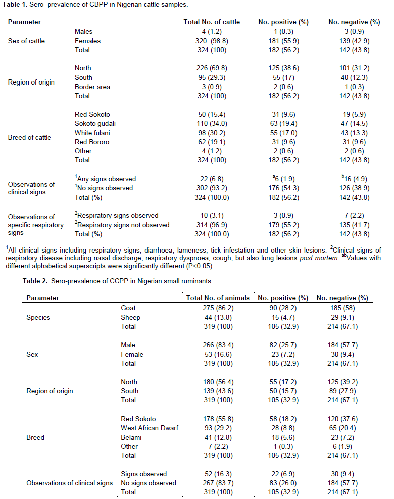
The sero-prevalence of CBPP amongst the sampled cattle was 56.2% (182 of 342). Table 1 shows the cattle sero-prevalence data disaggregated by test result, sex, breed and geographical origin. 320 of the cattle sampled were female while only four were male. Although the prevalence of CBPP was numerically higher in cattle originating from the northern region (38.6%) compared to the south (17.0%) this was not statistically significant (p≥0.05). 320 of the 324 cattle were from the 4 breeds Red Sokoto (50), Sokoto gudali (110), White Fulani (98) and Red Buroro (62), with no significant differences in prevalence between breeds (Table 1) (p≥0.05). The age range recorded for the sampled cattle was between 1 and 7 years; 310 were within the age range of 1 to 3 years while 14 were within the 4 to 7 years age range. The live weights of the cattle varied between 100 and 200 kg. The clinical observation data are also shown in Table 1. In all, 22 of the 324 cattle were recorded as showing clinical signs, 10 of which showed specific respiratory signs associated with possible CBPP infection, that is, dyspnoea, nasal discharge and cough with or without lung lesions seen subsequently at post mortem.
Small ruminants (SR)
The SR seroprevalence data are shown in Table 2. Of the 319 SR, 275 (86.2%) were caprine while 44 (13.8%) were ovine. In total, 32.9% (105) tested positive while 67.1% (214) were negative (Table 2). Their respective ages ranged from 5 to 35 months while their weights ranged from 5 to 45 kg. Of the 319 SR, 126 (39.5%) were brown in colour, 92 (28.8%) were black, 67 (21.0%) were white while 34 (10.7%) contained mixed colours. Most SR originated from Northern Nigeria (56.4%). There were no significant differences (p≥0.05) in the proportion of seropositives between the breeds (Table 2). Of the 319 SR, 52 (16.3%) showed signs of unspecified clinical disease while 267 (83.7%) did not.
Since it was first reported in 1924, CBPP has been endemic in Nigeria (Foluso, 2003). Reasons have been associated with transhumance, nomadism, and other inadequate control measures (Egwu et al., 1996). For instance, Billy et al. (2017) reported an overall sero-prevalence of 26.0%. The herd level prevalence of 54.7% with 30.2% seropositivity to CBPP within agro-pastoral areas was documented by Suleiman et al. (2015). Danbirni et al. (2010) reported 47% sero-prevalence in a herd of cattle with combined infection with TB. Okaiyeto et al. (2011) reported a sero-prevalence of 16.7 and 17.5% for adults and young cattle, respectively, in a herd of cattle with CBPP outbreak. Musa et al. (2016) also reported 3.33% prevalence of CBPP from lung samples from abattoirs in North Eastern Nigeria. The result of this study also differ from the work of Nawathe (1992) and Adamu and Aliyu (2006) who in their separate studies recorded a lower sero-prevalence of 0.52 and 0.33%, respectively in Borno State and Aliyu et al. (2000) who recorded a sero-prevalence of 0.29% in 5 other states in northern part of Nigeria.
In this survey, the overall seroprevalence of CBPP was 182 (56.2) in cattle, CCPP was 90 (28.2) in goats, 15 (4.7) in sheep, and overall seroprevalence of 105 (32.9) in small ruminants, this make our reported prevalence not only higher than others, but our survey also captured the small ruminants. The higher seroprevalence rate recorded in this study could be as a result of the inadequate prevention and control measures that resulted in absence or irregular vaccination programmes for cattle over the years, as well as the introduction of infected cattle into the areas (particularly through transhumance and nomadism) that were initially thought to be free of the disease (Aliyu et al., 2000). It could also be as a result of epidemiological trend of the disease with the presence of carriers in some herds which might not have been detected clinically and hence the maintenance and gradual spread of the disease (Egwu et al., 1996).
Reliable diagnosis of these two diseases by serology is complex, as the performance of available tests varies with the stage of infection and/or disease (Muuka et al., 2011). Thus the fact that the presence of antibodies did not coincide well with the detection of clinical signs is not surprising. A complement fixation test (CFT) and competitive ELISA test for CBPP are approved by OIE (2018a, b) for screening purposes but are generally not considered sufficiently reliable for diagnosis in individual animals because of variable sensitivities and/or specificities.
The CFT is reported as being highly specific at 98% (Bellini et al., 1998) but much lower sensitivity at between 55 and 60%. According to March et al. (2003), CBPP-CFT-false negative results may occur but LAT is cheaper, highly specific and easy to apply in the field, without any specialist training or equipment. The LAT used here specifically targets antibodies to a capsular polysaccharide (CPS) and is reported to have equivalent specificity to the CFT, that is, in the high 90%s, but with lower sensitivity (Ayling et al., 1999a, b). Therefore, in interpreting the present data, an apparent seroprevalence of 56.2% is likely to have under-estimated the true prevalence of M. mycoides subsp. mycoides CPS antibodies. Nevertheless, a value of 56.2% in cattle and 32.9% in small ruminants reported in this study represents a very significant prevalence which suggests that M. mycoides subsp. mycoides infection is very common in these regions of Nigeria.
In the case of CCPP, the LAT used was as described by March et al. (2002) which is based on recognition of the M. capricolum subsp. capripneumoniae CPS antigen and is a recognised test by OIE (2018b). This test has a high specificity (March et al., 2002) of approximately 99% (APHA, Weybridge, UK, personal communication). A close agreement of 96% was found between the LAT and the CFT in 54 sera from goats experimentally infected with M. capricolum subsp. capripneumoniae (Abdel-Hamid et al., 2016). The test is considered to be highly sensitive particularly in the early stages of infection (March et al., 2002; OIE 2018a, b).
In general, despite some limitations in sensitivity, the specific LATs for M. mycoides subsp. mycoides and M. capricolum subsp. capripneumoniae are regarded as useful tests for field screening and have the advantage that they can be used relatively easily in the field with little need for specialist training or equipment. The speed of agglutination reactions was consistent with those previously published (Bahir et al., 2017).
CBPP in cattle
The seroprevalence rate of 56.2% reported here, is generally higher than previous abattoir seroprevalence studies of CBPP reported in Nigeria and other parts of Africa of between 10 and 32% (Griffin and Laing, 1966, Aliyu et al., 2003; Okaiyeto et al., 2011). This suggests an increasing trend over time, which is in agreement with the annual report by OIE (2013) which stated that CBPP is gradually spreading across Nigerian states from the northern region to other regions because of constant migration of cattle herders. It is known that there is significant migration of cattle herders between regions and in particular, there is evidence that the disease is increasingly moving from the north towards the south (Foluso, 2003; Mailafia et al., 2010). In the present study, the majority of the cattle were from the north and the seroprevalence was numerically higher in these cattle (38.6%) compared to the southern (17.0%) but this was not statistically significant.
In this study, the majority of the cattle were female with just 1.2% male. This is probably because females are normally kept in higher numbers for rearing than males. Because of the small numbers of males any gender differential in seroprevalence could not be determined. Egwu et al. (2012) reported a lack of gender predisposition to CBPP infection.
Just 22 of the 324 cattle showed clinical signs of disease but only 10 of these included respiratory signs indicative of possible CBPP, which included dyspnoea, nasal discharge and cough with or without emaciation. More than half (54.3%) of the animals certified as clinically healthy when inspected pre-slaughter, were sero-positive to M. mycoides subsp. mycoides. This could be attributed to late disease manifestation, high immunity levels of the animal or as a result of being an asymptomatic carrier or latent carrier; the latter state usually occurring when an affected animal partially recovers after 3 to 4 weeks (OIE, 2011).
CCPP in small ruminants
The overall serological prevalence of M. capricolum subsp. capripneumoniae in small ruminants was 32.9% and this suggests that there is significant circulation of M. capricolum subsp. capripneumoniae in Nigeria. These results are consistent with previous reports of CCPP prevalence in the north of Nigeria (Egwu et al., 2012; Chinedu et al., 2014). Also, significant prevalence rates of CCPP have been reported in other parts of Africa. For example, Hadush et al. (2009) reported a CCPP prevalence rate of 32.68% and Mekuria et al. (2008), reported a CCPP prevalence rate of 18.62%, both in Ethiopia.
In the present study there was a higher prevalence of CCPP in goats (28.2%) compared to sheep (4.7%). This is to be expected because M. capricolum subsp. capripneumoniae has most often been associated with goats whereas infection in sheep is often associated with their close proximity to goats, goats being regarded as more susceptible than sheep to mycoplasmas (Akwuobu et al., 2014). CCPP seroprevalence tended to be higher in males than females (25.7% vs. 7.2%) although this was not statistically significant. This effect has been previously reported by Akwuobu et al. (2014) who suggested that males may be more susceptible than females. This is totally at variance to the submission of Torsson et al. (2017) who argued that females are more susceptible to these diseases due to the length of time they are kept for the purpose of reproduction.
More (56.4%) of the SR originated from the north than from the south (43.6%), a similar finding to the studies of Lawal-Adebowale and Alarima (2011) and Akwuobu et al. (2014). The geography of the northern region is more conducive to SR production due to the larger areas of suitable agroclimatic conditions in terms of adequate rainfall, a longer dry season and lighter sandy soil. Highest seropositivity was noted amongst the Red Sokoto which was also the largest population, followed by the WAD and Balami. 52 of the total 319 SR (16.3%) showed evidence of clinical signs of disease but only 22 of these were sero-positive for CCPP. Thus the majority of sero-positives did not show signs of clinical disease again suggesting a carrier state as in cattle.
In conclusion, the present study demonstrated the presence of both M. mycoides subsp. mycoides and M. capricolum subsp. capripneumoniae in animals originating from both the north and south of the country. This suggests that the two mycoplasma species are well established in both cattle and small ruminants in Nigeria. In the past, efforts to control these diseases have not been effective due both to lack of sustained and effective control strategies, doubts about vaccine safety and efficacy and, an antipathy to the use of antibiotics for CBPP in cattle. An assessment of the economic impact of these diseases would be appropriate to determine the cost effectiveness of control programmes to rid Nigeria of these potentially devastating diseases.
The authors have not declared any conflict of interests.
REFERENCES
|
Abdel-Hamid N, Abdel-Mortada O, Abd Elhady H, Farouk R (2016). Bacteriological examination and diagnostic performance of some serological tests used in diagnosis of small ruminant brucellosis. Veterinary Medical Journal Giza 62(4):11-21.
|
|
|
|
Ajuwape ATP, Adetosoye AI, Ikheloa JO, Alaka OO, Taiwo O, Talabi OA, Otesile EB, Ojo MO (2004). Pathogenicity of Mycoplasma capricolum subsp. capripneumoniae for cattle immunesuppressed by Trypanasoma congolence. Tropical Veterinarian 22(1):29-36.
Crossref
|
|
|
|
|
Akwuobu CA, Ayling RD, Chah KF, Oboegbulem SI (2014). Studies into the prevalence of Mycoplasma species in small ruminants in Benue State, North-central Nigeria. Springer: Tropical Animal Health and Production 46(6):1087-1092.
Crossref
|
|
|
|
|
Aliyu MM, Obi, TU, Egwu GO (2000). Prevalence of Contagious Bovine Pleuropneumonia (CBPP) in Northern Nigeria. Preventive Veterinary Medicine 47:263-269.
Crossref
|
|
|
|
|
Aliyu MM, Obi TU, Oladosu LA, Egwu GO, Ameh JA (2003). The use of competitive enzyme linked immuno-sorbent assay in combination with abattoir survey for CBPP surveillance in Nigeria. Tropical Animal Health and Production 21:35-41.
Crossref
|
|
|
|
|
Ayling RD, Regalla J, Nicholas RAJ (1999a). A field test for detecting antibodies to Mycoplasma mycoides subsp.small colony type using the latex slide agglutination test. In: COST 826 Agriculture and biotechnology. Mycoplasmas of ruminants: pathogenicity, diagnostics, epidemiology and molecular genetics Vol III Stipkovits, L., Rosengarten, R., and Frey,J., eds. Luxembourg; Office for official publications of the European Communities 3:155-158.
|
|
|
|
|
Ayling RD, Miles RJ, Regalla J, Nicholas RAJ (1999b). Development of improved serological tests for the diagnosis of CBPP. Proceedings of the International Symposium on Mycoplasma of Ruminants. Toulouse, France June, 2nd-4th, P 35.
View
|
|
|
|
|
Babalobi OO (2011). Participatory Epizootiology Research of Settled Pastoralists in Igangan Grazing Reserve, Southern Guinea Agro-Pastoral Zone, Oyo State, Nigeria: First Report Nigerian Veterinary Journal 32(1):16-20.
Crossref
|
|
|
|
|
Bahir W, Omar O, Rosales RS, Hlusek M, Ziay G, Schauwers W, Whatmore AM, Nicholas RAJ (2017). Search for OIE-listed ruminant mycoplasma diseases in Afghanistan. BMC Veterinary Research 13(1):149.
Crossref
|
|
|
|
|
Bellini S, Giovannini A, Di Francesco C, Tittarelli M, Caporale V (1998). Sensitivity and specificity of serological and bacteriological tests for contagious bovine pleuropneumonia. Revision Science Technology 17:654-670.
Crossref
|
|
|
|
|
Billy IL, Balami AG, Sackey AKB, Tekdek LB, Sa'idu SNA, Okaiyeto SO (2017). Sero-Prevalence of Contagious Bovine Pleuropneumonia in Three Senatorial District of Kaduna State, Nigeria Using Latex Agglutination Test. World Veterinary Journal 7(2):65-73.
Crossref
|
|
|
|
|
Chinedu AA, Roger DA, Kennedy FC, Stephen IO (2014). Studies into the prevalence of Mycoplasma species in small ruminants in Benue State, North-central Nigeria. Springer: Tropical Animal Health Production 46:1087-1092
Crossref
|
|
|
|
|
Danbirni S, Okaiyeto SO, Pewan SB, Kudi AC (2010). Concurrent infection of Contagious Bovine Pleuropneumonia and Bovine tuberculosis in Bunaji nomadic cows. Research Journal of Animal Science 4(1):23-25.
Crossref
|
|
|
|
|
Egwu GO, Nicholas RAJ, Ameh JA, Bashiruddin JB (1996). Contagious bovine pleuropneumonia. An update. Veterinary Bulletin 66:875-888.
|
|
|
|
|
Egwu GO, Adamu M, Mshelia GD, Bukar-Kolo YM (2012). A sustainable laboratory approach for contagious bovine pleuropneumonia (CBPP) monitoring in Nigeria: comparison between two serological tests in an endemic area complimented with post mortem lesions. African Journal of Microbiology Research 6(30):5890-5895.
Crossref
|
|
|
|
|
Foluso EF (2003). Status of contagious bovine pleuropneumonia (CBPP) in Nigeria with emphasis on control strategies. Proceedings of the FAO-OIE-AU/IBAR-IAEA Consultative Group on CBPP 3rd Meeting, Towards Sustainable CBPP Control Programmes in Africa, Nov. 12-14, 2003, Rome P 29.
View
|
|
|
|
|
Food and Agricultural Organization (FAO) (2004). Statistical Yearbook of the Food and Agricultural Organization for the United Nations FAO. 2004. The State of Food and Agriculture Source: FAO, Statistics Division
View.
|
|
|
|
|
Griffin RM, Laing DF (1966). Contagious bovine pleuropneumonia in Northern Nigeria. Bulletin of Epizootic Disease in Africa 14:255-279.
|
|
|
|
|
Hadush B, Eshetu L, Mengistu W, Hailesilassie M (2009). Seroprevalence of contagious caprine pleuropneumonia in Kefta Humera, Alamata (Tigray) and Aba-'ala (Afar), Northern Ethiopia. Tropical Animal Health and Production 41:803-806.
Crossref
|
|
|
|
|
Lawal-Adebowale OA, Alarima CI (2011). Challenges of small ruminants' production in selected urban communities of Abeokuta, Ogun State. Agriculturale Conspectus Scientificus 76(2):129-134. Published by Faculty of Agriculture, University of Zagreb, Croatia. Available online at
View
|
|
|
|
|
Manso-Silván L, Vilei EM, Sachse K, Djordjevic SP, Thiaucourt F, Frey J (2009). Proposal to assign Mycoplasma leachii sp. nov. As a new species designation for Mycoplasma sp. bovine group 7 of leach, and reclassification of Mycoplasma mycoides subsp. mycoides LC as a serovar of Mycoplasma mycoides subsp. Capri. International Journal of Systematic and Evolutionary Microbiology 59(6):1353-1358.
Crossref
|
|
|
|
|
March JB, Harrison JC, Borich SM (2002). Humoral responses following experimental infection of goats with Mycoplasma capricolum subsp. capripneumoniae. Veterinary Microbiology 84:29-45.
Crossref
|
|
|
|
|
March JB, Kerr K, Lema B (2003). Rapid detection of contagious bovine pleuropneumonia by a Mycoplasma mycoides subsp. mycoides small colony capsular polysaccharide-specific antigen detection latex agglutination test. Clinical Diagnostic Laboratory Immunology 10:233-340.
Crossref
|
|
|
|
|
Mailafia S, Onakpa MM, Dandam KP (2010). A ten year study on the prevalence of ruminant diseases encountered at the Ministry of the Federal Capital Territory veterinary clinics Gwagwalada, Abuja, Nigeria. Sahel Journal of Veterinary Science 9(2):4-6.
|
|
|
|
|
Mariner JC, McDermott J, Heesterbeek JAP, Thomson G and Martin SW (2005). A model of contagious bovine pleuropneumonia transmission dynamics in East Africa. Preventive Veterinary Medicine 73(1):55-74.
Crossref
|
|
|
|
|
Masiga WN, Domenech J, Windsor RS (1996). Manifestation and epidemiology of contagious bovine pleuropneumonia in Africa. Revue Scientifique et Technique 15(4):1283-1308.
Crossref
|
|
|
|
|
Mekuria S, Zerihun A, Gebre-Egziabher B, Tibbo M (2008). Participatory investigation of Contagious Caprine Pleuropneumonia (CCPP) in goats in the Hammer and Benna-Tsemay districts of southern Ethiopia. Tropical Animal Health and Production 40:571-82.
Crossref
|
|
|
|
|
Muuka G, Hang'ombe BN, Nalubamba KS, Kabilika S, Mwambazi L, Muma JB (2011). Comparison of complement fixation test, competitive ELISA and LppQ ELISA with post-mortem findings in the diagnosis of contagious bovine pleuropneumonia (CBPP). Tropical Animal Health and Production 43:1057-1062.
Crossref
|
|
|
|
|
Nawathe DR (1992). Resurgence of Contagious Bovine Pleuropneumonia in Nigeria. Revue Science Technical Office International Epizootics 11:799-804.
Crossref
|
|
|
|
|
Nicholas R, Churchward C (2012). Contagious caprine pleuropneumonia: new aspects of an old disease Transboudary Emerging Disease 59(3):189-196.
Crossref
|
|
|
|
|
Nicholas RAJ, Santini FG, Clark KM, Palmer NM, DeSantis P, Bashiruddin JB (1996). A comparison of serological tests and gross lung pathology for detecting contagious bovine pleuropneumonia in two groups of Italian cattle. Veterinary Record 139:89-93.
Crossref
|
|
|
|
|
Office International Des Epizooties (OIE) (2011). General disease information sheet: Contagious bovine pleuropneumonia.
View
|
|
|
|
|
Office International Des Epizooties (OIE) (2018a). Manual of Diagnostic Tests and Vaccines for Terrestrial Animals Chapter 3.4.8 pp. 1097-2018.
|
|
|
|
|
Office International Des Epizooties (OIE) (2018b). Manual of Diagnostic Tests and Vaccines for Terrestrial Animals Chapter 3.7.4. pp. 1441-1455.
|
|
|
|
|
Okaiyeto SO, Danbirni S, Allam L, Akam E, Pewan SB, Kudi AC (2011). On-farm diagnosis of contagious bovine pleuropneumonia in nomadic herds using latex agglutination test. Journal Veterinary Medicine and Animal Health 3(5):62-66.
|
|
|
|
|
Olorunshola ID, Andrew RP, Scacchia M, Nicholas RAJ (2017). Contagious bovine pleuropneumonia - never out of Africa? CAB Reviews 12(019):1-7.
Crossref
|
|
|
|
|
Pan African Control of Epizootics (PACE) (2004). Newsletter No. 6:2.
View
|
|
|
|
|
Suleiman A, Bello M, Dzikwi AA, Talba AM, Grema HA, Geidam YA (2015). Serological prevalence of contagious bovine pleuropneumonia in agro-pastoral areas of Nigeria. Tropical Animal Health and Production 47(6):1033-1042.
Crossref
|
|
|
|
|
Thiaucourt F, Manso-Silvan L, Salah W, Barbe V, Vacherie B, Jacob D, Breton M, Dupuy V, Lomenech AM, Blanchard A, Sirand-Pugnet P (2011). Mycoplasma mycoides, from "mycoides small colony" to"capri". A micro evolutionary Perspective. BioMed Central Genomics 12:114.
Crossref
|
|
|
|
|
Torsson E, Berg M, Misinzo G, Herbe I, Kgotlele T, Päärni M, Roos N, Blomström A, Ståhl K, Wensman JJ (2017). Seroprevalence and risk factors for peste des petits ruminants and selected differential diagnosis in sheep and goats in Tanzania. Infection Ecology and Epidemiology 7:1368336
Crossref
|
|
|
|
|
Office International Des Epizooties (OIE) (2013). Manual of Diagnostic Tests and Vaccines for Terrestrial Animals. OIE, Paris.
View (Accessed 16th January 2020)
|
|