ABSTRACT
A cross-sectional study was conducted from December 2016 to May 2017 in and around Sebeta town with the aim of assessing the prevalence of mastitis, isolation of aerobic bacterial and fungal causal agents and assessing antimicrobial resistance pattern of isolated Staphylococcus species in dairy cows. From a total of 383 dairy cows, 220 (57.4%) were found to be positive for mastitis of which 10.4% were affected by clinical mastitis and 47% by subclinical mastitis. Mastitis was more likely to occur in cows above 8 years of age (OR = 16.9, 95% CI = 7.8 - 36.00) and in those cows washed by hand before milking (OR =7.8, 95% CI = 4.2-14.6) as compared to those subjected to udder washing and drying using towels. Staphylococcus aureus was the most frequently isolated bacterial species (25%) followed by Streptococcus agalactiae (12.3%) and coagulase negative Staphylococcus species (10.5%). Yarrowia lipolytica (10.9%) and Candida etchellsii (7.3%) were the major yeast species isolated, while Aspergillus (6.8%), Mucor (5.9%), Penicillium (3.6%) and Fusarium (3.6%) were the major filamentous fungi species identified from the cultured milk samples. The results of antimicrobial susceptibility testing revealed that the isolated Staphylococcus species were highly resistant to penicillin G (93.1%) and oxytetracycline (79.3%) but were susceptible to vancomycin (100%), sulphamethoxazole/trimethoprim (96.6%), ampicillin (89.7%) and erythromycin (86.2%). It could be concluded that bovine mastitis is a major challenge to the dairy producers in and around Sebeta towns. Appropriate control and preventative measures must be instituted and dairy farmers and workers must be trained on proper milking and hygiene practices in order to reduce the prevalence of mastitis in this region. The penicillin resistant S. aureus could be a source of serious infection in humans as well and hence comprehensive studies including molecular characteristics of drug resistance gene of S. aureus especially of methicillin-resistant should be conducted as farm animals, primarily dairy cattle might serves as a reservoir of infection for humans. In 2% of the cases, only fungal species were identified as causes of mastitis, hence further investigation regarding their pathogenicity and contribution to bovine mastitis is needed.
Key words: Antimicrobial resistance, bovine mastitis, bacteria, Ethiopia, fungus, prevalence, Sebeta.
Ethiopia has the largest cattle population in Africa with an estimated population of 57.83 million (CSA, 2016). Development of the dairy sector in the country can contribute significantly to poverty alleviation and nutrition (Shapiro et al., 2015). However, currently milk production do not satisfy the countries milk requirement due to multitude of factors such as mastitis and other diseases that lead to significant loss in production (Biffa et al., 2005).
Mastitis occurs worldwide among dairy animals and it has been described to have an extreme economic impact (Al-majali et al., 2008). According to Bhikane and Kawitkar (2000), dairy cattle mastitis contributes up to 70% of reduced milk production, 9% of discarded milk after treatment, 7% of the cost of veterinary services and 14% of premature culling. The disease can be classified as clinical or subclinical (Eriskine, 2001).
The disease is caused by a multitude of etiological agents that includes bacteria, virus, fungi and algae (Wellenberg et al., 2002; Kivaria et al., 2004). The most common bacterial pathogens are Staphylococcus aureus, Streptococcus agalactiae, other Streptococcus species and coliforms (Sumathi et al., 2008). Other organisms may also include Arcanobacterium pyogenes, Pseudomonas aeruginosa, Nocardia asteroides, Clostridium perfringens, Mycobacterium, Mycoplasma, Pastuerella and Prototheca species and yeasts (Radostits et al., 2007). Fungal infections account for up to 2 to 13% of all cases of mastitis in cows (Krukowski et al., 2006). Usually mycotic mastitis is unnoticed by clinician in the first attempt of treatment and administration of antibiotics may aggravate fungal mastitis as some of the antibiotics like penicillin and tetracycline act as a source of nitrogen for various species of fungi (Tarfarosh and Purohit, 2008).
In Ethiopia, available information indicates that bovine mastitis is a serious challenge in dairy farms. Based on one meta-analysis report that evaluated 39 different studies, the overall prevalence of bovine mastitis was 47.0% (95% confidence interval [CI] = 42.0-52.0) of which 8.3% (95% CI = 6.5-10.3) was clinical mastitis and 37% (95% CI = 32.9-40.7) was subclinical mastitis (Getaneh and Gebremedhin, 2017).
Prevalence of bovine mastitis and predisposing factors were assessed 12 years ago in Sebeta towns (Sori et al., 2005). Since then, many dairy farms have been introduced into the area. Moreover, there are many complaints on poor response to treatment of mastitis using common antimicrobial drugs. Furthermore, to the author’s knowledge, there are no published literatures on major fungal pathogens isolated from bovine mastitis in Ethiopia. Based on the aforementioned facts, this study was conducted to assess risk factors, causative agents and to determine antimicrobial resistance patterns of isolated Staphylococcus species isolated from bovine mastitis in and around Sebeta town.
Study area and population
Sebeta town is located around 25 km from Addis Ababa city at 8°55′N latitude and
38°37′E longitude and 2,356 m above sea level. The climate is warm with the average annual temperature of 17.4°C and 1073 mm averages annual rainfall.
The study population comprises of lactating dairy cows that are managed under extensive, semi intensive and intensive farming systems. A total of 383 lactating dairy cows in and around Sebeta town were examined to determine the overall prevalence of mastitis in the area.
Study design and sample size determination
A cross-sectional study was conducted from December 2016 to May 2017 and study cows were randomly selected from extensive, semi intensive and intensive dairy farms in the area. The sample size was determined based on the formula given by Thrufield (2007) considering 5% absolute precision, 95% confidence interval and 52.78% expected prevalence from previous studies in the area (Sori et al., 2005). Therefore, the calculated sample size was 383.
Study methodology
Physical examination of the udder and milk
The udders were first examined visually and then palpated to detect any possible fibrosis, inflammatory swelling and atrophy of the tissue. The size and consistency of the mammary quarter was inspected for the presence of any abnormalities such as disproportional symmetry, swelling, firmness and blindness of the teat canal. In addition, two streaks of milk from each quarter in a strip cup were inspected visually for the presence of any flakes, clots, pus, watery appearance, blood and color change (Radostits et al., 2007).
California mastitis test (CMT) screening
After physical examination, milk samples were tested by California mastitis test (CMT Kit Lot number 67467, ImmuCell, USA). Briefly, a squirt of milk from each quarter of the udder was placed in each of four shallow cups in the CMT paddle and an equal amount of the reagent was added. A gentle circular motion was applied in a horizontal plane. Positive samples show gel formation within a few seconds. The California mastitis test was conducted to diagnose the presence of subclinical mastitis and based on the thickness of the gel formed by CMT reagent and milk mixture 1:1 test results were scored as 0 (negative), 1 (weak positive), 2 (distinct positive) and 3 (strong positive). Milk samples with test result of CMT 1 to 3, were classified as evidence of subclinical mastitis (Radostits et al., 2007). If at least one quarter was positive by the CMT then the cow was considered positive for mastitis.
Data and sample collection
A semi-structured questionnaire was used to collect data on risk factors which include age, parity and hygiene of udder, farm owners and milker’s. The age of the animals was determined from birth records and asked from owner and categorized as young adult (3 to 5 year), adult (6 to 8 year) and old (8 years and above); parity as few (1 to 2 parities), moderate (3 to 4 parities), and many (4 and above) (Umer et al., 2015). Udder hygiene was evaluated by observing the presence of any cow dung stain or spot on the udder and hind legs and through asking milker’s. The practice for keeping the hygiene of udder was divided into four categories; those that do not practice udder washing at all wash by hand without drying, wash using towel without drying, and wash and dry by using towels.
Milk samples were collected following the standard procedures by the national mastitis council (NMC, 2004). After a quarter had been washed with tap water and dried (in cases when there was a considerable amount of dirt), the teat end was swabbed with cotton soaked in 70% ethyl alcohol. Approximately 10 ml of milk sample was then collected aseptically from clinically and sub clinically (CMT positive) mastitic cows into sterile universal bottles after discarding the first milking streams (squirt). The bottles were labeled with permanent marker and transported on packed ice box to National Animal Health and Diagnostic Investigation Center (NAHDIC), where they were immediately cultured or stored at 4°C until processed for bacterial and fungal isolation.
Laboratory work
Bacterial identification
Collected milk samples were inoculated on sheep blood agar (OXOID) and MacConkey agar (OXOID) and incubated aerobically at 37°C and cultured plates were examined after 24 to 48 h of incubation for any visible growth. The colonies were identified using primary and secondary biochemical tests at least to determine the genus of the suspected isolate. Then, OMNILOG/BIOLOG (fully automated coated microplate based bacterial identification system) using GEN III micro plate (Lot number 3003241, BIOLOG, USA) with protocol A method was used to further confirm the species of suspected colonies. A single colony grown in Biolog Universal growth (BUG) agar medium was selected and emulsified into ‘inoculating fluid A’ (IF A). According to the manufacturer's instructions, cell density of the bacterial inoculum was measured and adjusted for a specified transmittance (90 to 98%) using a turbidiometer. For each isolate, 100 μl of the bacterial suspension was inoculated into each of the 96 well coated micro plates, using 8 channel pipette and incubated aerobically at 33°C for 22 h. The Omni Log identification system automatically reads each microplate and provides species/sub-species identification (ID), and then the results were printed out (OMNILOG, 2010).
Fungal identification
Milk samples were also simultaneously inoculated onto sabouraud dextrose agar (SDA) and cultured at 26°C for up to 4 weeks (if no visible fungal growth was observed within this period, no growth was recorded). Isolates were examined macroscopically and identified based on colony shape, size, color and growing pattern. For filamentous fungi, slides were prepared from each colony using scotch tape method where transparent scotch tape was lightly pressed to colony and then the tape was fixed to slide that had a drop of lactophenol cotton blue stain. The slides were observed under microscope in X10 and X40 magnification power and were identified at genus level using fungal identification key (Quinn et al., 2002). Yeast colonies were examined microscopically using Gram staining and species level identification was conducted by OMNILOG identification system using yeast microplates.
Antimicrobial susceptibility test
Three to five well isolated colonies of isolated Staphylococcus species were transferred to 5 ml nutrient broth and incubated at 35°C for 4 h. The turbidity of the broth culture was adjusted to obtain turbidity optically comparable to 0.5 McFarland standard solution. After adjusting the turbidity, sterile cotton swab was dipped into the suspension and then Mueller Hinton agar plate was inoculated by rotating 60°. Antimicrobials disc were applied on the media using disc dispenser and then incubated for 24 h. Measurement of zone of inhibition was done by using digital caliper. Six antimicrobials were used: Ampicillin, erythromycin, penicillin G, oxytetracycline, vancomycin, sulphamethoxazole/trimethoprim (OXOID discs) (CLSI, 2010).
Data analysis
Statistical analysis was performed using ‘STATA, version 12. Mastitis prevalence was calculated by dividing the number of CMT positive cows by the total number of cows tested. Multivariate logistic regression was used to see the association of the potential risk factors with occurrence of mastitis. The strength of the association was measured using odds ratio (OR). Factors with odds ratio greater than one were considered as risk factors and those with odds ratio less than one were protective factor. In all the analysis, P- value lower than 0.05 was considered as significant.
Prevalence of clinical and subclinical mastitis
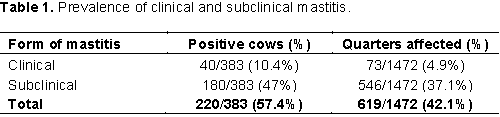
From a total of 383 dairy cows examined for mastitis, 220 (57.4%) of them were found positive. The details of the types of mastitis and quarter level mastitis were indicated in Table 1. From the total of 1532 quarters examined, 60 (3.9%) were blind (inactive quarters) and 619 (42.1%) were affected by mastitis (Table 1).
The association of risk factors with mastitis
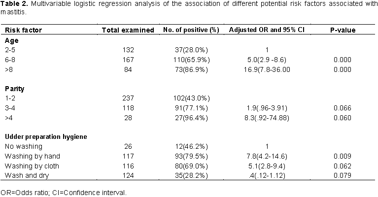
The prevalence of mastitis was higher in older cows than young and adults and the difference was statistically significant (P<0.05). The disease was more likely to occur in cows above 8 years of age in comparison to younger animals (OR = 16.9, 95% CI = 7.8- 36.00). Similarly, the prevalence was statistically higher (P<0.05) in cows which their udders were washed by hand only before milking (OR =7.8, 95% CI = 4.2-14.6) as compared to those cows which their udders were washed and dried using towels. The details of factors considered and association with mastitis are summarized in Table 2.
Bacteria and fungi agents isolated from mastitic milk
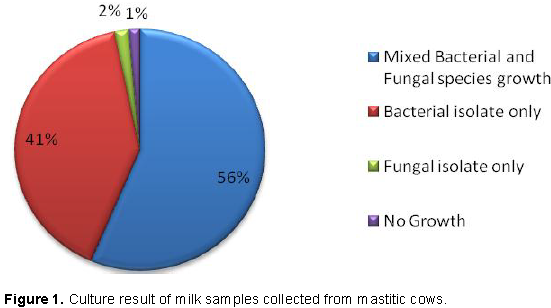
From 220 milk samples cultured for bacterial and fungal species identification, 41% were positive for bacterial isolates and 2% for fungal species. Mixed bacterial and fungal isolates were observed in 56% of the samples (Figure 1).
The predominant bacterial isolates were Staphylococcus aureus with isolation rate of 25% followed by Streptococcus agalactiae (12.3%) and coagulase negative Staphylococcus species (10.5%). Yarrowia lipolytica (10.9%) and Candida etchellsii (7.3%) were the major yeast species observed while Aspergillus (6.8%), Mucor (5.9%), Penicillium (3.6%) and Fusarium (3.6%) were filamentous fungi species identified from the cultured milk samples (Table 3).
In vitro antimicrobial susceptibility testing
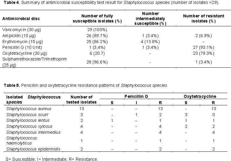
Antimicrobial susceptibility test was carried on 29 randomly selected S. species isolates. Staphylococcus species were found to be resistant to penicillin G (93.1%) and oxytetracycline (79.3%) but were highly susceptible to vancomycin (100%), sulphamethoxazole/Trimethoprim (96.6%), ampicillin (89.7%) and erythromycin (86.2%) (Tables 4 and 5).
The overall prevalence of mastitis observed in the present study (57.4%) was higher than the reported prevalence 12 years ago (52.78%) from the same study area (Sori et al., 2005). Some eight years ago, Mekibib et al. (2009) reported 71.1% prevalence of bovine mastitis from Holeta, a town which was located close to the present study site. This indicated that bovine mastitis remains a serious problem to the dairy producers in and around these neighborhood towns which costs the farmers from losses associated with reduced production, increased replacement cows, drug costs, veterinary fees and labour costs. It might be due to lack of coordinated actions on prevention and control of bovine mastitis. The numbers of dairy farms has increased in the study areas as compared to previous years. However, most of these farms have poor housing facilities and this might contribute to the contamination and exposure of teats to environmental pathogens and could be reason for increased prevalence of bovine mastitis.
Region to region variations on prevalence of both clinical and subclinical mastitis were wide in Ethiopia. Lakew et al. (2009) reported 64.6% overall prevalence from Assela, a town which has similar agro-ecology with the present study site. Mungube et al. (2004) and Delelesse (2010) reported 46.6 and 44.1% overall prevalence while Tolosa et al. (2009) reported 9.5%, Bedada and Hiko (2011) 12.1%, Sori et al. (2005) 16.11% and Workineh et al. (2002) 25.1% subclinical mastitis from different localities in Ethiopia. This similarities and differences might be due to complex nature of the disease involving interactions of various factors such as management and husbandry, environmental conditions, animal risk factors, and causative agents (Radostits et al., 2007). The variation in the prevalence of mastitis might also be due to management differences like hygienic condition during milking process practiced by each farm, or individual cow’s defense mechanism (Suriyasathaporn et al., 2000).
The higher prevalence of subclinical mastitis than that of clinical mastitis in the present as well previous studies could be attributed to the little attention given to subclinical mastitis as subclinical mastitis is not clinically visible while treating clinical cases. Moreover, dairy farmers might not be well informed about the silent nature of subclinical mastitis (Karimuribo et al., 2006; Almaw et al., 2008). Likewise, the predominance of subclinical mastitis and its serious economic relevance compared to clinical mastitis was underscored elsewhere out of Ethiopia (Kaliwal and Kurjogi, 2011; Awale et al., 2012; Shittu et al., 2012; Elbably et al., 2013; Katsande et al. , 2013).
Quarter level prevalence of mastitis (42.14%) was lower than finding of Kifle and Tolosa (2008) who reported prevalence rate of 63.1%, but higher than the report made by Zelalem (2001) in Ethiopia. The teat canal is the first barrier against invading pathogens, and the efficiency of teat defense mechanisms depends on the integrity of teat tissue; its impairment leads to an increase in the risk of intra-mammary infection. The observed high number of inactive quarter 60(3.9%) may be an indication of a serious mastitis problem on the respective farms and the absence of culling chronically infected cows that can serve as a means to prevent and control the disease within a farm.
In the present study the prevalence of mastitis was higher in old adult cows than young adults. This might be due to older cows have largest teats and more relaxed sphincter muscles, which increase the accessibility of infectious agent in the cow’s udder (Radostits et al., 2007). Cows with many parities were also at greater risk than moderate and few parities which might be due to physical alterations of udder, and is in line with findings reported by other authors (Carlen et al., 2004; Zwald et al., 2004; Abdel-Rady and Sayed, 2009; Belayneh et al., 2013; Katsande et al., 2013, Abrahmsen et al., 2014; Mureithi and Njuguna, 2016).
Cows in farms with poor milking hygiene were severely affected than those with good milking hygiene practices. Similar findings were reported by Sori et al. (2005); Lakew et al. (2009) and Moges et al. (2011). The reason most probably might be due to cross contamination from infected teat to others, or from infected to non infected cows during milking. The milkers’ hand and washing towels might also facilitate pathogens spread. It was also documented that udder preparation both before and after milking influence the prevalence of mastitis.
In this study, the dominant bacterial pathogens isolated from milk samples were Staphylococcus species (25%) however; this was lower than that of the 42.1% reported by Abera et al. (2010). Similarly, S. aureus was isolated as main etiological agent of mastitis in cattle in many African and Asian countries (FAO, 2014). S. aureus is considered as typical contagious pathogen causing bovine mastitis. Accordingly, the wide spread S. aureus mastitis might be cows positive in herd which act as primary reservoir and infected others especially during milking. Radostits et al. (2007) asserted that S. aureus is well adapted to survive in the udder and usually establishes a mild sub clinical infection of long duration from which it shed in milk facilitating transmission to healthy animals mainly during milking. Generally, S. aureus has been designated as a causative agent of both clinical and subclinical mastitis.
The isolation rate of S. agalactiae (12.3%) in this finding coincides with that of Bitew et al. (2010) at Bahir Dar who reported 13.9%. However, the finding was higher than the 4% report by Lakew et al. (2009) from Asella, and 6.4% isolation rate reported by Sori et al. (2005) in and around Sebeta town.
E. coli was identified from 5% of the samples in this study and this proportion was lower than reports by Sori et al. (2005), Mekebib et al. (2009), Bitew et al. (2010) who reported an isolation rate of 26.57, 43.13 and 20.3%, respectively. This lower isolation rate of environmental mastitis causal agents might be partly associated with effective and good sanitation of the barns with immediate removal of feaces practices. Moreover, the proportion of Micrococcus species in this study was lower than the finding of Mekonnen et al. (2005) and Bedada and Hiko (2011), who reported 10.2 and 5.6%, respectively. It was also reported that Micrococcus species causes mastitis only occasionally.
Mixed (fungal and bacterial) and fungal infection alone in this study represented 56 and 2% respectively. The overall 56% mixed fungal infection were comparable with the result of Pachauri et al. (2013) who found 64%. However, this was lower than the results of Al-Ameed (2013) in Iraq who reported 80% and was higher than the 13% fungal mastitis prevalence reported by Sukumar and James (2012). This might be due to unhygienic condition of the animal sheds and high humidity along with favorable environmental conditions supporting growth of fungal spores. Hence favorable conditions increase the chances of fungal spore to enter into the udder which provide suitable environment to these fungi (Williamson and Di Menna, 2007). Under immunosuppressive conditions, the dynamics of microorganisms may be disrupted, and the fungi together with the other microorganisms are able to overcome the udder defense mechanisms.
The overall isolation rate of yeast from the current study was 38.18% of which Candida species accounted for 11.8%, Yarrowia lipolytica for 10.9%, Rhodotorula species for 3.2%, Rhodosporidium diobovatum (2.3%), Galactomyces geotrichum (3.6%), Geotrichum terreste (1.4%), Trichosporon species (3.2%) and Saccharomyces species (1.8%). This isolation rate was lower than that reported by Andreia et al. (2008) on Candida (37.9%), Cryptococcus (10.3%) and Rhodotorula (10.3%). Although the distribution of Candida species shows diversity in several countries, it is important to note the increase in number of mammary gland infections caused by Candida species in the recent years (Krukowski et al., 2001). Filamentous fungi was isolated from 20% of the tested samples, the isolated fungal species were Aspergillus (6.8%), Mucor (5.9%), Penicillium (3.6%) and Fusarium (3.6%). The isolation rate of Aspergillus species was lower than the 38% reported by Mdegela et al. (2005) and Blowey and Edmondson (2010). The management practices adopted on dairy cows , like discarding first few strips of milk on ground while milking of animals as well as during treatment of mastitic animals and reluctance to disinfect hand between milking by milkers may contribute as potent source of lateral transmission of fungal and yeast infections (Pachauri et al., 2013). There are also reports in which yeasts like Candida spp. utilizes nitrogen from penicillin and tetracycline antibiotics, antibiotic therapy leads to perturbation in udder homeostasis, inhibition of T cells and neutrophil activity and in consequence this may also stimulates yeast growth (Corti et al., 2003; Noris et al., 2007).
In the present study, Staphylococcus species were found resistant to penicillin G (93.1%) and oxytetracycline (79.3%) and this is comparable with many previous reports in the country. The resistance of Staphylococcus species to penicillin may be attributed to the production of beta lactamase, an enzyme that inactivates penicillin and closely related antibiotics. The development of antibiotic resistance probably is a result of repeated therapeutic use or indiscriminate use of these antibiotics (Jaims et al., 2002). The uses of antimicrobials have, overtime, increased the number of antimicrobial-resistant microbes globally, and any use of antimicrobial agents will to some extent facilitate the development of resistant strains (Williams, 2000). The majority of authors have noted the development of antimicrobial resistance by Staphylococcus species isolated from mastitis cases (Pitkala et al., 2004; Turutoglu et al., 2006; Pyorala and Taponen, 2009; Sori et al., 2011).
It could be concluded that bovine mastitis is a major challenge to the dairy producers in and around Sebeta towns. Large numbers of microorganism were isolated from milk of CMT positive cows with Staphylococcus, Streptococcus, Candida species and Y. lipolytica being the predominant. The demonstrated resistance pattern of Staphylococcus species to penicillin and oxytetracycline may alarm on the repeated use of these drugs for mastitis treatment in the country. Hence, comprehensive studies including molecular characteristics of drug resistance gene of S. aureus especially of methicillin-resistant should be conducted in farm animals. In 2% of the cases, fungal species were identified as causes of mastitis, hence further investigation regarding their pathogenicity and contribution to bovine mastitis is needed.
CI, Confidence interval; CLSI, Clinical Laboratory Standards Institute (CLSI); CMT, California mastitis test; FAO, Food and Agriculture Organization; NAHDIC, National Animal Health Diagnostic and Investigation Center; OR, odds ratio.
ETHICS APPROVAL AND CONSENT TO PARTICIPATE
This work is a part of sub-thematic research “Bovine mastitis: Udder morphometrical traits, common bacterial isolates, histopathological changes and predisposing factors to clinical and subclinical mastitis in local zebu and crossbreed dairy cattle in central Ethiopia “RD/LT-038/15” for which the investigators have received ethical clearance referenced with VM/ERC/005/08/2015 from ethical clearance and animal welfare committee of Addis Ababa University college of Veterinary Medicine and Agriculture. After briefing the purpose of the study consent was requested from all participating dairy farm owners for collecting samples. All the procedures used were non invasive and in addition all the results were communicated to animal owners.
The study was financially supported by Addis Ababa University Research and Technology transfer and Thematic Research Project, Grant No. RD/LT-038/15. This fund was used to cover the cost for field sample collection and all the consumables used for sample collection and laboratory analysis was covered by National Animal Health Diagnostic and Investigation Center. The funding body had no role in study design, data collection, analysis, interpretation, or writing of the manuscript.
The authors have not declared any conflict of interests.
REFERENCES
|
Abdel-Rady A, Sayed M (2009). Epidemiological studies on subclinical mastitis in dairy cows in Assiut governorate Search Results Web results Veterinary World 2:373.
|
|
|
|
Abera M, Demie B, Aragaw K Regassa F, Regassa A (2010): Isolation and identification of Staphylococcus aureus from bovine mastitic milk and their drug resistance patterns in Adama town, Ethiopia. Journal of Veterinary Medicine Animal Health 2(3):29-34.
|
|
|
|
|
Abrahmsen M, Persson Y, Kanyima BM, Bage. R (2014): R. Prevalence of subclinical mastitis in dairy farms in urban and peri-urban areas of Kampala, Uganda. Tropical Animal Health Production 46:99-105.
Crossref
|
|
|
|
|
Al-Ameed AI (2013). Isolation and identification of fungi from infected milk samples obtained from cattle with mastitis and studying the antifungal activity of Rosemary Ethanolic extract agaimst the main strains. Diyala Journal of Agricultural Science 5(2):1-13.
|
|
|
|
|
Al-Majali AM, Al-Qudah KM, Al-Tarazi YH, Al-Rawashdeh OF (2008): Risk Factors Associated With Camel Brucellosis In Jordan. Tropical Animal Health Production 40:193-200.
Crossref
|
|
|
|
|
Almaw G, Zerihun A, Asfaw Y (2008): Bovine mastitis and its association with selected risk factors in smallholder dairy farms in and around Bahir Dar, Ethiopia. Tropical Animal Health Production 40:427-432.
Crossref
|
|
|
|
|
Andreia S, Elsio A, Daniela I, Juliana A, Edna C, Patricia V, Laerte F (2008). Diversity of yeasts from bovine mastitis in southern Brazil. Revista iberoamericana de micologia 25:154-156.
|
|
|
|
|
Awale MM, Dudhatra GB, Avinash K, Chauhan,BN, Kamani DR (2012) Bovine mastitis, a threat to economy. Open Access Scientific Reports 1:295.
|
|
|
|
|
Bedada BA, Hiko A (2011). Mastitis and antimicrobial susceptibility test at Asella, Oromia Regional state, Ethiopia. Journal of Microbiology and Antimicro 3(9):228-232.
|
|
|
|
|
Belayneh R, Belihu K, Wubete A (2013). Dairy cows mastitis survey in Adama town, Ethiopia. Journal of Veterinary Medicine and Animal Health 5:281-287.
|
|
|
|
|
Bhikane AV, Kawitkar SB (2000). Handbook for veterinary clinician. Venkateh Book. Udgir, India pp. 453-564.
|
|
|
|
|
Biffa D, Debela E, Beyene F (2005). Prevalence and risk factors of mastitis in lactating dairy cows in southern Ethiopia. International Journal of Veterinary Science Medicine 3:189-198.
|
|
|
|
|
Bitew M, Tafere A, Tolosa T (2010): Study on bovine mastitis in dairy farms of Bahir Dar town and its environment. Journal of Animal Veterinary Advances 9:2912-2917.
Crossref
|
|
|
|
|
Blowey R, Edmondson P (2010). Mastitis control in dairy herds, 2nd edition. CAB international, Cambridge MA, USA P 55.
Crossref
|
|
|
|
|
Carlen E, Strandberg E, Roth A (2004). Genetic parameters for clinical mastitis, somatic cell score, and production in the first three lactations of Swedish Holstein cows. Journal of Dairy Science 87:3062-3070.
Crossref
|
|
|
|
|
Central Statistical Authority (CSA) (2016): Federal Democratic Republic of Ethiopia Central Statistical Agency agricultural Sample Survey Volume II Report on livestock and livestock Characteristics Statistical Bulletin P583 Addis Ababa.
|
|
|
|
|
Clinical Laboratory Standards Institute (CLSI) (2010): Performance Standards For Antimicrobial Disk Susceptibility Tests. M100-S20.
|
|
|
|
|
Corti S, Sicher D, Regli W, Stephan R (2003). Current data on antibiotic resistance of the most important bovine mastitis pathogens in Switzerland. Schweiz Arch Tierheilkd 145:571-575.
Crossref
|
|
|
|
|
Delelesse GD (2010). Study on prevalence of bovine mastitis on Cross breed dairy cow around Holeta areas, West Shoa Zone of Oromia, Ethiopia. Global Veterinary 5(6):318-323.
|
|
|
|
|
Elbably MA, Emeash HH, Asmaa NM (2013). Risk factors associated with mastitis occurrence in dairy herds in Beni-Suef Governorate. World Veterinary Journal 3:05-10.
|
|
|
|
|
Eriskine RJ (2001). Intramuscular administration of ceftiofur sodium versus intrammamary infusion of penicillin/novobiocin for treatment of streptococcus agalactiae mastitis in dairy cows. Journal of the American Veterinary Medical Association 208:258-260.
|
|
|
|
|
Food and Agriculture Organization (FAO) (2014): Impact of mastitis in small scale dairy production systems. Animal Production and Health Working P 13. Rome.
|
|
|
|
|
Getaneh AM, Gebremedhin EZ (2017). Meta-analysis of the prevalence of mastitis and associated risk factors in dairy cattle in Ethiopia. Tropical Animal Health and Production 49((4):697-705.
|
|
|
|
|
Jaims E, Montros L, Renata D (2002). Epidemiology of drug resistance. The case of S. aureus and CNS infection Epidemiology. Drug Research 44:108-112.
|
|
|
|
|
Kaliwal BB, Kurjogi MM (2011). Prevalence and antimicrobial susceptibility of bacteria isolated from bovine mastitis. Advances in Applied Science Research 2:229-235.
|
|
|
|
|
Karimuribo ED, Fitzpatrick JL, Bell CE, Swai ES, Kambarage DM, Ogden NH, MBryant J, French NP (2006). Clinical and subclinical mastitis in smallholder dairy farms in Tanzania: Risk, intervention and knowledge transfer. Preventive Veterinary Medicine 74:84-98.
Crossref
|
|
|
|
|
Katsande S, Matope G, Ndengu M, Pfukenyi DM (2013). Prevalence of mastitis in dairy cows from smallholder farms in Zimbabwe. Onderstepoort Journal of Veterinary Research 80:523.
Crossref
|
|
|
|
|
Kifle A, Tolosa T (2008). Prevalence of sub clinical mastitis in small holder dairy farms in Selale, north shewa zone, Central Ethiopia. International Journal of Veterinary Science Medicine 5(1):1-4.
|
|
|
|
|
Kivaria FM, Noordhuizen J.P, Kapaga AM (2004). Risk indicators associated with subclinical mastitis in smallholder dairy cows in Tanzania. Tropical Animal Health Production 36:581-592.
Crossref
|
|
|
|
|
Krukowski H, Tietze M, Majewski T, Rozanski P (2001). Survey of yeast mastitis in dairy herds of small-type farms in the Lublin region, Poland. Mycopathologia 150:5-7.
Crossref
|
|
|
|
|
Krukowski H, Lisowski A, Rozanski P, Skorka A (2006). Yeasts and algae isolated from cows with mastitis in the south-eastern part of Poland. Polish Journal of Veterinary Sciences 9(3):181-184.
|
|
|
|
|
Lakew M, Tolosa T, Tigre W (2009). Prevalence and major bacterial causes of bovine mastitis in Asella, South Eastern Ethiopia. Tropical Animal Health and Production 41:1525-1530.
Crossref
|
|
|
|
|
Mdegela RH, Karimuribo E, Kusiluka LJM, Kabula B, Manjurano A, Kapaga AM, Kambarage DM (2005). Mastitis in smallholder dairy and pastoral cattle herds in the urban and peri-urban areas of the Dodoma municipality in Central Tanzania. Livestock Research for Rural Development 17:123.
|
|
|
|
|
Mekibib B, Furgasa M, Abunna F, Megersa B Regassa A (2009). Bovine Mastitis: Prevalence, Risk Factors and Major Pathogens in Dairy Farms of Holeta Town, Central Ethiopia. Veterinary World 3(9):97-403.
|
|
|
|
|
Mekonnen H, Workineh S, Bayleyegne M, Moges A, Tadele K (2005). Antimicrobial susceptibility profile of mastitis isolates from cows in three major Ethiopian dairies. Medicine Veterinary 176(7):391-394.
|
|
|
|
|
Moges N, Asfaw Y, Belihu K (2011). A Cross Sectional Study on the Prevalence of Sub Clinical Mastitis and Associated Risk Factors in and around Gondar, Northern Ethiopia. International Journal of Animal Veterinary 3(6):455-459.
|
|
|
|
|
Mungube ED, Tenhagen BA, Kassa T, Regessa F, Kyule MN, Greiner M, Baumann MPO (2004). Risk factors for dairy cows in the central highland of Ethiopia. Tropical Animal Health Production 36:463-472.
Crossref
|
|
|
|
|
Mureithi DK, Njuguna MN (2016). Prevalence of subclinical mastitis and associated risk factors in dairy farms in urban and peri-urban areas of Thika Sub County, Kenya. Livest. Research for Rural Development 28:13.
|
|
|
|
|
National Mastitis Council NMC (2004): Microbiological procedures for the diagnosis of udder infection. 3rd ed., National Mastitis Council Inc. Arlington, VA.
|
|
|
|
|
Noris M, Casiraghi F, Todeschini M, Cravedi P, Cugini D, Monteferrante G, Aiello S, Cassis L (2007). role of immunosuppressive drugs. Journal of American Society 18:1007-1018.
|
|
|
|
|
Omnilog (2010). OMNILOG data collection software, bacterial and fungi identification system, user guide part no.90311, version 2.3.
|
|
|
|
|
Pachauri S, Varshney P, Dash SK, Gupta MK (2013). Involvement of fungal species in bovine mastitis in and around Mathura, India. Veterinary World 6(7):393-395.
Crossref
|
|
|
|
|
Pitkala A, Haveri M, Pyorala S, Myllys V, Hankonen-Buzalski T (2004). Bovine mastitis in Finland prevalence, distribution of bacteria and antimicrobial resistance. Journal of Dairy Science 87:2433-2441.
Crossref
|
|
|
|
|
Pyorala S, Taponen S (2009): Coagulase negative staphylococci-emerging mastitis pathogens. Veterinary Microbiology 134:3-8.
Crossref
|
|
|
|
|
Quinn PJ, Markey BK, Carter ME, Donnelly WJ. Leonard FC (2002). Veterinary Microbiology and Microbial Disease. Blackwell Science Ltd, Blackwell Publishing Campany pp. 465-474.
|
|
|
|
|
Radostits OM, Gay CC, Hinchcliff KW, Constable PD (2007). Mastits. In: Veterinary Medicine: A Text book of disease of cattle, sheep, pigs, goats, and horses 10th edition, Ballier, Tindall, London pp. 674-762.
|
|
|
|
|
Shapiro BI, Gebru G, Desta S, Negassa A, Nigussie K, Aboset G, Mechal H (2015). Ethiopia livestock master plan. A contribution to the Growth and Transformation Plan II (2015-2020), ILRI Project Report.Nairobi, Kenya: International Livestock Research Institute (ILRI).
|
|
|
|
|
Shittu M, Abdullahi J, Jibril A, Mohammed AA, Fasina FO (2012). Sub-clinical mastitis and associated risk factors on lactating cows in the Savannah Region of Nigeria. BMC. Veterinary Research 8:134.
Crossref
|
|
|
|
|
Sori T, Zerihun A, Abdicho S (2005). Dairy cattle mastitis in and around Sebeta, Ethiopia. International Journal of Applied Research Veterinary Medicine 3(4):332-338.
|
|
|
|
|
Sori T, Hussien J, Bitew M (2011). Prevalence and susceptibility assay of Staphylococcus aureus isolated from bovine mastitis in Dairy Farms of Jimma Town, South West Ethiopia. Journal of Animal Veterinary Advances 10(6):745-749.
Crossref
|
|
|
|
|
Sukumar K, James PC (2012). Incidence of fungal mastitis in cattle. Tamilnadu. Journal of Veterinary Animal Sciences 8(6):356-359.
|
|
|
|
|
Sumathi BR, Veeregowda BM, Amitha R (2008): Prevalence and antibiogram profile of bacterial isolates from clinical bovine mastitis. Veterinary World 1:237-238.
|
|
|
|
|
Suriyasathaporn W, Schukken YH, Nielsen M, Brand A (2000) Low somatic cell count: a risk factor for subsequent clinical mastitis in dairy herd. Journal of Dairy Science 83:1248-1255.
Crossref
|
|
|
|
|
Tarfarosh MA, Purohit SK (2008). Isolation of Candida Spp. From mastitic cows and milkers. VetScan 3:14-18.
|
|
|
|
|
Thrufield M (2007). Veterinary Epidemiology, 3rd Edition, London: Blackwell Science, A Blackwell Publishing Company pp. 214-265.
|
|
|
|
|
Tolosa T, Geberetsadik Z, Regassa F (2009). Bovine mastitis and its associated risk factor in lactating cow in Wolayta Sodo, Southern Ethiopia. Animal Health Production 57(4):311-319.
|
|
|
|
|
Turutoglu H, Ercelik S, Ozturk D (2006): Antibiotic resistance of Staphylococcus aureus and coagulase-negative staphylococci isolated from bovine mastitis. Bulletin of Veterinary Institute in Pulawy 50:41-45.
|
|
|
|
|
Umer S, Tilahun Z, Gizat A, Abdela, E., Haimanot, D, Tadele, K, Firmaye G, Girma,K (2015). Prevalence, risk factors and major bacterial causes of bovine mastitis in west Arsi zone of Oromia Region, Southern Ethiopia. Natural science 13(8):19-27.
|
|
|
|
|
Wellenberg GJ, Van Der Poel WHM, Van Oirschot JT (2002). Viral Infections And Bovine Mastitis: A Review Veterinary Microbiology 88:37-45.
Crossref
|
|
|
|
|
Williams R (2000). The impact of antimicrobial resistance. Acta Veterinaria Scandinavica Supplementum 93:17-20.
|
|
|
|
|
Williamson JH, Di Menna ME (2007). Fungi isolated from bovine udders, and their possible sources. NZ. Veterinary Journal 55:188-190.
Crossref
|
|
|
|
|
Workineh SM, Bayleyegne H, Mekonnen L, Potgieter ND (2002). Prevalence and aetiology of mastitis in cow from two major Ethiopian dairies. Tropical Animal Health Production 34:19-25.
Crossref
|
|
|
|
|
Zelalem G (2001). Prevalence of mastitis and identification of major isolates in Walaita Sodo. DVM Thesis, Jimma University, Ethiopia.
|
|
|
|
|
Zwald NR, Weigel KA, Chang YM, Welper RD, Clay JS (2004). Genetic selection for health traits using producer recorded data. II. Genetic correlations, disease probabilities, and relationships with existing traits. Journal of Dairy Science 87:4295-4302.
Crossref
|
|