Full Length Research Paper

ABSTRACT
The authors investigated the growth of hydromycobiota on the eggs of rainbow trout (Oncorhynchus mykiss) in waters of different trophicity. Of the O. mykiss eggs that were investigated, 16.6% were found to be infected by mycotal species. Thirty mycotal species were found on eggs obtained from adult female representatives. Achlya polyandra, A. radiosa, Aphanomyces laevis, Leptomitus lacteus, Saprolegnia ferax and Saprolegnia parasitica belonged to the species that were most commonly encountered. Aphanomyces frigidophilus, Candida albicans and Zoopage phanera were rarely found in salmonid fishes. Amino acid, carbohydrate and urease tests were used, and all analyses of species from the Achlya, Aphanomyces, Leptolegnia, Pythium and Saprolegnia genera showed that they assimilate glucose and starch. However, they did not assimilate glycine, leucine, lysine, ornithine, and arabinose. Urease was assimilated only by species from the Leptolegnia, Pythium and Saprolegnia genera.
Key words: Oncorhynchus mykiss, rainbow trout, eggs, mycotal species, infection, hydrochemistry.
INTRODUCTION
Rainbow trout were classified as part of the Salmo genus until 1988 when the use of the generic name Oncorhynchus was adopted for all Pacific trout and salmon, to distinguish them as different (Smith and Stearley, 1989). Analyses of mitochondrial DNA showed that the mtDNA of rainbow trout had more similarity to Pacific salmon than to brown trout and Atlantic salmon (Berg and Farris, 1984; Thomas et al., 1986; Gyllensten and Wilson, 1987). This was confirmed through osteological analysis (Smith and Stearley, 1989). Today, all forms of rainbow trout belong to the Oncorhynchus genus, as the O. mykiss species (derived from the Kamchatkan name "mikizha" or "mykiz") (Ethier and Starnes, 1993).
Both marine and freshwater fish species deliver not only protein and fat, but also biologically active substances that are important for human organisms. Therefore, the consumption of fish is increasing from year to year (Food and Agricultural Organization of the United Nations (FAO), 2012). However, in recent years freshwater fishing has increased much more than fishing in seas and oceans. The growing number of freshwater fish farms may account for this. An important factor to consider in investigating what limits the fish populations in some cultures is the mycotal pathogens that can result in reduced breeding, sometimes affecting from 50% (Hatai and Hoshiai, 1992) to 75% (Lartzeva, 1986) of the incubated eggs. For example, Chien (1981) has described the mass death of the rainbow trout that occurred off the coast of Taiwan during their reproduction period and which were caused by the Aphanomyces laevis. As a result of this, we became interested in the extent to which mycotal species affect the eggs of the rainbow trout in waters of different trophicity
MATERIALS AND METHODS
Occurrence of rainbow trout
Oncorhynchus mykiss (Walbaum, 1792) (syn. Salmo gairdneri Richardson, 1836; Salmo irideus Gibbons, 1855) (English name: rainbow trout).
O. mykiss is native to the Pacific Slope, extending from the Kuskokwin River, in Alaska, through Rio Santo Domingo, in Baja, California, to rivers in Mexico, to the upper Mackenzie River (Arctic Basin), in Alberta and British Columbia, Canada, and to the endorheic basins of southern Oregon, U.S.A. This species has also been widely introduced to coastal waters elsewhere in North America and in rest of the world, including South Africa (Page and Burr, 1991; ITIS, 2010).
In the Eastern Pacific, O. mykiss occurs on the Kamchatkan Peninsula and has been recorded on the Commander Islands (east of Kamchatka) and sporadically in the far south of the Okhotsk Sea where the mouth of the Amur River flows along the mainland (ITIS, 2010).
This species inhabits cold headwaters, creeks, small-to-large rivers and lakes (usually not stocked in water that reaches summer temperatures above 25°C) and ponds with very low oxygen concentrations. It feeds on a variety of aquatic invertebrates and on small fishes. O. mykiss is cultured in many countries (including some in the south of Africa) and is often hatched and stocked in rivers and lakes, especially to attract recreational fish species (Frimodt, 1995; ITIS, 2010). With the exception of the common carp, the rainbow trout is one of the oldest species that is being cultured (Gall and Crandell, 1992). According to Wales (1939), the first transfer of eggs took place from McCloud River (northern California) to a hatchery in Caledonia, New York, in 1874. Rainbow trout eggs were also shipped from North America to Japan in 1877 and to England in 1885. The European rainbow trout farming industry began in Denmark (Laird and Needham, 1988; Gall and Crandell, 1992).
Characteristics of water bodies
Three water bodies located in the north-eastern region of Poland (53°07’N, 23°10’E to 53°13’N, 23°20’E) were chosen for this study: Pond Fosa: an area of 2.5 hectares (ha); maximum depth of 1.75 m; breeding site of wild ducks; a culture of crucian carp intended for anglers; Pond Komosa: an area of 12.1 ha; maximum depth of 2.25 m; surrounded by coniferous trees of the dense Knyszynska Forest; River Suprasl: a length of 106.6 kilometers (km); a right-bank tributary of the middle part of the Narew River, flowing through the Knyszynska Forest.
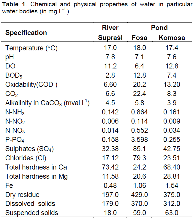
Nineteen (19) parameters of those water samples were measured (Table 1) according to generally accepted methods (APHA, 2005).
Isolating and identifying mycotal species
Eggs were collected (after fertilization) at the end of April from the hatchery at the Gawrych Ruda Farm. 150 - 450 eggs for each of the water bodie were investigated.
Water samples from specific water bodies 800 ml each were placed into 1000-ml vessels and 50 eggs were transferred to each vessel in accordance with the general principles of culture (Watanabe, 2000). The vessels were stored at temperature of 7± 0.5°C, with access to daylight that resembled natural conditions and following the recommended instructions (Seymour and Fuller, 1987). The pH of the water was analysed separately for every vessel (Peterson and Brindge, 1994). The water analysis and experiments were done in three parallel repetitions. Eggs were taken for each vessel, and the eggs that were covered with fungal mycelia were observed every 3 - 4 days under a light-microscope. The presence of any morphological structures, such as zoospores, antheridia and oogonia, belonging to aquatic fungi were recorded. Fungal species were identified using the keys of Johnson et al. (2005), Pystina (1998) and Petrini and Petrini (2013). The systematics of straminipiles species according to Dick (2001) were used in this experiment. The experiments were carried out for one month, and the results were then tested for significance using ANOVA and evaluated by the Scheffe test (Winer, 1997).
Determination of the amino acid, carbohydrate and urease assimilation tests
Amino acid, carbohydrate and urease tests were performed on the Achlya, Aphanomyces, Leptolegnia, Pythium and Saprolegnia genera, based on Yuasa and Hatai (1996). For the carbohydrate utilization test, Yeast Nitrogen Base agar (Difco) was the medium used for the cultures of the fungal isolates. GY agar (Difco) was used for the urease test. The basal medium used in the amino acid assimilation test was the same as that used for the carbohydrate assimilation test. Bromo thymol blue and phenol red that was added to the yeast nitrogen-based broth and the GY broth, respectively, were used as indicators. These methods are described in detail in our previous paper (Czeczuga et al., 2011b).
RESULTS
Table 1 shows the hydrochemical parameters of the water that was used in the experiment. The water from Pond Fosa was the most eutrophic. It showed heightened oxidability and alkalinity and higher levels of CO2, N (NO3), phosphates and other parameters. The lowest indices of the parameters that have been mentioned, as well as the lowest amounts of chlorides and iron, were found in water from Pond Komosa and River Supra?l.
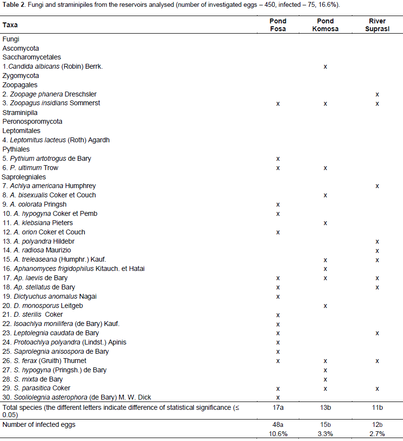
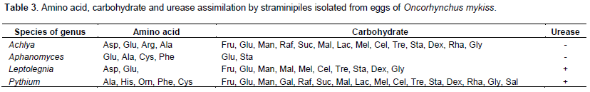
Thirty mycotal species, including twenty four (24) belonging to the Saprolegniales, two to the Zoopagales, two to the Pythiales and one each to the Leptomitales, and Saccharomycetales, were found to be growing on the eggs of the O. mykiss (Table 2). The highest number of species was growing in water samples from Pond Fosa (the most eutropic), while the lowest number occurred in water from River Supra?l and Pond Komosa (the lowest trophicity). It is worth making a special note that Aphanomyces frigidophilus, Candida albicans and Zoopage phanera have rarely been found in fish living in Polish waters. Table 3 shows the assimilation tests for species from the Achlya, Aphanomyces, Leptolegnia, Pythium and Saprolegnia genera. All analysed species from the Achlya, Aphanomyces, Leptolegnia, Pythium and Saprolegnia genera showed that they assimilated glucose and starch. However, they did not assimilate glycine, leucine, lysine, ornithine or arabinose. The urease was assimilated by species from the genera of Leptolegnia, Pythium and Saprolegnia only.
DISCUSSION
The present study has proven that the growth of aquatic mycotic species on the eggs of rainbow trout depends on the chemical characteristic of the water bodies from which the water samples are obtained for the experiment. Chemical analyses of the water samples that were collected enabled water differentiation with respect to the content of chemical compounds. Water from Pond Fosa contained more biogenic compounds, mainly phosphorus. This confirms once again our earlier assumptions (Czeczuga and Woronowicz, 1993) that the degree of infection of the fish eggs in hatcheries depends largely on the state of cleanliness and trophicity of the water that supplies the hatchery.
As shown in Table 2, the most commonly encountered species included Saprolegnia parasitica, S. ferax, Achlya polyandra, Laptomitus lacteus and Aphanomyces laevis. All these species belong to the group of opportunistic sapro- and necrotrophic pathogens (Bruno and Wood, 1999). S. parasitica, which has been described as a pathogen in the eggs of various fish species (Hatai et al., 1990) and in the fish fry in Pacific salmon breeding farms, causes the death of almost the entire population (Neitzel et al., 2004; van West, 2006). It is also responsible for considerable losses in fish populations in lakes. S. ferax kills the eggs of sterlet (Lartzeva, 1986) and cyprinids (Czeczuga and Muszynska, 1999). A. polyandra was observed quite frequently on the eggs of four lampreys that were examined (Czeczuga, 1997) and on Atlantic salmon (Czeczuga et al., 2011a), and L. lacteus infects many fish species in different water bodies. Aphanomyces frigidophilus was described as occurring on the eggs of the Japanese char Salvelinus leucomaenis (Kitancharoen and Hatai, 1997) and, for the first time in Europe, on the eggs of Coregonus lavaretus (Czeczuga et al., 2004). Aphanomyces frigidophilus also grows on some species from the Salmo genus (Czeczuga et al., 2011a), including sturgeonid fishes (Czeczuga et al., 2012b), Chinook salmon (Czeczuga et al., 2012a) and African catfish (Czeczuga et al., 2013), and on the alevins of the Nile tilapia (Czeczuga et al., 2014b), and on the eggs of Stenodus species (Czeczuga et al., 2014a).
The immune response of rainbow trout to Aphanomyces invadans has also been examined (Thompson et al., 1999), and according to Khan et al. (1998) and Oidtmann et al. (2008), the rainbow trout is moderately susceptible to Aphanomyces invadans through intramuscular infection.
Rainbow trout is one of the main species that is bred on a large scale in fish farms, not only in Europe (Backiel, 1964; Goryczko, 2000) but also on other continents (MacCrimmon, 1972), including Africa (FAO, 2012). Therefore, from the 1930s to the present, mycosis has caused huge losses in the populations of this species and has been studied intensely. Members of this species can be infected by A. laevis, L. lacteus, Saprolegnia delica, S. ferax, S. monoica and S. parasitica (Tiffney, 1939a, b; Scott and O’Bier, 1962; Scott, 1964; Chien, 1981; Noland-Tintigner, 1970; Hatai et al., 1990). In addition, A. laevis, Aphanomyces sp., L. lacteus, S. delica, S. monoica, S. parasitica and S. asterophora have been observed in rainbow trout eggs (Scott and O’Bier, 1962; Scott, 1964; Florinskaja, 1971; Czeczuga and Woronowicz, 1993). Czeczuga and Muszynska (1996) also revealed the presence of such straminipiles as Achlya polyandra and Achlya radiosa. Experimental infection with Saprolegnia spp. in the eggs of rainbow trout has been investigated by Kitancharoen and Hatai (1996), Kitancharoen et al. (1997), Fregeneda-Grandes et al. (2001), Hussein et al. (2001) and Hussein and Hatai (2002).
It is also worth noting that the fungi Zoopage phanera that was found on eggs that were examined from water in the Biala River have been described as predacious fungus-catching soil amoebae (Drechsler, 1935). The growth of Z. phanera in fish has been reported on peled eggs (Czeczuga and Woronowicz, 1993). Candida albicans yeast has also been seen very rarely as a fish parasite, although it was found on eggs from water samples taken from Pond Komosa. The growth of yeast- like fungi has now been found on coregonid and salmonid fry. Bauer et al. (1973) reported yeast infections on salmon fry, while Nagornaya et al. (1996) observed the growth of several species of the genus Candida on the eggs of rainbow trout. C. albicans growth was observed on the eggs of Coregonus albula in a hatchery (Czeczuga and Woronowicz, 1993), and species of fungi belonging to Candida, Rhodotorula and Torulopsis have been isolated in fish from the African continent (Refai et al., 2010).
Finally, the amino-acid, carbohydrate and urease assimilation by straminipiles on the eggs of rainbow trout in the water bodies that have been mentioned differ from the assimilation by straminipiles in the waters of Japan (Yuasa and Hatai, 1996; Kitancharoen and Hatai, 1998). Perhaps it is related to huge biological variety of straminipiles species.
CONCLUSIONS
Examination of the growth of fungi and straminipiles organisms on the eggs of rainbow trout (O. mykiss Walbaum), in three trophically different water bodies was performed. Thirty species of mycotal organisms, developing and growing on the eggs of rainbow trout (24 belonging to the Saprolegniales, 2 to the Pythiales, 2 to the Zoopagales, 1 to the Leptomitales and 1 to the Saccharomycetales), were found. The greatest number of mycotal organisms was found in water from the most eutrophic Pond Fosa (17 species) and the lowest was identified in water from the less eutrophic River Supra?l (11) and Pond Komosa (13). Also, the greatest number of infected was found in water from Pond Fosa (48 eggs – 10.6% out of 450 investigated) and the lowest in water from River Supra?l (12 – 2.7%) and Pond Komosa (15 – 3.3%). Achlya and Saprolegnia were the most prevalent genera. The most commonly encountered species were: Z. insidians, A. laevis, S.ferax and S. parasitica. C. albicans, Z. phanera and A. frigidophilus were rarely found.
Species of Achlya, Aphanomyces, Leptolegnia, Pythium and Saprolegnia genera did not assimilate methionine, lysine, ornithine, leucine and glycine. All species of Achlya, Aphanomyces, Leptolegnia, Pythium and Saprolegnia genera assimilated glucose and starch, but did not assimilate arabinose. Urease was only assimilated by species from the Leptolegnia, Pythium and Saprolegnia genera.
The investigations showed that different trophicity of respective water bodies increases the prevalence of mycotal infections of the eggs of the rainbow trout.
CONFLICT OF INTERESTS
The authors did not declare any conflict of interest.
REFERENCES
|
APHA (American Public Health Association) (2005). Standard Methods for the Examination of Water and Wastewater. APHA, Washington, DC. |
|
|
Backiel T (1964). PstrÄ…gi [Trout]. PWRiL, Warszawa. (In Polish). |
|
|
Bauer ON, Trilenko WL, Semenova NW (1973). Candidamycosis of the juvenile salmon. Ryb. Choz. 10:23-25. |
|
|
Berg WT, Farris SD (1984). Restriction endonuclease analysis of salmonid mitochondrial DNA. Can. J. Fish. Aquat. Sci. 41:1041-1047. |
|
|
Bruno DW, Wood BP (1999). Saprolegnia and other Oomycetes. In: Woo PTK, Bruno DW (Eds.) Fish Diseases and Disorders. Viral, Bacterial and Fungal Infections, vol. 3. CABI Publishing, Wallingford, UK. pp. 599-659. |
|
|
Chien CY (1981). Observations on the growth and morphology of saprolegniaceaus fungi isolated from rainbow trout (Salmo gairdneri). Fish Pathol. 15: 241-247. |
|
|
Czeczuga B (1997). Aquatic fungi growing on lamprey eggs (Petromyzontidae). Bull. Lampetra 3: 7-19. |
|
|
Czeczuga B, Bartel R, Semeniuk A, Czeczuga-Semeniuk E, Muszyńska E, Godlewska A, Mazalska B, Grochowski A (2011a). Straminipilous organisms (Mycota) growing on the eggs of Atlantic salmon (Salmo salar L.) entering Polish rivers for spawning or reared in fresh water. Trends Comp. Biochem. Physiol. 15:73-81. |
|
|
Czeczuga B, Czeczuga-Semeniuk E, Semeniuk A (2011b). Microfungi – like organisms developing on the eggs of pink salmon Oncorhynchus gorbusha. Curr. Trends Microbiol. 7: 21-29. |
|
|
Czeczuga B, Czeczuga-Semeniuk E, Semeniuk A (2012a). Aquatic fungi developing on eggs of Chinook salmon Oncorhynchus tshawytscha and some their biochemical characteristics. Trends Comp. Biochem. Physiol. 16: 85-92. |
|
|
Czeczuga B, Czeczuga-Semeniuk E, Semeniuk A, Semeniuk J (2013). Straminipiles (Oomycota) developing on the eggs of an African catfish Clarias gariepinus Burchell in water bodies of Poland. Afr. J. Microbiol. Res. 7(20):2378-2384. |
|
|
Czeczuga B, Kiziewicz B, Godlewska A (2004). Zoosporic fungi growing on eggs Coregonus lavaretus holsatus Thienemann, 1916 from Lake Wdzydze in Kaszuby. Pol. J. Environ. Stud. 13: 355-359. |
|
|
Czeczuga B, Muszyńska E (1996). Growth of zoosporic fungi on the eggs of North Pacific salmon of the genus Oncorhynchus in laboratory conditions. Acta Ichthyol. Piscat. 26: 25-37. |
|
|
Czeczuga B, Muszyńska E (1999). Aquatic fungi growing on the eggs of fish representing 33 cyprinid taxa (Cyprinidae) in laboratory condition. Acta Ichthyol. Piscat. 29:53-72. |
|
|
Czeczuga B, Semeniuk A, Czeczuga-Semeniuk E (2012b). Aquatic fungi developing on eggs of six sturgeon species from Far East. Curr. Trends Microbiol. 8:51-59. |
|
|
Czeczuga B, Semeniuk A, Czeczuga-Semeniuk E (2014b). Straminipiles fungi growing on the olevins on the Nile tilapia in limnologically and trophically different water bodies. Afr. J. Agric. Res. 9(18):1346-1353. |
|
|
Czeczuga B, Semeniuk A, Czeczuga-Semeniuk E (2014a). Effect of the different thropically water bodies on the straminipiles fungal infection of Stenodus species (Coregonidae) eggs. Afr. J. Microbiol. Res. 8(6): 503-510. |
|
|
Czeczuga B, Woronowicz L (1993). Aquatic fungi developing on the eggs of certain freshwater fish species and their environments. Acta. Ichthyol. Piscat. 23: 39-57. |
|
|
Dick MW (2001). Straminipilous Fungi: Systematics of the Peronosporomycetes Including Amounts of the Marine Straminipilous Protists, the Plasmodiophorids and Similar Organisms. Kluwer, Dordrecht. NL. |
|
|
Drechsler C (1935). Some conidial Phycomycetes destructive to terricolous amoebae. Mycologia 27:6-40. |
|
|
Ethier DA, Starnes WC (1993). The fishes of Tennessee. The University of Tennessee Press, Knoxville, Tennessee, USA. |
|
|
FAO (2012). The State of World Fisheries and Aquaculture. FAO Fisheries and Aquaculture Department Food and Agriculture Organization of the United Nations, Rome. |
|
|
Florinskaja AA (1971). On finding Saprolegnia on eggs and fish during artificial cultivation in the region of Leningrad. Tr. UNTTPRCh 18: 222-226. |
|
|
Fregeneda-Grandes JM, Fernandez-Diez M, Aller Gancedo JM (2001). Experimental pathogenicity in rainbow trout Oncorhynchus mykiss (Walbuum), of two distinct morphotypes of long – spined Saprolegnia isolates obtained from wild broun trout Salmo trutta L., and river water. J. Fish Dis. 24:351-359. |
|
|
Frimodt C (1995). Multilingual illustrated guide to the world's commercial warmwater fish. Fishing News Books, Osney Mead, Oxford, England. |
|
|
Gall GAE, Crandell PA (1992). The rainbow trout. Aquaculture 100:1-10. |
|
|
Goryczko K (2000). Pstrąg tęczowy [The rainbow trout Oncorhynchus mykiss Walbaum 1792]. In: Brylińska M. (Eds.) Freshwater Fishes of Poland. Wyd. Nauk. PWN, Warszawa, pp. 428-431.[In Polish]. |
|
|
Gyllensten U, Wilson AC (1987). Mitochondrial DNA of salmonids. In: Ryman N & Utter F. (Eds.) Populations Genetics and Fishery Management. University of Washington Press, Seattle, WA. pp. 301-318. |
|
|
Hatai K, Hoshiai G (1992). Mass mortality in cultured coho salmon (Oncorhynchus kisutch) due to Saprolegnia parasitica Coker. J. Wild Dis. 28: 532-536. |
|
|
Hatai K, Willoughby LG, Beakes GW (1990). Some characteristics of Saprolegnia obtained from fish hatheries in Japan. Mycol. Res. 94:182-190. |
|
|
Hussein MMA, Hatai K (2002). Pathogenicity of Saprolegnia species associated with outbreaks of salmonid saprolegniosis in Japan. Fish. Sci. 68:1067-1072. |
|
|
Hussein MMA, Hatai K, Nomura T (2001). Saprolegniosis in salmonids and their eggs in Japan. J. Wildl. Dis. 37:204-207. |
|
|
ITIS – Integrated Taxonomic Information System (2010). Report. Retrieved from http://www.fishegase.org./Summary/speciesSummary/php?ID=2692&genusname |
|
|
Johnson TW Jr, Seymour LR, Patgett DE (2005). Systematics of Saprolegniaceae: New combination. Mycotaxon 92:11-32. |
|
|
Khan MH, Marshall L, Thompson KD, Campbell PA, Lilley JH (1998). Susceptibility of five fish species (Nile tilapia, rosy barb, rainbow trout, stickleback and roach) to intramuscular injection with the oomycete fish pathogen, Aphanomyces invadans. Bull. Eur. Assoc. Fish Pathol. 18:192-197. |
|
|
Kitancharoen N, Hatai K (1996). Experimental infection of Saprolegnia spp. in rainbow trout eggs. Fish Pathol. 31:49-50. |
|
|
Kitancharoen N, Hatai K (1997). Aphanomyces frigidophilus sp. nov. from eggs of Japanese char, Salvelinus leucomaensis. Mycoscience 38:135-140. |
|
|
Kitancharoen N, Hatai K (1998). Some biochemical characteristics of fungi isolated from salmonid eggs. Mycoscience 39:249-255. |
|
|
Kitancharoen N, Hatai K, Yamamoto A (1997). Aquatic fungi developing on eggs of salmonids. J. Aquat. Anim. Health 9:314-316. |
|
|
Laird LM, Needham T (1988). The farmed salmonids. In: Laird LM & Needham T (Eds.) Salmon and Trout Farming. Ellis Horwood, Chichester. pp. 15-31. |
|
|
Lartzeva LV (1986). Saprolegnia on the spawn of sturgeons and salmon. Hydrobiol. J. 22:103-107. |
|
|
MacCrimmon HR (1972). World distribution of rainbow trout (Salmo gairdneri): further observations. J. Fish. Res. Board. Can. 29:1788-1791. |
|
|
Nagornaya SS, Ignatova EA, Isaeva NM, Dovydov ON, Podgorsky VS (1996). Yeast, contaminating salmon eggs. Mikrobiol. Jurn. 58:9-12. |
|
|
Neitzel DA, Elston RA, Abernethy CS (2004). DOE Report. Prevention of Prespawning Mortality: Cause of Salmon Headburns and Cranial lesions. pp. 1-326. |
|
|
Noland-Tintinner N (1970). Deux epidemies de Saprolegniose des poisons par Saprolegnia ferax (Smith) et par Saprolegnia declina (Humphrey). A Parasit. (Paris) 45: 761-770. |
|
|
Oidtmann B, Steinbauer P, Geiger S, Hoffmann RW (2008). Experimental infection and detection of Aphanomyces invadans in European catfish, rainbow trout and European eel. Dis. Aquat. Org. 82: 195-207. |
|
|
Page LM, Burr BM (1991). A field guide to freshwater fishes of Notrh America, north of Mexico. Houghton Mifflin Company, Boston. |
|
|
Peterson RRM, Bridge PD (1994). Biochemical Techniques for Filamentous Fungi. CAB International, UK. |
|
|
Petrini LE, Petrini O (2013). Identifying Moulds. A Practical Guide. J. Cramer, Stuttgart. |
|
|
Pystina KA (1998). Genus Pythium Pringsh. Nauka, Sankt Petersburg. |
|
|
Refai MK, Mohamed LA, Kenawy ANM, Shimaa El-SMA (2010). The assessment of mycotic settlement of freshwater fishes in Egypt. J. Am. Sci. 6: 594-602. |
|
|
Scott WW (1964). Fungi associated with fish diseases. Dev. Ind. Microbiol. 5: 109-123. |
|
|
Scott WW, O'Bier AH (1962). Aquatic fungi associated with diseased fish and fish eggs. Prog. Fish Cult. 24: 3-15. |
|
|
Seymour RL, Fuller MS (1987). Collection and isolation of water molds (Saprolegniaceae) from water and soil. In: Fuller MS, Jaworski A (Eds.) Zoosporic Fungi in Teaching and Research. Southeastern Publishing Athens. pp. 125-127. |
|
|
Smith GR, Stearley RF (1989). The classification and scientific names of rainbow and cutthroat trout. Fisheries 14: 4-10. |
|
|
Thomas WK, Withler RE, Reckenback AT (1986). Mitochondrial DNA analysis of Pacific salmonid evolution. Can. J. Zool. 64: 1058-1064. |
|
|
Thompson KD, Lilley JH, Chen SC, Adams A, Richards RH (1999). The immune response of rainbow trout (Oncorhynchus mykiss) against Aphanomyces invadans. Fish Shellfish Immun. 9:195-210. |
|
|
Tiffney WN (1939a). The identity of certain species of the Saprolegniaceae parasitic to fish. J. Elisha Mitchell Sci. Soc. 55:134-151. |
|
|
Tiffney WN (1939b). The host range of Saprolegnia parasitica. Mycologia 31: 310-321. |
|
|
van West P (2006). Saprolegnia parasitica, an oomycete pathogen with a fishy appetite: new challenges for an old problem. Mycologist 20: 99-104. |
|
|
Wales JH (1939). General report of investigations of the McCloud River drainage in 1938. Calif. Fish Game 25:272-309. |
|
|
Watanabe T (2002). Pictorial Atlas of Soil and Seed Fungi: Morphologies of Cultured Fungi and Key to Species. CRL Press, Boca Raton, Florida. |
|
|
Winer BJ (1997). Statistical Principles in Experimental Design. McGraw Hill, New York. |
|
|
Yuasa K, Hatai K (1996). Some biochemical characteristics of the genera Saprolegnia, Achlya and Aphanomyces isolated from fishes with fungal infection. Mycoscience 37:477-479. |
|
Copyright © 2024 Author(s) retain the copyright of this article.
This article is published under the terms of the Creative Commons Attribution License 4.0


