ABSTRACT
The emergence of antibiotic resistance as well as the recent undesirable side effect of some of the commercially available antibiotics has led to the screening of plant extract in order to discover new drug that could serve as alternative therapy for the treatment of various infections and diseases. Fresh leaf of Ocimum gratissimum (scent leaf) sample was collected, air-dried at room temperature and blended to powder using electric blender. The extraction was done using reflux extraction method and methanol as solvent. The phytochemical analysis and the antibacterial activity of O. gratissimum were determined to ascertain the different phytochemicals present in the plant extract. The extract was also tested against some selected Gram negative intestinal pathogenic bacteria; Escherichia coli, Shigella and Salmonella species, by reconstituting the extract in dimethyl sulphoxide (DMSO) to obtain different concentration (0.2, 0.1, 0.05 and 0.025 g/ml) and agar well diffusion techniques were used to evaluate the antibacterial susceptibility of the leaf extract. The qualitative phytochemical analysis of the extract revealed the presence of alkaloid, anthraquinone, flavonoid, glycoside, phenol, saponin, steroid and tannins. The result of antibacterial analysis showed that the extract of O. gratissimum has antibacterial activity against E. coli. This could be as a result of the presence of various phytochemicals or the interaction of one or more of the identified metabolites against the test organisms. However, there was no zone of inhibition (antibacterial effect) recorded on Salmonella and Shigella spp. as they were resistant to the extract. The results obtained from this research, suggest that Escherichia coli was susceptible to the leaf extract and the plant could be used as potential source of natural product for the treatment of infection.
Key words: Antibacterial activity, scent leaf, gastrointestinal bacteria, phytochemicals, plant extract.
Pathogenic gastrointestinal bacteria are bacteria that cause gastroenteritis (Okigbo and Igwe, 2007). They infect the gut leading to inflammation of the stomach and intestines (Ishiwu et al., 2014). This leads to vomiting, severe abdominal cramps, and diarrhea. They include Escherichia coli, Shigella species, and Salmonella species. Bacterial gastroenteritis commonly occurs as a result of poor hygienic practices (Russell and Jarvis, 2011). However, infections can also occur after close contact with infected animals or consuming food or water contaminated with bacteria or the toxic substances produced by bacteria (Opara et al., 2014).
E. coli are commonly found in fecaloid matters and can cause serious food poisoning in their hosts (Kotloff et al., 2013).
Shigella spp. causes shigellosis, commonly referred to as bacterial dysentery (Ram et al., 2008).
Salmonella spp. are facultative intracellular pathogens. They are two serotypes; the non-typhoidal and typhoidal serotypes. The non-typhoidal serotype invades only the gastrointestinal tract and cause Salmonella food poisoning while the typhoidal serotype spreads throughout the body, invades organs, and secretes endotoxins (Su and Chiu, 2007).
Antibacterial are forms of antimicrobial agent used especially against bacteria, for the treatment of bacterial infections (Prabhu et al., 2009). The discovery of antibiotics (a substance produced by microbes which inhibit or kill another microorganism at a very low concentration) has helped in the control of pathogenic bacteria until the recent development of resistance by most pathogens. Antibiotic resistance is becoming a worldwide problem posing danger to humanity as various diseases and infections that are formerly treated with this substance are now difficult to control (Suree and Pana, 2015).
In addition, some synthetic available antibacterial agents are becoming ineffective due to their side effects such as tendonitis, seizure, and Steven-Johnson syndrome (WHO, 2002). The emergence of antibiotic resistance as well as the recent undesirable side effect of some of the commercially available antibiotics has led to the screening of plant extracts in search for new drug that could serve as alternative therapy for the treatment of various infections and diseases (Effraim et al., 2013).
Ocimum gratissimum popularly referred to as scent leaf because of its aroma is commonly used as spices for food or soup preparation in Nigeria (Akinjogunla et al., 2009). It is a medicinal plant which has been used traditionally for the treatment of various infections (Abdullahi, 2012). The plant is cultivated in abundant in different part of Nigeria and it contains some bioactive substances such as tannis, saponins, alkaloids, glycosides, phenols and flavonoids, also referred to as phytochemicals.
These phytochemicals when consumed served as medicine for protection and treatment of human or animal disease (Abdullahi, 2012). The in-vitro antimicrobial screening of O. gratissimum against Staphylococcus aureaus, E. coli, Streptococcus fecalis, Psudomonas aeruginosa and Lactobacilli showed that the leaf extract is effective against human pathogens (Prabhu et al., 2009).
Hence, the ‘Green’ Movement in Western Society has established that naturally derived substances are safer and more desirable than synthetic chemicals products (Opara et al., 2014). Therefore, this study aimed at determining the phytochemical constituent and antibacterial activity of O. gratissimum leaf extract on these pathogenic gastrointestinal bacteria; Escherichia coli, Shigella spp., and Salmonella spp.
Collection and preparation of plant
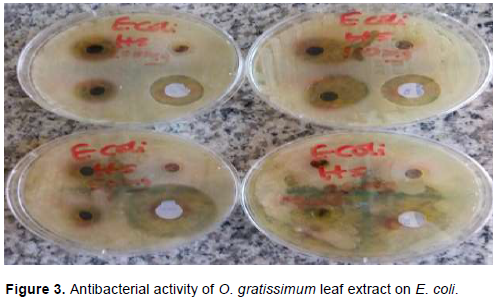
Fresh samples of O. gratissimum leaves were collected from Niger State Polytechnic Staff Quarter, Wushishi Local Government, Niger State, on coordinate 9° 48’N 6° 9’ E and elevation 149 m (489 ft) on 16 October, 2017. The plant was identified and authenticated in the Department of Biological Sciences, Niger State Polytechnic, Zungeru. The fresh leaves were dried completely for two weeks at room temperature (Figure 2) and were blended into powder form using an electric blender.
Extraction process
The plant extraction was obtained using reflux extraction technique as described by Abdullahi (2012) and Sofowora (1993). 250 g of the blended plant leaf powder was weighed and wrapped in Whatman No. 1 filter paper and placed in the holding chamber of the reflux extractor. 600 ml of methanol was used for the extraction at 40°C for 48 h. Thereafter, the extract was concentrated by evaporating to dryness using water bath. The dark green coloured solid extract of the O. gratissmum was stored in an airtight container at 4°C in a refrigerator.
Qualitative phytochemical analysis of the methanolic extract of O. gratissmum
The qualitative and quantitative phytochemcial screening of the methanolic leaf extract were carried out following standard procedures according to the method described by Abdullahi (2012) and Okwu (2005) at the Centre for Genetic Engineering and Biotechnology Laboratory, Federal University of Technology Minna, Niger State.
Determination of antibacterial activity of the plant extract
Reconstitution of the plant extract
The methanol plant extracts were reconstituted by weighing 0.2, 0.1, 0.050 and 0.025 g each into different sterile test tubes containing 1 ml of dimethyl sulphoxide (DMSO), respectively to obtain the following concentration of the extract: 0.2, 0.1, 0.05, and 0.025 g/ml.
Test for sterility of the plant extract
The plant extracts were tested for sterility by introducing 1 ml of the reconstituted plant extract into 5 ml of sterile nutrient broth and were incubated at 37°C for 24 h. The absence of turbidity of the broth (as compared to MacFarland standard) after incubation indicated that the extract was sterile (Preethi et al., 2010).
Collection and confirmation of test organisms
The pure clinical isolates of some pathogenic gastrointestinal bacteria belonging to the family Enterobacteriaceae: Escherichia coli, Salmonella spp. and Shigella spp. were obtained from the Department of Medical Microbiology, Kaduna State University, Kaduna State, Nigeria. All the isolates were checked for purity and confirmed by Gram staining and sub culturing them on selective media (Salmonella shigella Agar and MacConkey Agar); thus, observing the colony characteristics and morphology of the cells.
Antibacterial susceptibility assay
Agar well diffusion techniques were employed for the antimicrobial testing of the plant extracts. The 24 h old cultures were transferred into nutrient broth and incubated at 37°C for 5 h and standardized to Macfarland standard. Each of the test organisms from the broth cultures were streaked on 4 different Mueller Hinton agar plates under aseptic condition and were labeled accordingly.
Wells of approximately 5 mm in diameter were made on the surface of the inoculated agar medium using a sterile cork borer no. 1 and the wells labeled with a marker based on the concentration of the plant extract (0.2, 0.1, 0.05, and 0.025 g/ml) and the wells were filled with the different concentration of the extract. DMSO and ciprofloxacin were used as the negative and positive control, respectively as described by Lino and Deogracious (2006).
The plates were incubated at 37°C and the susceptibility of the test organisms to the plant extract were recorded after 24 h by measuring the average diameter of the clear zone of inhibition in millimeters (mm).
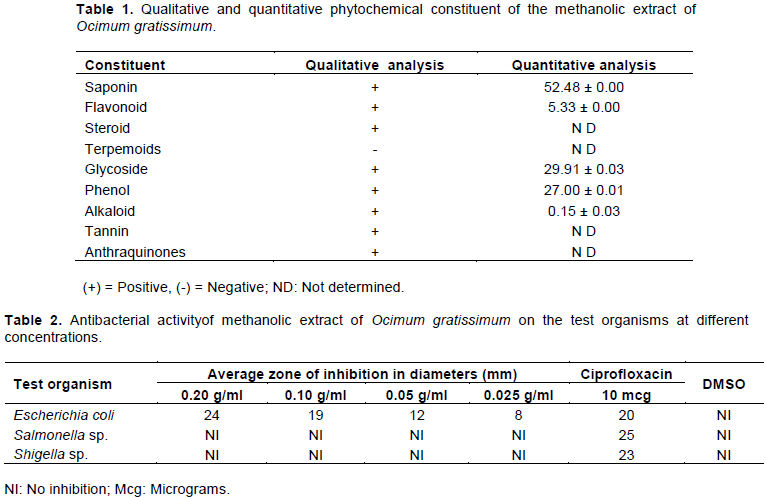
The result of qualitative phytochemical analysis of methanolic extract of O. gratissimum leaves revealed the presence of eight classes of secondary metabolites. However, the quantitative phytochemical analysis conducted on five classes of the identified metabolites showed that saponin has the highest phytochemical constituent (52.48 mg/g) of the extract, followed by tannin (29.91 mg/g), phenol (27.00 mg/g), flavonoid (5.33 mg/g) and alkaloid (0.15 mg/g) shown in Table 1. However, quantitative phytochemical analysis for anthraquinones, tannin, terpemoids and steroid were not determined.
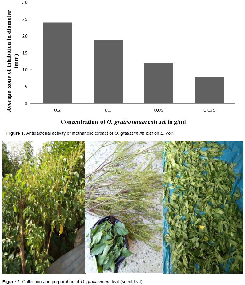
The result of antibacterial activity of methanolic extract of O. gratissimum on E. coli, Salmonella spp. and Shigella spp. shown in Table 2, E. coli was sensitive to the extract while Salmonella spp. and Shigella sp. were resistant to the plant extract and there was no zone of inhibition of DMSO against the test organisms. However, the lower the concentration of the extract (from 0.20 to 0.025 g/ml), the lower the average zone of inhibition (from 24 to 8 mm) against E. coli.
The result of antibacterial activity of methanolic extract of O. gratissimum on E. coli shows decreased average zone of inhibition with decreased concentration of the extract (Figure 1), that is, the lower the concentration, the lower the antibacterial activity.
The extraction of O. gratissimum leaf was obtained using methanol as solvent and the phytochemical analysis of the leaf extract revealed the presence of alkaloid, anthraquinone, flavonoid, glycoside, phenol, saponin, steroid and tannins which agrees with the finding of Ladipo et al. (2010) that shows that O. gratissimum contains all the phytochemicals mentioned earlier.
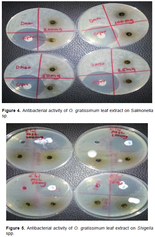
The antibacterial analysis of the methanolic extract of O. gratissimum (Figure 3) revealed that the extract has antibacterial activity against E. coli shown in Table 2. This could be as a result of the presence of one of the phytochemicals or the interaction of two or more of the bioactive compounds against the test organism which agreed with the work of Abdullahi (2012) and also supported the traditional uses of the plant in the treatment of variou s bacterial enteric diseases such as diarrhea, dysentery and other gastrointestinal infections (Nwinyi et al., 2009). Hence, from the result obtained, there was decreased in antibacterial activity with decreased in concentration of the extract as shown in Figure 1; as the concentration of the extract decreases from 0.20l to 0.025 g/ml, the average zone of inhibition also decreased from 24 to 8 mm. This also agreed with the finding of Ishiwu et al. (2014) who demonstrated that increase in the concentration of O. gratissimum extract reduces the number of viable E. coli from 36 to 5 cfu/ml.
However, there was no antibacterial effect of the extract on Salmonella (Figure 4) and Shigella spp. (Figure 5). This resistance may be due to the lipid content on the membranes of these bacteria that prevented the permeability of the active phytochemicals into the cell or the low quantity of alkaloid (0.15±0.03) extracted with methanol (Abdullahi, 2012). However, this disagreed with the work of Ladipo et al. (2010), who reported that the methanol extract of O. gratissimum leaf has antibacterial activity against Salmonella, Shigella and Klebsiella spp. But Justina and Solomon (2017) suggested that difference in phytochemical constituent of the leaf of O. gratissimum could be as a result of the planting location, seasonal and environmental variations. This could also have effect on the antibacterial activity of the leaf extract on Salmonella and Shigella spp. Therefore, the leaf extract of O. gratissimum could serve as natural antibacterial agent and herbal drug against gastroenteritis.
The results obtained in this study, suggest that Escherichia coli was susceptible to the plant leaf extract and the methanolic extract of O. gratissimum leaf contains phytochemicals that possess antibacterial property and could be used as potential source of natural product in industrial manufacturing of drugs, for the treatment of infections/diseases caused by E. coli. However, the toxicity and the side effects of the plant leaf extract should be determined even if the plant is consumed locally.
The authors have not declared any conflict of interests.
REFERENCES
|
Abdullahi M (2012). Phytochemical constituents and antimicrobial and grain protectant activities of Clove Basil (Ocimum gratissimum L.) grown in Niger. Journal of Plant Research 2(1):51-58.
|
|
|
|
Akinjogunla OJ, Adegoke AA, Udokang IP, Adebayo-Tayo B (2009). Antimicrobial potential of Nymphaea lotus (Nymphaeaceae) against wound pathogens. Journal of Medicinal Plants Research 3(3):138-141.
|
|
|
|
|
Effraim KD, Jacks TW, Sopio OA (2013). Histopathological Studies on the Toxicity of Ocimum Gratissimum Leaves Extracts on Some Organs of Rabbits. African Journal of Biomedical Research 6:21-25.
|
|
|
|
|
Ishiwu CN, Umenwanne CP, Obiegbuna JE, Uchegbu N (2014). In vitro assessment of antibacterial effect of extracts of Ocimum gratissimum and carica papaya leaves. International Journal of Applied Science and Technology 1(4).
|
|
|
|
|
Justina YT, Solomon AM (2017). Proximate, Phytochemical, and In-Vitro Antimicrobial Properties of Dried Leaves from Ocimum gratissimum. Nutrition and Food Science 22(3):191-194.
|
|
|
|
|
Kotloff KL, Nataro JP, Blackwelder WC (2013). Burden and aetiology of diarrhoeal disease in infants and young children in developing countries): a prospective, case-control study. The Lancet 382(9888):209-222.
Crossref
|
|
|
|
|
Ladipo MK, Doherty VF, Kanife UC (2010). Phytochemical Screening and antibacterial Investigation of The Extract of Ocimum gratissimum (Scent Leaf) On Selected Enterobacteriaceae. PAT 6(2):75-84.
|
|
|
|
|
Lino A, Deogracious O (2006). The in vitro antibacterial activity of Annonnas enegalensis, Sacuridecae longipendiculata and Steganotaema araliacea. The Journal of African Health Sciences 6(1):31-35.
|
|
|
|
|
Nwinyi OC, Chinedu NS, Ajani OO, Ikpo CO, Ogunniran KO (2009) Antibacterial effects of extracts of Ocimum gratissimum and Piper guineenseon on Escherichia coli and Staphylococcus aureus. African Journal of Food Science 3(3):071-081.
|
|
|
|
|
Okigbo RN, Igwe M (2007). The antimicrobial effects of Piperguineense uziza and Phyllantusamarus ebe-benizo on Candida albicans and Streptococcus faecalis. Acta microbiologica et immunologica Hungarica 54(4):353-366.
Crossref
|
|
|
|
|
Okwu DE (2005). Chemical Composition of Spondias Mombia Linn Plant Parts. Journal of Sustainable Agriculture and the Environment 6:140-147.
|
|
|
|
|
Opara A, Egbuobi R, Dike J, Ndudim E, Onyewuchi C, Nnodim J (2014). Antibacterial Activity of Ocimum gratissimum (Nchu-Anwu) and Vernonia amygdalina (Bitter-Leaf). British Biotechnology Journal 4(10):1115-1122.
Crossref
|
|
|
|
|
Prabhu KS, Lobo R, Shirwaikar AA, Shirwaikar A (2009). Ocimum gratissimum: A Review of its Chemical, Pharma-cological and Ethnomedicinal Properties. The Open Complementary Medicine Journal 1:1-15
Crossref
|
|
|
|
|
Preethi RM, Devanathan VV, Loganathan M (2010). Antimicrobial and antioxidant efficacy of some medicinal plants against food borne pathogens. Advanced Biomedical Research 4:122-125.
|
|
|
|
|
Ram PK, Crump JA, Gupta SK, Miller MA, Mintz ED (2008). Analysis of Data Gaps Pertaining to Shigella Infections in Low and Medium Human Development Index Countries 1984–2005. Epidemiology and Infection 136(5):577-603.
Crossref
|
|
|
|
|
Russell JB, Jarvis GN (2011). Practical mechanisms for interrupting the oral-fecal life cycle of Escherichia coli. Journal of Molecular Microbiology and Biotechnology 3(2):265-272.
|
|
|
|
|
Sofowora LA (1993). Medicinal plants and traditional Medicinal Medicine in African Spectrum books Ltd, Ibadan. pp. 55-71.
|
|
|
|
|
Suree N, Pana L (2015). Antibacterial activity of crude ethanolic extracts and essential oil of spices against Salmonella and other Enterobacteriacea. Science Technology Journal 5(3):527-538.
|
|
|
|
|
Su L, Chiu CH (2007). Salmonella: Clinical Importance and Evolution of Nomenclature. Chang Gung Medical Journal 30(3):210-219.
|
|
|
|
|
World Health Organization (WHO) (2002). Traditional Medicine: Growing Needs and Potential. WHO Policy Perspectives on Medicines. World Health Organization, Geneva pp. 1-6.
|
|