Full Length Research Paper

ABSTRACT
Drug resistant and opportunistic organisms are a problem to medical health due to the fact that most of the drugs that were used are now not effective. Currently, there is a need to search for new drugs that can enhance the control of these organisms. Actinomycetes and their secondary metabolites can be used as such drugs. This study was designed to isolate actinomycetes producing novel anti-fungal compounds from waste dump soil in Western Uganda. Fifty six (56) actinomycetes were isolated from 22 waste dump soil samples. All isolates were screened using modified spektra - plak method against Candida albicans ATCC1023, Penicillium sp., Aspergillus sp., Fusarium sp. and Rhizopus sp. Eight 8(14.29%) isolates showed antifungal activity to at least one test fungi during primary screening. Two isolates [KBRWDSa (FR) and KBMWDSb6] showed activity to all test fungi. Secondary screening was carried out by growing all 56 isolates in broth and their supernatant was tested for antifungal activity using agar well diffusion method. 11(19.64%) of these isolates showed activity against at least one test fungi with mean zone of inhibition 5.33 to 29.69 mm. Isolate KBMWDSb6 showed a broad spectrum activity against all test fungi. The remaining broths were extracted using ethanol. The ethanol extract at 2.5 mg/ml concentration was also tested for antifungal activity using agar well diffusion method. 13 (23.21%) isolates showed activity against at least one test fungi with mean zone of inhibition 6.33 to 30.67mm. The findings showed that some of these isolates had antifungal activity.
Key words: Waste dump soil, actinomycetes, novel anti-fungal compounds, Western Uganda.
INTRODUCTION
Infections caused by drug resistant and opportunistic fungi especially among immuno-compromised patients are now a global concern; this has caused a substantial morbidity and mortality (CDC, 2016). Most of these fungi developed resistance to commonly prescribed drugs (CDC, 2016; Howard et al., 2009). Chowdhary et al. (2013) reported that, Candida auris was the new emerging drug resistant fungus found in Japan (Satoh et al., 2009) and South Korea (Oh et al., 2011). This fungus causes fungemia and was reported to be resistant to fluconazole (Chowdhary et al., 2013). However, other fungi like Aspergillus, Fusarium, and Cryptococcus species have been reported to be resistant to the azole group of antifungal compounds (Arendrup, 2014). Patients infected by these drug resistant fungi have poorer outcomes than those affected by drug susceptible fungi (Baddley et al., 2008; Lortholary et al., 2011). It was also reported that patients with fungemia stay longer in the hospital and consumed more healthcare facilities (Morgan et al., 2005; CDC, 2016). The long stay in hospitals affects the economy of the patient’s family specifically and the concerned nation generally. When the population of Uganda was 35 million, 1.1 million had HIV and about 9.2% (101,000) had a CD4 count <200 cells/μL out of which, 2783 developed Cryptococcal meningitis per year with approximated mortality rate of 2086 per year (Parkes-Ratanshi et al., 2013). Parkes-Ratanshi et al. (2013) also added that, fungal infections were estimated to be 1 million cases per year with exclusion of Tinea capitis. There are reports on increasing rate of motility due to fungal infections which was related to emergence of antifungal resistance (Smith et al., 2015). These put more demands for search of new antifungal compounds to face these global challenges. Isolation and characterization of microorganisms from the most extreme habitat and unstudied areas or geographical location is one of the ways to discover novel antimicrobial agents as recommended by action-mycetologist (Monisha et al., 2011; Jagan et al., 2014). Actinomycetes are considered to be one of the golden microbes in the 20th century due to their ability to produce different kinds of bioactive compounds which includes antifungal agents (Lakshmipathy and Krishnan, 2010; Ensieh and Maryam, 2016; Rotich et al., 2017). Actinomycetes have produced 80% of the discovered antibiotics (Masna et al., 2016). Actinomycetes are Gram positive bacteria, widely distributed in soil and most abundant microbes in the soil (Jemimah et al., 2012). In Uganda, there is paucity of data on production of antifungal agents from soil actinomycetes (Nalubega et al., 2016). Therefore, this study was designed to isolate and screen actinomycetes from waste dump soil, from Western Uganda for novel antifungal compound(s).
MATERIALS AND METHODS
Study area and design
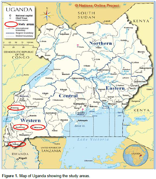
The samples were collected from two different temperate sites, cold areas (Bushenyi and Kabale districts) and warm areas (Kasese and Mbarara districts) Western Uganda (Figure 1). The study was experimental laboratory research that involved isolation, Identification and screening of actinomycetes for production of novel antifungal compounds.
Sample collection
A total of 22 waste dump soil samples were collected from market waste dump soil, residential waste dump soil and manure plantation farm soil. Samples were collected from two different temperate areas: cold (Bushenyi 6 samples and Kabale 6 samples) and warm (Kasese 4 samples and Mbarara 6 samples) regions of Uganda.
Two plots (120 × 120 cm) were mapped out from each sampling sites and three samples (3 to 15 cm depth) were collected randomly from each of the plots using a sterile stainless spoon with hand core and sterile gloves to avoid contamination. The collected soils were mixed to have one representative sample per plot (Rinoy et al., 2012; Ekeke and Okonwu, 2013). The samples were placed in sterile polythene bags (Ismail et al., 2015) and transported to the Microbiology laboratory at Department of Microbiology and Immunology Kampala International University Western campus, Uganda for further study. The Geographical location, soil temperature of each sampling sites were recorded during samples collection while moisture content and pH of each sample was determined immediately using oven dried method and digital pH meter, respectively (Rinoy et al., 2012).
Isolation of actinomycetes
Isolation of actinomycetes was carried out according to the method described by (Arifuzzaman et al., 2010; George et al., 2012). Two grams (2 g) of each soil sample was air dried for 10 days at room temperature, and approximately 1g of air dried soil sample was suspended in 9 ml of sterile distilled water supplemented with 0.9% of NaCl and incubated in a Gas bath thermostats oscillator (THZ-82B) for 1 h at 200 rpm and 55°C.
The suspension was serially diluted (10-1 to 10-7). Hundred microliter (100 mL) from dilution 10-2 was spread on starch casein nitrate agar (Composition in media g/L: Starch 10, Casein 0.3, KNO3, NaCl2, K2HPO4 2, MgSO4.7H2O 0.5, CaCO3 0.02, FeSO4.7H2O Agar 18 pH adjusted 7 ± 0.2) (Sengupta et al., 2015), glycerol casein agar (Kuster agar) (Composition in media g/L: Glycerol 10, casein 0.3, KNO3 2, K2HPO4 2, MgSO4 0.05, CaCO3 0.02, Fe2 (SO4)3.6H2O 0.01, Agar 15 and pH was adjusted to 7 ± 0.2 ) (Lakshmanaperumalsamy et al., 1984) and yeast extract starch casein agar (YSCA) (Composition in media g/L: yeast extract 3, peptone 3, casein 3, starch 8, K2HPO4 0.5, MgSO4.7H2O 0.5, NaCl 2, agar 15 and pH 7.0 to 7.6 ) (Mincer et al., 2002).
The inoculated plates were incubated at 28°C for 7 to 14 days. Colonies with limiting growth, appeared dry powdery or velvety, tough leathery or chalky texture; dry or folded and branching filamentous with or without aerial mycelia and clear zone of inhibition were chosen and sub-cultured on clean starch casein nitrate agar plates to obtain pure cultures (Oskay et al., 2004). The pure cultures were maintained on starch casein nitrate agar at 4°C for short storage and 30% glycerol at -80°C for long storage (Oskay et al., 2004; Madigan et al., 1997).
Production of anti-fungal compounds
Test organisms
The test fungi were standard for Candida albicans ATCC1023, which was obtained from the Department of Medical microbiology Mekerere University, Kampala Uganda, while Aspergillus sp, Fusarium sp., Rhizopus sp. and Penicillium sp. were obtained from the Department of Microbiology and Immunology, Kampala International University Western Campus, Uganda.
Primary screening
Antifungal activities of isolates were tested by modified spektra-plak method (Oskay et al., 2004; Madigan et al., 1997). Plates with potato dextrose agar supplemented with (g/l : K2HPO2 0.5, CaCO3 0.75, MgSo4.7H2O 0.5, NaCl 2 and pH adjusted to 7 ± 4) were inoculated with 7 days old actinomycetes cultures by forming a circle of two to four different actinomycetes isolates on a plate and incubated for 7 days.
72 h cell and spores of tests fungi were inoculated and incubated for another 3 days. Antifungal activity was observed by formation of inhibition zone.
Secondary screening
Fermentation: All isolates were subjected to fermentation; this was to confirm the ability to produce bioactive compounds in solid and liquid media (Ensieh et al., 2015). Fermentation was carried out by the submerged culture in Erlenmeyer flask (500 ml).
The 7 days old culture of actinomycetes was inoculated in yeast extract starch broth (g/l: yeast extract 3, Peptone 3, Casein 3, Starch 8, Glycerol 3, CaCO3 0.75, K2HPO2 0.5, MgSO4.7H2O 0.5, NaCl 12 and pH 7.4) and incubated in gas bath thermostats oscillator (THZ-82B) at 28°C and 200 ± 5 rpm for 7 days after which the broth was centrifuged at 3000 rpm for 20 min and filtered using filter paper (Whatman No 1) (Sohan et al., 2015).
Antifungal activity of fermented broth: Agar well diffusion method was employed to assess the antifungal activity of the fermented filtered broth. Cell concentration of C. albicans was adjusted at 0.5 McFarland turbidity standards and inoculated on potato dextrose agar plate while Aspergillus sp, Penicillium sp, Rhizopus sp and Fusarium sp were grown on Potato dextrose agar (PDA) for 72 h and spores were collected and inoculated on fresh PDA medium.
Wells were bored by sterile 1000 μl micropipette tip (Hotam et al., 2013). The wells were filled with 200 μl of supernatant of centrifuged broth and the plates were incubated at 28°C for 72 h. Amphotericin B at 50 μg/ml was used as positive control. All experiments were performed in triplicates.
Extraction of bioactive compounds: The fermented broths were centrifuged and filtered. The filtered broths were extracted using a solvent by adding equal volume (1:1) of ethanol (95%). The solution was shaken vigorously on a rotatory shaker for 24 h. The solvent phase was collected and evaporated in hot air oven at 40°C. The extracts were dried and stored at 4°C for further studies (Hotam et al., 2013; Raja and Prabakaran, 2011).
Antifungal activity of the solvent extract: The dried extracts were re-dissolved in 2.5% dimethyl sulphoxide (DMSO) at concentration of 2.5 mg/ml and antifungal activity was determined by agar well diffusion method. Cell concentration of C. albicans was adjusted at 0.5 McFarland turbidity standards and inoculated on PDA plates using sterilized cotton swabs. While Aspergillus sp, Penicillium sp, Rhizopus sp. and Fusarium sp. were grown on PDA for 72 h and spores were collected and inoculated on fresh PDA medium, wells were bored using sterile 1000 μl micropipette tip (Hotam et al., 2013).
The wells were filled with 200 μl of 2.5 mg /ml of the extract and the plates were incubated at 28°C for 72 h. Amphotericin B at 50 μg/ml was used as positive control. DMSO 2.5% was also used to serve as negative control. All experiments were performed in triplicates.
Identification of actinomycetes
The active actinomycetes isolates that showed activity during primary and secondary screening were identified using macroscopic, microscopic and biochemical methods.
Macroscopic study
The macroscopic features of the active isolates observed were colony colour, aerial mycelium, substrate mycelium, pigment production and texture.
Microscopic study
Surface appearances of the selected actinomycetes isolates were studied using dissecting microscope. While spores arrangements were studied using slide-culture method, blocks of Starch casein nitrate agar were cut and placed on sterile glass slides. The active actinomycetes isolate was inoculated on the block by streaking over the agar block surface, a cover slip was placed over the block, and the entire set up was incubated at 28°C for 7 days. The cover slip was removed and stained using Gram’s staining techniques.
Cover slip was covered with crystal violet for 60 s and washed off with water, followed by Gram's iodine for 60 s, decolorized with alcohol for 05 s, and washed with water. Finally cover slip was stained with safranin counter stain for 1 min. After washing and drying, the test was microscopically observed under high power using phase-contrast microscope (X100) (Hotam et al., 2013; Kekuda et al., 2012).
Biochemical characterization
Gelatin hydrolysis: Gelatin test was carried out using methods described by (Sundaramoorthi et al., 2011). Seven day old culture of active isolate was stabbed into nutrient gelatin tubes, using sterile inoculating needle. The tubes were incubated for 10 days at 30°C. Un-inoculated tube was used as a control.
After incubation, the tubes were placed in to refrigerator, at 4°C for 15 min. The refrigerated gelatin tubes that remained liquefied were considered positive test or solid to confirm negative test.
Starch hydrolysis: Starch hydrolysis test was carried out according to methods described by (Remya and Vijayakumar, 2008). Starch agar medium composed of (g/l): soluble starch, 20; yeast extract, 3; Peptone, 5; Agar 15 was prepared and autoclaved. Seven day old culture was inoculated in the medium and incubated at 30°C for 7 days. An un-inoculated plate was used as a control.
At the end of the incubation period, iodine solution was flooded on the plates to observe the clear zone of hydrolysis around the colony.
Esculin degradation: Esculin degradation test was conducted according to the method adopted by (Tiwarty, 2009). Seven day old culture was inoculated into Esculin agar slants (yeast extract, 0.3; ferric ammonium citrate, 0.05, agar 0.75, 0.1% of esculin and distilled water 50 ml) and incubated at 30°C for seven days. The test tube was observed for blackening the medium.
Methyl red – Voges – Proskauer test (MR-VP): Methyl red – Voges – Proskauer test (MR-VP) was carried out according to the method described by (Cheesebrough, 2006). It was used to determine the ability of the organism to ferment glucose with production of acid. Five millilitres (5 ml) of MR-VP broth was inoculated with the test organism and incubated for 48 to 72 h at 37°C.
After incubation, 2 to 3 drops of methyl red test was added to 1ml of the broth. A red colour signified a positive methyl red test while yellow colour signified negative test. To what remained, five drops of 4% potassium hydroxide (KOH) was added followed by fifteen drops of 5% α –naphthol in ethanol. The development of red colour within 1 h indicates VP positive test while no colour change indicated VP negative test.
Catalase test: Catalase test was carried out according to the method described by Cheesebrough (2006), to determine the ability of the isolate to produce the enzyme, catalase. A drop of 3% hydrogen peroxide was added to a loop full of the test organisms. Presence of bubbles indicates catalase activity.
Indole test: Indole test was carried out according to the method described by Cheesebrough (2006), to determine the ability of the isolate to degrade amino acid tryptophan and produce tryptophanase, enzyme was tested. 1% tryptophan broth in a test tube was inoculated with 7 days isolate and incubated at 37°C for 48 h.
After 48 h, 1 ml of chloroform was added to the broth. The test tube was shaken gently, and 2.1 ml of Kovac’s reagent was added and again shaken gently, this was allowed to stand for 20 min. The formation of red coloration at the top layer, indicate positive while yellow coloration indicate negative.
Urease test: Urease test was carried out according to the method described by Cheesebrough (2006) to determine the ability to hydrolyse urea to produce ammonia and carbon dioxide. Test organism was inoculated into urease broth and incubated at 30°C for 72 h. Purplish pink coloration of the medium indicates positive reaction.
Nitrate test: Nitrate test was carried out according to the method described by Cheesebrough (2006), to determine the ability to hydrolyse nitrate to nitrite. Nitrate broth was inoculated with a loopful of active isolate and incubated at 28°C for 7 days. Un-inoculated test tube was used as a control. Two drops of sulphanilic acid and α – napthylamine solution was added to the broth. Presence of red colour indicates positive reaction.
Triple sugar iron test: Triple sugar iron test was carried out according to the method described by (Vlab, 2011); the test determines the ability of the organism to ferment the three sugar component of the medium: glucose, lactose and sucrose. The medium contains a pH indicator (phenol red) and a detection system (thiosulphate and ferrous sulphate) for hydrogen sulphide (H2S). The medium was prepared as an agar slant.
The test organism was inoculated by stabbing the medium using sterilized straight wire loop and the surface of the slope was also streaked with the test organism. The test was incubated at 37°C for 3 days. After incubation, gas production was determined by observing the cracking of the medium, and production of H2S was observed by the blackening of the butt (bottom) of the medium.
Glucose fermentation was determined by yellowing of the butt, the fermentation of lactose or sucrose or both was determined by the yellowing of both butt and slant.
Citrate utilization: This was carried out by inoculating the test organism in test tube containing Simon’s citrate medium and incubated for 24 to 72 h. The development of deep blue colour after incubation indicated a positive result (Cheesebrough, 2006).
Ethical approval
Ethical approval of the study was obtained from Kampala International University (KIU), Institutional Research and Ethics Committee (IREC). All experiments were performed in accordance to the ethical standards of the microbiology laboratory operation.
Data analysis
Data was analysed using past software (Version 3.14). Pearson’s correlation coefficient was used to compare between environmental factors (temperature, percentage moisture content and pH) with percentage colonies distribution, value at p ≤ 0.05 was considered to be significant. One way analysis of variance (ANOVA) was used to compare between antifungal activity of fermented broth and ethanol extract, p ≤ 0.05 was considered to be significant.
RESULTS AND DISCUSSION
Fifty six (56) actinomycetes isolates were isolated from 22 waste dump soil samples collected from four studied areas in Western Uganda. The samples were collected from market waste dump soil, residential waste dump soil and manure plantation farm. Three media (Starch casein nitrate agar, Glycerol casein agar and Yeast extract starch casein agar) were used for the isolation of actinomycetes (Figure 2). Glycerol casein agar was found to support high growth of actinomycetes followed by yeast extract starch casein agar (Result not shown). Although starch casein nitrate agar showed less growth, it which was chosen for the storage of pure isolates due to its ability to support the growth of both actinomycetes isolated from different media.
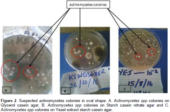
Table 1 below showed the results of pH, temperature, percentage moisture, colonies and percentage colonies distribution from the studied areas. The pH value ranged from 7.32 to 8.63 which are slightly alkaline, temperature was 18 to 57°C, percentage moisture content was 33 to 93% and colony and percentage colonies distribution ranged from 1 to 6 and 1.79 to 10.71%, respectively. Although the studied areas have high percentage moisture content and lower temperature, significant number of actinomycetes isolates (56) were isolated. There was positive weak correlation between percentage colonies distribution with moisture and negative weak correlation with temperature, but it was not statistically significant (r = 0.146, p ≤ 0.05 and r = - 0.053, p ≤ 0.05, respectively). This was in accordance to the findings of Moselio and Joshua (2004) and George et al. (2012), who reported that environmental factors like temperature and moisture can affect the distribution of actinomycetes and tend to be abundant in wasteland than the moist soil. However, this was contrary to the findings of Rinoy et al. (2012), who reported negative correlation between actinomycetes loads and moisture content and positive correlation between actinomycetes loads and temperature.
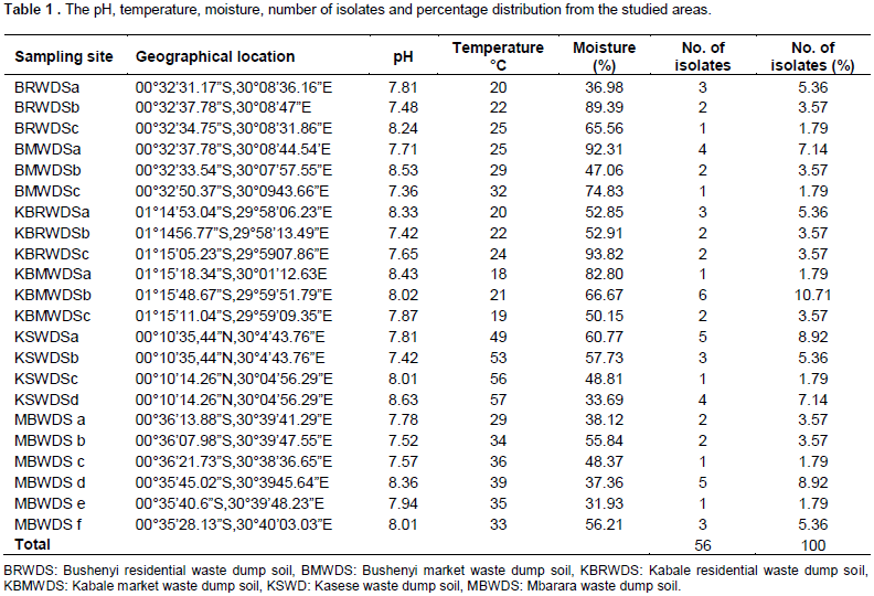
The colonies and percentage colonies distribution were found to have a positive weak correlation with pH (which is slightly alkaline) of the samples but, was not statistically significant (r = 0.206, p ≤ 0.05). This was in line with findings of Basilio et al. (2003) and Rinoy et al. (2012), who reported that actinomycetes distribution is affected by pH and its desired alkaline pH condition than acidic environment. Basilio et al. (2003) also added that, actinomycetes loads drop at pH less than 5 which is acidic.
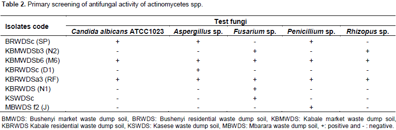
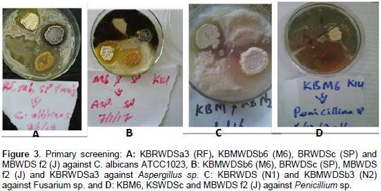
Eight actinomycetes isolates 8 (14.29%) showed antifungal activity to at least one test organism during primary screening. The isolates KBRWDSa (FR) and KBMWDSb6 showed antifungal activities to all test fungi (Table 2 and Figure 3). This result showed that 85.71% of the cultured actinomyctes isolates did not produce bioactive compounds in the solid medium. This could be due to the lacks of nutrients requirement or their cell structure as some actinomycetes have been reported not to produce bioactive compounds on the solid medium or the bioactive compound produced in the solid medium may be inactive due to presence of polar or nonpolar functional groups in the metabolites which may require particular amount of polar or nonpolar solvents to dissolve. This finding was similar to the previous studies carried out by different researchers where higher percentage number of the actinomycetes isolates subjected to primary screening failed to shows activity against test organism (Jemimah et al., 2012; Sohan et al., 2015; Alireza et al., 2010; Tara et al., 2009).
All actinomycetes isolates (56) were subjected to secondary screening involving two parts: Testing for antifungal activity of shake flask filtered broths and ethanol extracts. The results from the shake flask broth showed that some isolates that did not have activity during primary screening had activity after subjecting them into liquid broth culture and vice versa. Eleven actinomycetes isolate (19.64%) showed activity on at least one tested fungus (Table 3). The actinomycetes isolate KBMWDSb (M6) maintained its activity to all test fungi. The mean standard error antifungal activity of shake flask culture broth ranged from 5.33 ± 0.88 to 29.67 ± 0.88 mm. Although isolate KBMWDSb (M6) produced activity to all test organisms, isolates KBRWDSc (D1) produced the highest mean standard error zone of antifungal activity against C. albicans ATCC1023 (29.67 ± 0.88 mm).
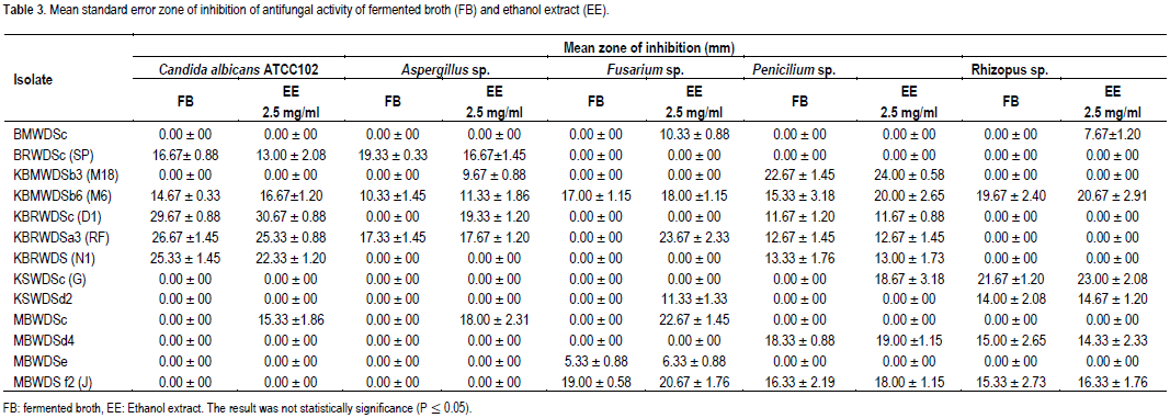
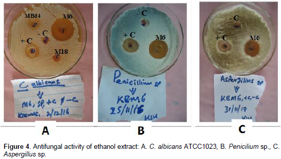
The inability of isolate KBRWDSa (FR) to maintain its broad spectrum antifungal activity could be as result of cell structures or the bioactive compound produced in the liquid broth, contained non polar functional group which could not be dissolved in polar solvent (Tara et al., 2009). The result of solvent extract showed that some shake flask culture broth that failed to shows activity during screening, had activity when solvent was used for extraction of bioactive compounds. Thirteen (23.21%) actinomycetes isolates produced activity to at least one test organism (Table 3 and Figure 4).
The mean standard error zones of inhibition value of ethanol extract ranged from 6.33 ± 0.88 to 30.67 ± 0.88 mm. Although some fermented broth showed activity after solvent was used for extraction, there was a decreased in the zone of inhibition of antifungal activity of some fermented broth after ethanol was used for extraction. This could be as result of extraction processes, inability of solvent (ethanol) to extract the bioactive compounds or bioactive compounds were missing from ethanol segment due to presence of nonpolar compounds which could not dissolve in ethanol which is a polar solvent (Tara et al., 2009; Jirayut et al., 2012). Comparison between fermented broth and ethanol extract mean zone of inhibition using one way ANOVA showed no significant difference at (p ≤ 0.05) between the two extracts.
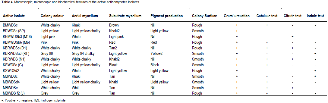
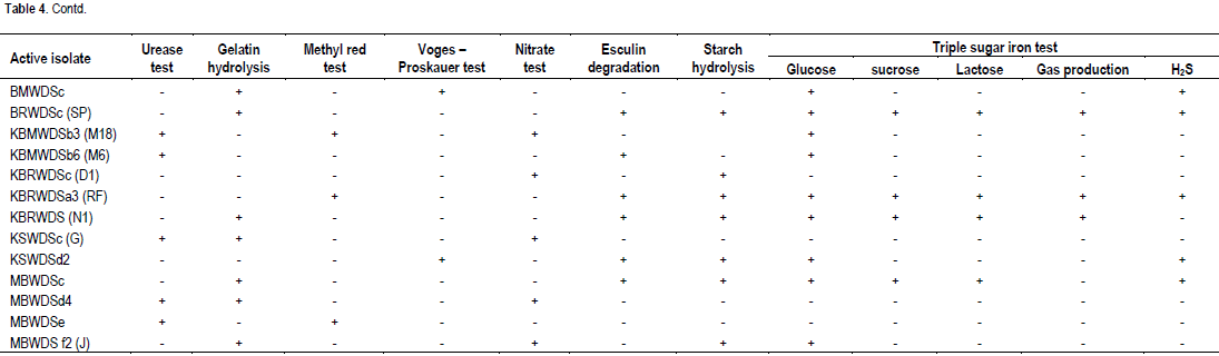
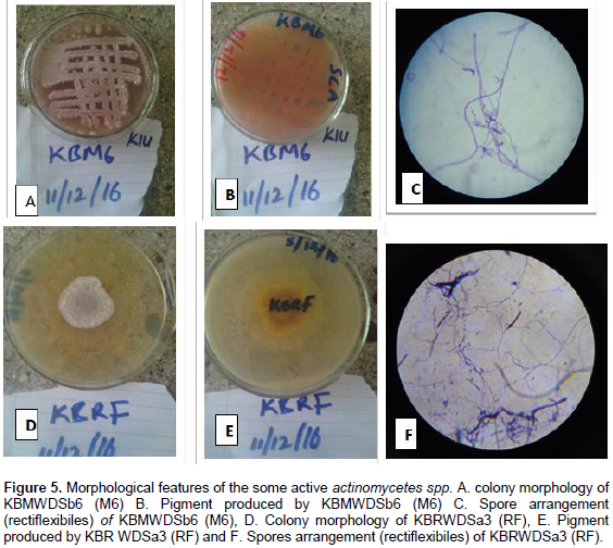
Macroscopic, microscopic and biochemical features of the active actinomycetes isolates was presented in Table 4 and Figure 5. The macroscopic features of the isolates showed that majority were white chalky, grey and pink in colour, smooth to rough surface colonies with aerial and substrate mycelium and pigment production. Micro-scopically, the isolates were all Gram positive and had filamentous structures. The biochemical and phenotypical features of the isolates showed similarity to the genera Actinomycetes as described in Bergey’s manual of determinative bacteriology 8th edition (Buchanan et al., 1974).
CONCLUSION
This finding shows that, waste dump soil of Western Uganda could be used as a rich source to explore novel actinomycete strains producing antifungal compounds. Among the isolates obtained, isolate KBMWDSb6 (M6) produced broad spectrum activity against all test fungi. This organism will be subjected to further studies in order to discover its novelty as an antifungal agent.
CONFLICT OF INTERESTS
The authors have not declared any conflict of interests.
REFERENCES
|
Alireza D, Laleh PY, Rouhollah B, Ahad M, Samad A, Ali RM, Gasanova S, Rahib A (2010). Investigation antibacterial activity of Streptomycetes isolates from soil samples, West of Iran. Afr. J. Microbiol. Res. 4(16):1685-1693. |
|
|
Arendrup MC (2014). Update on antifungal resistance in Aspergillus and Candida. Clin. Microbiol. Infect. 20(s6):42-48. |
|
|
Arifuzzaman M, Khatun MR, Rahman H (2010). Isolation and screening of actinomycetes from Sundarbans soil for antibacterial activity. Afr. J. Biotechnol. 9:4615-4619. |
|
|
Baddley JW, Patel M, Bhavnani SM, Moser SA, Andes DR (2008). Association of fluconazole pharmacodynamics with mortality in patients with candidemia. Antimicrob. Agents Chemother. 52:3022-3028. |
|
|
Basilio A, Vicente MF, Gorrochategui J, Gonzalez I, Cabella A, Gonzalez A, Genilloud O (2003). Patterns of antimicrobial activities from soil actinomycetes isolated under different conditions of pH and salinity. J. Appl. Microbiol. 95(4):814-823. |
|
|
Buchanan RE, Gibbons NE, Cowan ST, Holt JG, Liston J, Murary RGE, Niven CF, Ravin AW, Stainer eds. Bergey's RY (1974). Manual of Determinative Bacteriology, eighth edn, Williams &Wilkins Company, Baltimore, U.S.A. pp. 659-881. |
|
|
CDC-Centre for Disease Control and Prevention (2016). Fungal diseases: Antifungal resistant. Available at: View Accessed 16/11/2016 |
|
|
Cheesebrough M (2006). District laboratory practice in tropical countries. Part 2. Low Price edition. Cambridge University Press, London. |
|
|
Chowdhary A, Agarwal K, Kathuria S, Singh PK, Roy P, Gaur SN, de Hoog GS, Meis JF (2013). Clinical significance of filamentous Basidiomycetes, illustrated by the novel opportunist Ceriporialacerata isolated from the human respiratory tract. J. Clin Microbiol. 51:585-590. |
|
|
Ekeke C, Okonwu K (2013). Comparative Study on Fertility Status of Soils of University of Port Harcourt, Nigeria. Res. J. Bot. 8:24-30. |
|
|
Ensieh S, Maryam N (2016). Isolation of biologically active Actinomycetes from untouched soils: a case study from Karaj district, Iran. Prog. Biol. Sci. 6(1):65-74. |
|
|
Ensieh S, Mona S, Vida T (2015). Antibacterial activity of some actinomycetes isolated from soils of Alborz province, Iran. Prog. Biol. Sci. 5(2):159-167. |
|
|
George M, Anjumol A, George G, Mohamed HAA (2012). Distribution and bioactive potential of soil actinomycetes from different ecological habitats. Afr. J. Microbiol. Res. 6(10):2265-2271. |
|
|
Hotam SC, Jayprakash YAR, Shrivastava SS, Anil KS, Natrajan G (2013). Antibacterial activity of actinomycetes isolated from different soil samples of Sheopur (A city of central India). J. Adv. Pharm. Technol. Res. 4(2):118-123. |
|
|
Howard SJ, Cerar D, Anderson MJ, Albarrag A, Fisher MC, Pasqualotto AC, Laverdiere M, Arendrup MC, Perlin DS, Denning DW (2009). Frequency and evolution of Azole resistance in Aspergillus fumigatus associated with treatment failure. Emerg. Infect. Dis. 15:1068-1076. |
|
|
Ismail S, Ban A, Racha A (2015). Testing of Production of Inhibitory Bioactive Compounds by Soil Streptomycetes as Preliminary Screening Programs in UAE for Anti-Cancer and Anti-Bacterial Drugs. Int. J. Curr. Microbiol. Appl. Sci. 4:446-459. |
|
|
Jagan MY, Sirisha B, Prathyusha K, Pola SR (2014). Isolation, Screening and Characterization of Actinomycetes from Marine Sediments for their Potential to Produce Antifungal Agents. Glob. J. Biol. Agric. Health Sci. 3(4):131-137. |
|
|
Jemimah NS, Nasimunislam N, Vaishnavi B, Mohanasrinivasan V, Subathra DC (2012). Isolation of Soil actinomycetes inhabiting Amrithi forest for the potential source of bioactive compounds. Asian J. Pharm. Clin. Res. 5:189-192. |
|
|
Jirayut E, Arjaree N, Srisurang T, Takuya N, Yusuhiro I, Watanalai P (2012). Identification and characterization of soil-isolated Streptomyces SJE177 producing actinomycin. Southeast Asian J. Trop. Med. Public Health 41(5):1177. |
|
|
Kekuda PTR, Shobha KS, Onkarappa R, Gautham SA, Raghavendra HL (2012). Screening biological activities of a Streptomyces species isolated from soil of Agumbe, Karnataka, India. Int. J. Drug Dev. Res. 4(3):104-114. |
|
|
Lakshmanaperumalsamy P, Chandramohan D, Natarajan R (1984). Seasonal variation of microbial population from sediments of Vellar estuary, South India. In. 2. Colloque International de Bacteriologie Marine, Brest (France), pp. 1-5. |
|
|
Lakshmipathy D, Krishnan K (2010). Antagonistic activity of Streptomyces VITDDK1 spp. (GU223091) isolated from the coastal region of Tamil Nadu, India. Pharmacologyoline 1:17-29. |
|
|
Lortholary O, Desnos-Ollivier M, Sitbon K, Fontanet A, Bretagne S, Dromer F, French Mycosis Study Group (2011). Recent exposure to caspofungin or fluconazole influences the epidemiology of candidemia: a prospective multicenter study involving 2441 patients. Antimicrob. Agents Chem. 55:532-538. |
|
|
Madigan MT, Martiko JM, Parker J (1997). Antibiotics: Isolation and characterization. In. Brook Biology of Microorganisms, 8th edn. Prentice-Hall International Inc. New Jersey, pp. 440-442. |
|
|
Masna R, Nisha B, Nisha D, Pappu KM (2016). Isolation of antibiotic producing Actinomycetes from soil of Kathmandu valley and assessment of their antimicrobial activities. Int. J. Microbiol. Allied Sci. 2(4):22-26. |
|
|
Mincer TJ Jensen PR, Kauffman CA, Fenical W (2002). Widespread and persistent of population of a major actinomycetes taxon in ocean sediments. Appl. Environ. Microbiol. 68(10):5005-5011. |
|
|
Monisha K, Renu S, Rup L (2011). Selective isolation of rare actinomycetes producing novel antimicrobial compounds. Int J. Adv Biotechnol. Res. 2:357-375. |
|
|
Morgan J, Meltzer MI, Plikaytis BD, Sofair AN, Huie-White S, Wilcox S, Harrison LH, Seaberg EC, Hajjeh RA, Teutsch SM (2005). Excess mortality, hospital stay, and cost due to candidemia: a case-control study using data from population-based candidemia surveillance. Infect. Control Hosp. Epidemiol. 26:540-547. |
|
|
Moselio S, Joshua L (2004). The desk Encyclopedia of Microbiology, Elsevier Academic. Press, California, Amsterdam. |
|
|
Nalubega Fatuma, Meklat Atika, Mellouk Imène, Chaabane Chaouch Fawzia, Verheecke Caro, Bouras Noureddine, Mokrane Salim, Mathieu Florence and Sabaou Nasserdine (2016). Taxonomy and antagonistic activity of A12, a new Streptomyces strain isolated from Ugandan soil. ElWahat pour les Recherches et les Etudes 9 (1):1 - 11 |
|
|
Oh BJ, Shin JH, Kim MN, Sung H, Lee K, Joo MY, Shin MG, Suh SP, Ryang DW (2011). Biofilm formation and genotyping of Candida haemulonii, Candida pseudohaemulonii, and a proposed new species (Candida auris) isolates from Korea. Med. Mycol. 49:98-102. |
|
|
Oskay M, Tamer A, Azeri C (2004). Antibacterial activity of some actinomycetes isolated from farming soils of Turkey. Afr. J. Biotechnol. 3:441-446. |
|
|
Parkes-Ratanshi RB, Achan B, Kambugu A, Meya D, Denning D (2013). Estimated burden of fungal disease in Ugand. 23rd European Congress of Clinical Microbiology and Infectious Diseases. Berlin, Abstract 3635. |
|
|
Raja A, Prabakaran P (2011). Preliminary screening of antimycobacterial effect of psychrophilic actinomycetes isolated from Manali ice point Himachal Predesh. J. Microbiol. Antimicrob. 3(2):41-46. |
|
|
Remya M, Vijayakumar R (2008). Isolation and Characterization of marine antagonistic actinomycetes from west coast of India. Med. Biol. 15:13-19. |
|
|
Rinoy V, Nishamol S, Suchithra R, Jyothy S, Mohamed HD AA (2012). Distribution and Antibacterial Activity of Actinomycetes from Shola Soils of Tropical Montane Forest in Kerala, South India. J. Environ. 1(3):93-99. |
|
|
Rotich MC, Magiri E, Bii C, Maina N (2017). Bio-Prospecting for Broad Spectrum Antibiotic Producing Actinomycetes Isolated from Virgin Soils in Kericho County, Kenya. Adv. Microbiol. 7:56-70. |
|
|
Satoh K, Makimura K, Hasumi Y, Nishiyama Y, Uchida K, Yamaguchi H (2009). Candida auris sp. nov., a novel ascomycetous yeast isolated from the external ear canal of an inpatient in a Japanese hospital. Microbiol. Immunol. 53:41-44. |
|
|
Smith KD, Achan B, Hullsiek KH, McDonald TR, Okagaki LH, Alhadab AA, Akampurira A, Rhein JR, Meya DB, Boulware DR, Nielsen K, ASTRO-CM/COAT Team (2015). Increased antifungal drug resistance in clinical isolates of Cryptococcus neoformans in Uganda. Antimicrob Agents Chemother. Am. Soc. Microbiol. 59:7197-7204. |
|
|
Sengupta S, Pramanik A, Ghosh A, Bhattacharyya M (2015). Antimicrobial activities of actinomyceties isolated from unexflored regions of Sundarbans mangrove ecosystem. BMC Microbiol. 15(1):170. |
|
|
Sundaramoorthi C, Vengadesh PK, Gupta S, Karthic K, Tamilsehli V (2011). Production and characterization of antibiotics from soil–isolated actinomycetes. Int. Res. J. Pharm. 2:114-118. |
|
|
Tara DG, Chringma S, Vishwanath PA, Binod L (2009). Isolation and Characterization of Antibacterial Actinomycetes from Soil Samples of Kalapatthar, Mount Everest Region. Nepal J. Sci. Technol. 10:173-182. |
|
|
Tiwarty KB (2009). Protocol for actinomycetes studies in RLABB. 2008/9. Central department of microbiology, institution of science and technology. Tribhuven University, kiripur. P 21. |
|
|
Vlab.amrita.edu. (2011). Triple sugar Iron agar. Available View.Retrieved 12 November, 2016. |
|
Copyright © 2024 Author(s) retain the copyright of this article.
This article is published under the terms of the Creative Commons Attribution License 4.0


