Full Length Research Paper

ABSTRACT
Seasonal outbreaks of swine erysipelas have been reported in back yard pig farms in the Phek district of Nagaland, India. The alpha haemolytic isolate of Erysipelothrix rhusiopathiae was recovered on blood agar from the clinical samples. The organisms were confirmed microscopically, biochemical analysis as well as by polymerase chain reaction (PCR) amplification of 16S rRNA gene and sequence analysis. These Nagaland isolates (KT160358, KT160359) were closely related to the type spp. E. rhusiopathiae in phylogenetic analysis and forms the same clad with Chineese isolates of swine and murine origin indicating an epidemiological link. The isolates were found to be most sensitive to oxytetracycline and responded to treatment. Swine erysipelas occurred in Phek district in a season due to sudden change of weather and temperature. Pigs exposed to such predisposing factors probably favoured to propagation of already persisted organisms in pigs. This is the first confirmed case of E. rhusiopathiae infection from the NE states of Nagaland, India.
Key words: Swine erysipelas, Erysipelothrix rhusiopathiae, pig, polymerase chain reaction (PCR) Nagaland, India, Oxytetracycline.
INTRODUCTION
Erysipelothrix rhusiopathiae, belonging to the family Erysipelotrichaceae, is a non-motile, Gram-positive, non-sporulating, non-acid-fast organism distributed worldwide affecting wide variety of vertebrate and invertebrate species including man (Reboli and Farrar, 1989). Organisms in many occasions harbour by pigs in lymph nodes and shed along with feces, urine, saliva and nasal secretions (Lee et al., 2011). Affected pigs manifest the disease as (i) acute septic form, (ii) subacute urticarial form marked by reddish-purple rhomboid spots or "diamonds" in the skin, (iii) joint or arthritic form, and (iv) chronic cardiac form (endocarditis) (Reboli and Farrar, 1989).Various predisposing factors, change of environmental conditions and parasitic infestation lead to reappearance of swine erysipelothrix (SE) infection in that population. Seasonal outbreaks of swine erysipelas were investigated in Phek district of Nagaland, India during 2013-2015.
MATERIALS AND METHODS
Outbreaks of Swine erysipelas – samples collection
The disease of swine erysipelas was reported from Porba village (altitude 1985 MSL), District Phek of Nagaland during the summer rainy season from 2013 to 2015. The village had a pig population of 552 cross bred and 200 local doom pigs as per the livestock census report 2012 (GOI, 2012). Farmers keep pigs as a back yard small unit mainly for meat purpose and fed them on household as well as hotel waste. In every rainy summer season (May – July) there was disease outbreaks in pigs. Affected animals (150) clinically were anorexic and recumbent with high fever (106°F) for 2-3 days. Some animals developed erythematous patches. Postmortem examination revealed haemorrhages in intestine, congestion of liver, spleen and kidney. During the course of investigation 3 more animals died within 12 days. All affected pigs were also reported to be infested with pig louse Haematopinus suis. Clinical sample like swabs from the wound and tissue biopsy from the affected areas were collected in sterile containers for isolation of the causative organism. Samples also included sloughed off tissues and biopsy samples preserved in 10% formalin for histopathological study.
Virological investigation
Tissue samples were processed for demonstration of classical swine fever using single step Reverse transcription polymerase chain reaction (RT-PCR) (Hoffmann et al., 2005) and swine pox as per the method of Medaglia et al. (2011).
Identification and antibiotic activity of Erysipelothrix rhusiopathiae
The swab samples (N=51) from the affected areas were inoculated into nutrient broth and incubated aerobically at 37°C for 48 h. Sub-culturing was done on blood agar plates, incubated at 37ËšC in presence of 5% CO2 for 24 h. Colony morphologies were studied. Gram’s staining and biochemical analysis were done to confirm the organism. A panel of antibiotic discs containing amikacin (30 µg), amoxicillin (30 µg), ampicillin (10 µg), cefotaxime (30 µg), chloramphenicol (30 µg), ciprofloxacin (5 µg), cloxacillin (30 µg), co-trimoxazole (25 µg), enrofloxacin (5 µg), gentamicin (10 µg), neomycin (30 µg), norfloxacin (10 µg), oxytetracycline (30 µg), streptomycin (25 µg) and tetracycline (30 µg) was used to study the antibiotic sensitivity pattern of the isolates. The zone of inhibition was measured, recorded and interpreted according to the clinical and Laboratory Standards Institute criteria (CLIS-MIC).
Detection of nucleic acid of Erysipelothrix rhusiopathiae and sequencing
For detection of E. rhusiopathiae nucleic acid tissue samples were processed using DNA Sure® Tissue Mini Kit (Nucleo-pore, cat.#NP-61305) and polymerase chain reaction (PCR) amplification for 16S rRNA gene primers (MO101 F AGATGCCATAGAAACTGGTA and M0102 R CTGTATCCGCCATAACTA) of E. rhusiopathiae (Makino et al., 1994) was used. The PCR conditions were optimized with a final volume of 25 µl at 94°C for 5 min followed by 30 cycle of 94°C for 30 s, 54ËšC for 2 min and 72ËšC for 45 s and final extension was carried out at 72°C for 5 min. In PCR reaction 28 ng/ µl total genomic DNA was taken along with positive and negative control. Products of PCR were visualized in 2% agarose gel electrophoresis under Geldoc (Kodak, USA). Further PCR products were purified by QIAquick PCR purification kit protocol and sequenced.
Analysis of gene sequence and phylogenetic studies on Erysipelothrix rhusiopathiae
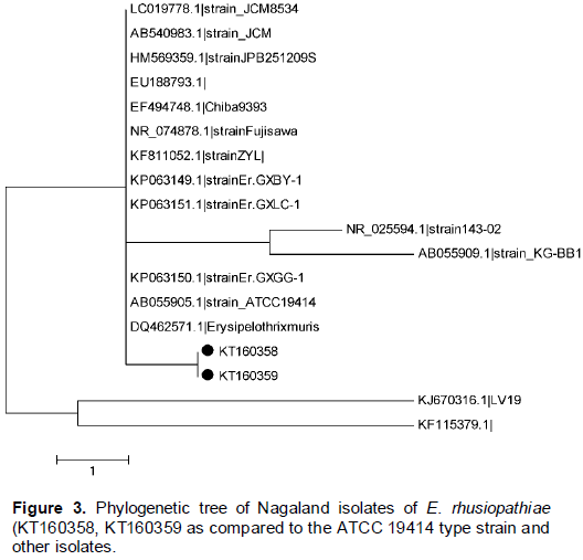
To determine the relationship of the Nagaland isolates of E. rhusiopathiae with other isolates of this species, 16S rRNA gene was amplified and sequenced (GenBank accession number KT160358 and KT160359). Sequence identity at nucleotide level was determined by Clustal W method of Meg-Align program in DNASTAR package (DNASTAR Inc., USA). The phylogenetic tree was constructed using other 16S rRNA gene sequences available at NCBI (viz. strain ZYL(KF811052.1), Isolate EU188793.1, strain:KG-BB1(AB055909.1), strain Fujisawa(NR_074878.1), Isolate DQ462571.1, strain: ATCC 19414(AB055905.1), strain JPB251209S(HM569359.1), Chiba9393(EF494748.1), sp. T127_5(JQ739693.1), sp. LV19(KJ670316.1), strain Er.GXLC-1(KP063151.1), inopinata strain 143-02 (inopinata strain 143-02) strain: JCM 8534(LC019778.1) and strain Er.GXBY-1(KP063149.1). Sequences were aligned by ClustalX version 2.1 (www.clustal.org), and the concatenated alignments were used for phylogeny inference (MEGA5; www.megasoftware.net) opting for the Maximum parsimony and Poisson correction. Computed replicates for bootstrap support was done and values were observed.
Histopathology
Formalin fixed tissues were processed as per standard protocol for histopathological studies. Sections of 4-5 micron thickness were stained routinely with Haematoxylin and Eosin stain and observed under oil immersion objective of a low power light microscope.
RESULTS AND DISCUSSION
Tissue samples processed for detection of classical swine fever virus and swine pox virus were confirmed as negative for both viral agents. However, bacteriological investigation demonstrated association of E. rhusiopathiae infection in affected (5) as well as in dead (2) pigs. Prevalence of erysipelothrix in many animals, mostly in pigs and birds has been reported throughout the world including from India (Shankar et al., 2009; Arora et al., 2011). But there was a single report on swine erysipelothrix (SE) from Meghalaya (Das et al., 2014). North Eastern Region has the highest pig population of the country. Diverse geographical locations, varied climatic situations and frequent movement of pigs favour for spread of the disease through carrier pigs (Leslie et al., 2015). Present report is a thorough investigation on swine erysipelothrix occurred seasonally at Nagaland, another NE state of India.
Clinical signs
A total of 150 pigs during 2013-15 exhibited clinically high fever (105-1070F), anorexia, firm faeces, and animals lying down. Cutaneous lesions appeared after 5-7 days of illness initiating with erythematous patches followed by papules of 4-5 mm diameter, dark purple, raised, firm to touch giving square to rhomboid shape in entire body. No vesicular stage was noticed. Although scabs developed in the entire body, prominent lesions were identified on head, face back, belly, limbs, tail and on ear (Figure 1). In unattended cases scab lesions were sloughed off within 17-22 days leaving a large ulcerated area. These typical cutaneous lesions have been mostly seen in grower animals. In affected pigs it appears that cutaneous lesions were developed following acute stages of illness.
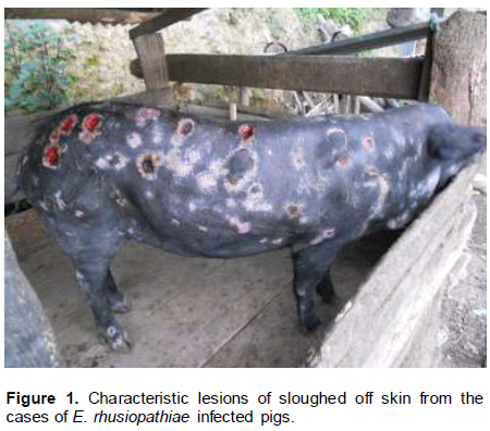
Considering the pathogenesis of E. rhusiopathiae, organisms gain access to the body, probably through the tonsils or other lymphoid tissue of the digestive tract and spread throughout the body. The bacteria produce neuraminidase, an enzyme that cleaves mucopolysaccharides in cell walls which may mediate the widespread vascular damage that accompanies SE. Vascular damage leads to thrombosis and interference with microcirculation in capillaries and venules at many sites. Classic cutaneous rhomboid urticaria (diamond skin) occurs in a percentage of pigs shortly after the acute febrile stages. In younger pigs with acute erysipelas, signs are similar, with cyanosis of extremities, ears and snouts pronounced and urticaria less common (Anonymous, 2016).
Seasonal occurrence of swine erysipelas in Nagaland justifies the stress due to sudden change of temperature. As stated by Amanda (2012)that stress factors such as overstocking, mixing pigs after weaning, and sudden changes in temperature can trigger clinical erysipelas.
Isolation and identification of organism
On blood agar the organism produced small, circular and transparent α- hemolytic colonies with a smooth glistening surface and edge. Biochemically all the isolates were catalase, oxidase and urease negative, produces H2S and ferments glucose and lactose. All 7 isolates were identified as E. rhusiopathiae based on the cultural, morphological and biochemical characteristics. The Nagaland isolates were sensitive in vitro to oxytetracycline, tetracycline, ampicillin, amoxicillin and cloxacillin; moderately sensitive to streptomycin, enrofloxacin, amikacin, co-trimoxazole, cefotaxime and ciprofloxacin; and resistant to gentamicin and norfloxacin, chloramphenicol and neomycin. Based on the antibiogram, survived ailing pigs were treated with oxytertacycline at 10 mg/kg body weight, intramuscularly for one week. Eight out of 13 treated animals responded promptly and recovered. Treatment of swine erysipelas cases with penicillin (Shankar et al., 2009) or other penicillin group of drugs such as the combination of amoxycillin and cloxacillin (Das et al., 2014) have been frequently reported.
Molecular confirmation and characterization
Tissue samples were negative for Swine fever virus and pox virus in PCR. A total of seven E. rhusiopathiae isolates recovered from tissue samples were subjected for PCR amplification using 16s rRNA gene specific primer set. All isolates were found to be positive for E. rhusiopathiae with amplification products of 407bp. (Figure 2).

Two Nagaland isolates KT160358 and KT160359 were sequenced and showed high sequence identity with the sequences of other E. rhusiopathiae available in the GenBank database. The Nagaland isolates (accession numbers KT160358, KT160359) were in the same clad (Figure 3) along with other strains originated from China. Phylogeny based on the nucleotide and amino acid sequences of the viruses provides a better understanding of the molecular epidemiology of the isolates. The state Nagaland shares international boundaries with Myanmar and having territorial link with other neighboring international countries like China, Bhutan and Bangladesh. The Chinese strains used in the construction of phylogenetic tree viz. swine isolates KPO 63150.1, strain Er.GXBY-1(KP063149.1), strain Er.GXLC-1(KP063151.1) and murine isolate DQ4625711are forming same clad with Nagaland isolates ( KT160358 and KT160359) indicated an epidemiological link. Movements of animals and animal products might spread the infection to this locality.
Histopathology
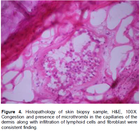
Histopathologic alteration of affected skin showed extensive damage to the capillaries and venules of dermis with infiltration of lymphoid cells and fibroblasts. Congestion and presence of microthrombi in the capillaries were consistent finding in all skin biopsy samples investigated in the present study (Figure 4). Similar observations were also recorded by Shankar et al. (2009).
Cultural characteristics, molecular confirmation and histopathological changes conclusively proved that swine erysipelas is prevailing in Nagaland. Pigs harbouring the infection manifest clinically at sudden change of climate. It is estimated that 30–50% of healthy swine harbour the organism in their tonsils and other lymphoid tissues (Stephenson and Berman, 1978). Again, trans border movement of pigs and their products could facilitate spreading of SE in this locality. Further study on prevalence of swine erysipelas and identification of carrier pigs can provide actual guidelines to control the disease in this part of India.
CONCLUSION
Unorganized pig farms in the Phek district of Nagaland experienced high mortality of grower pigs during rainy-summer season. A detail isolation and molecular investigation confirmed the association of swine erysipelas in this part of north eastern region of India. This is the only report on seasonal occurrence of swine erysipelas in the hilly low temperate climate of the NE states of Nagaland, India.
CONFLICT OF INTERESTS
The authors have not declared any conflict of interests.
REFERENCES
|
Amanda L (2012). Swine erysipelas. |
|
|
Anonymous (2016). Erysipelas. Veterinary Diagnostic and Production animal manual. College of vet. Med. Iowa State University. |
|
|
Arora N, Rajora VS, Prasad A, Misra S (2011). Urticarial form of swine erysipelas: a case report. Vet. Pract. 12(1):77. |
|
|
Das S, Ghatak S, Bhattacharya U, Puro K, Amarjit K, Kumar P, Deepak Verma KL, Ahuja A, Sen A (2014) . Identification of Erysipelothrix rhusiopathiae infection from a pig farm in Meghalaya, India. Vet. Pract. 15(1):52-54. |
|
|
Hoffmann B, Beer M, Schelp C, Schirrmeier H, Depner K (2005). Validation of a real-time RT-PCR assay for sensitive and specific detection of classical swine fever. J. Virol. Methods 130:36-44. |
|
|
Lee JJ, Kim DH, Lim JJ, Kim DG, Chang HH, Lee HJ, Kim SH, Rhee MH, EndaleM, Imada Y, Kim OJ, Kim S (2011). Characterization and Identification of Erysipelothrix rhusiopathiae isolated from an unnatural host, a cat, with a clinical manifestation of depression. J. Vet. Med. Sci. 73:149-154. |
|
|
Leslie EE, Christley RM, Geong M, Ward MP, Toribio JA (2015). Analysis of pig movement across eastern Indonesia, 2009-2010. Prev. Vet. Med. 118(4):293-305. |
|
|
Makino SI, Okada Y, Maruyama T, Ishikawa K, Takahashi T, Nakamura M, Ezaki T, Morita H (1994). Direct and rapid detection of Erysipelothrix rhusiopathiae DNA in animals by PCR. J. Clin. Microbiol. 32:1526-1531. |
|
|
Medaglia MLG, de Cassia Pereira A, Freitas TRP, Damaso CR (2011). Swine pox virus outbreak, Brazil. Emerg. Infect. Dis. 17(10):1976-1978. |
|
|
Reboli AC, Farrar WE (1989). Erysipelothrix rhusiopathiae: An Occupational Pathogen. Clin. Microbiol. Rev. 2:354-359. |
|
|
Shankar BP, Chandan S, Madhusudan HS, Ranjith D (2009). Pathology of erysipelas infection in piglet. Vet. World 2(6):234-235. |
|
|
Stephenson EH, Berman DT (1978). Isolation of Erysipelothrix rhusiopathiae from tonsils of apparently normal swine by two methods. Am. J. Vet. Res. 39:187-188. |
|
Copyright © 2024 Author(s) retain the copyright of this article.
This article is published under the terms of the Creative Commons Attribution License 4.0


