Full Length Research Paper

ABSTRACT
Bacterial isolates RP-1, RP-3 and RP-9 were isolated from agricultural soil using enrichment culture technique and screened positive for lindane degradation. RP-1, RP-3 and RP-9 were found to utilize and degrade higher concentrations (100 ppm) of lindane. RP-1 and RP-3 showed 69.5 and 65% lindane degradation after 10 days of inoculation where as RP-9 degraded 62% of lindane after 15 days. The estimated Cl- ion release was 49, 42 and 39 mg/mL, respectively for the three bacterial isolates. Gas chromatography was used for analysis of metabolite formed during lindane degradation and different parameters of degradation kinetics were calculated using first order kinetic equation. A drastic decrease in degradation rate was observed at initial lindane concentrations higher than 200 mg/l in the mineral media. The calculated half-life periods for RP-1, RP-3 and RP-9 were found to be 3.85, 2.77 and 4.00 days, respectively. All three isolates showed maximum degradation activity at: incubation period; 10-15 days, incubation temperatures; 30°C, pH; 7.0, shaking speed 120 rpm, initial substrate concentration; 100 mg/l. Galactose and succinate enhanced the degradation rate up to 10% whereas maltose, lactose and xylose decreased the degradation level up to 40%. Addition of glucose as a co-substrate was found highly favorable for enhancement of lindane degradation.
Key words: Enrichment culture, colorimetric assay, lindane, degradation, gas chromatography.
INTRODUCTION
The use of pesticides has increased dramatically during the last two decades at global level, due to their promising effects in agricultural and other related areas. Some of these are extremely resistant to biodegradation by native flora when compared with the naturally occurring compounds that are readily degraded upon introduction into the environment. Therefore, pesticides residues and their transformation products are frequently found in the environmental matrices. Despite concerns regarding their toxicity to humans and wildlife along with their relative stability to sediments and soil, they are still widely used (Diez, 2010). Lindane or γ- hexa-chlorocyclohexane (γ-HCH) has been used historically as a broad spectrum pesticide in agricultural, livestock, forestry, veterinary and human health applications because of its low production cost and effective pesticide properties. The HCH formulation consists of γ-(10–12%), α- (60–70%), β- (5–12%) and δ- (6–10%) isomers and out of these only γ-HCH possesses insecticidal activity (Li et al., 2003). Therefore γ-HCH is generally purified with 99% purity; the remaining four isomers are discarded and released as HCH muck. Its residues have been detected in drinking water sources, beverages and in different food stuffs. The half-life period for lindane in soil and water was reported as 708 and 2,292 days, respectively. It has been classified as a persistent organic pollutant (POP), potent carcinogen and hazardous, by Stockholm Convention in 2009. The production and agricultural use of lindane had been banned in more than 50 countries due to its toxicity and long persistence in soil but the pharmaceutical use is permitted till 2015. Large amounts of HCH still remain at the production sites even when the units have been closed. The use of γ-HCH for control of agricultural pests has been discontinued, run-offs from the already contaminated agricultural soils or from the dumping sites of adjoining regions can result in high levels of contamination. The use of lindane in agriculture leads to 12-30% volatilization into atmosphere and comes back in the form of rain.
The HCH degradation occurs by stepwise removal of chlorine atom known as dechlorination and influenced by temperature, pH, oxygen and biomass concentration. Nagata et al. (1996a) proposed the degradation pathway of lindane using Pseudomonas paucimobilis. The γ- HCH is transformed to 2,5-dichlorohydroquinone via sequential reactions catalyzed by enzymes LinA, LinB and LinC. The 2,5-dichlorohydroquinone in turn, is metabolized by enzymes LinD, LinE, LinF, LinGH and LinJ to succinyl-CoA and acetyl-CoA, that are further channeled and metabolized in the tricarboxylic acid cycle.
Though all HCH isomers are toxic, carcinogenic, endocrine disrupters are known to exert damaging effects on the reproductive and nervous systems in mammals, it is ubiquitously used in tropical countries to reduce vector-transmitted diseases, to protect livestock and to increase agricultural yields.
It produces histological alterations in cardiac tissue and cardiovascular dystrophy (Rajendran et al., 1999). Considering the various environmental impacts and persistence of lindane in the soil for a long time and its toxicity, threats of environmental contami-nation are of great concern. As lindane is highly recalcitrant and toxic compound which is degraded at a low rate, the present study is an attempt to isolate and characterize the potent bacterial strains from field soil, involved in biodegradation of lindane.
MATERIALS AND METHODS
Lindane (γ-HCH, 97.2% purity) technical grade was procured from Sigma Aldrich (USA). A stock solution of lindane, prepared at a concentration of 4 x 10³ mg/L in acetone (Fisher scientific India Pvt. Ltd.) was added to mineral salt medium according to the requirements. All other chemicals were of analytical grade, obtained from Hi-media, Merck and Qualikam India. Sphingomanas japonicum (MTCC No. 6362) procured from IMTECH, Chandigarh, was used as a reference culture for all the biodegradation studies.
Soil sampling
For isolation of lindane degrading bacteria, soil samples were collected from different agricultural field sites of Haryana, India which had a long history of pesticide application (more than 15 years). Soil cores (0-20 cm) taken from selected spots were collected in sterile plastic bags and stored at 4°C until microbial isolation. Physiological characterization of soil samples was carried out using Soil Testing Kit (K052), Hi-media, India.
Enrichment and isolation of bacterial
Bacterial isolation was carried out by enrichment culture technique using mineral broth (MSM) containing (per liter) potassium dihydrogen phosphate; 0.85 g, dipotassium hydrogen phosphate; 2.17 g, disodium hydrogen phosphate; 3.34 g, ammonium chloride; 0.1 g, magnesium sulphate; 0.5 g, calcium chloride; 0.5 g, ferrous sulphate; 0.01 g, sodium molybedate; 0.01 g at a pH of 7.2 ± 0 5 (Sahu et al., 1990). Two grams of collected soil sample was added to 100 mL of sterile medium supplemented with 10 mg/L of lindane, after sterilization. The flask were mixed thoroughly and incubated at 30°C for 7 days on rotary shaker at 120 rpm. Subsequently 1 mL of the inoculum having 5 x 10³ CFU/ mL, from the flasks were transferred to sterile medium (100 mL) containing same lindane concentration. The pesticide concentration was increased from 10 to 100 mg/L in a stepwise manner, transferring the inocula to fresh media each time. After acclimatization, bacterial colonies were isolated by serial dilution and spread plated onto mineral agar plates. These were incubated under aerobic conditions at 30°C for 24 h and colonies with different morphology were sub cultured on fresh agar plates in the form of single culture and preserved at 4°C.
Screening and selection of lindane degrading bacteria
γ-HCH utilization assay
For this, spray plates were prepared with 1.5% agar in mineral medium on Petri dishes and culture was streak on to the plates. The surface of the preset agar plates was sprayed with 0.5% of lindane in acetone. The plates were incubated for seven days in at 28 ± 2°C. The formation of lindane clearance zone surrounding bacterial colonies indicated utilization of lindane by that culture. The pure isolates were grown into mineral broth supplemented with 100 mg/L of lindane, for two to three days at 30°C.
Dechlorinase enzyme assay
For detecting haloalkane dehalogenase activity colorimetric assay was performed in a 96 well microtiter plate using the method given by Holloway et al. (1998), with slight modifications. The assay buffer contains 0.5 mM HEPES (pH 8.2), 10 mM sodium sulphate and 0.5 mM EDTA. Phenol red (dissolved in acetone) was added to the buffer before addition to 96 well plate to give a final concentration of 20 µg/mL. Each well was filled with 194 µL of buffer and 3 µL of lindane stock solution (12.5 mg/mL of acetone) prior to the addition of 6 µL of cell free extract. The organisms were also tested for dechlorination of dichloroethane (DCE). The microtitre plates were kept covered to prevent substrate volatilization and evaporation of the reaction mixture. A visual color change from red to yellow was indicative of lindane and DCE dechlorination which arises due to decreasing pH. Wells with blank samples were having only HEPES buffer instead of cell free extract.
Substrate tolerance
Different stocks solutions of lindane were prepared with varying concentrations. Then lindane utilizing cultures RP-1, RP-3 and RP-9 were inoculated in half strength mineral medium containing various concentrations of lindane as a sole carbon source (20, 40, 60, 80, 100, 120 ppm) and incubated at 30°C at 120 rpm. OD600 was taken periodically up to one month by taking inocula from all flasks having different concentration of lindane to check the viability count of bacteria which is directly related to the substrate tolerance. Subsequently chloride release was estimated to check degradation potential of the isolates at higher concentrations.
Morphological and biochemical study of important isolates
Different morphological (cell shape, cell size, gram stain, motility, capsule formation, colony morphology pigmentation) and biochemical studies (viz. gelatin liquefaction, starch and casein hydrolysis, catalase test, methyl red and Vogues Proskeur, sorbitol, adanitol, citrate, lysine, orinithine and urea utilization, phenylalanine deamination, nitrate reduction, H2S production, triple sugar iron test and various carbohydrate fermentation test like mannitol, xylose, galactose, sucrose glucose, rhamnose, lactose and arabinose) were carried out for the promising lindane utilizing isolates with the standard protocols given in Microbiology: A Laboratory Manual (Cappuccino and Sherman, 2010) (Table 1). For carbohydrate fermentation, 1% of each substrate was added to the medium as a carbon source.
Biodegradation studies
The potent isolates were inoculated into mineral broth supplemented with lindane (100 mg/L) and kept on rotary shaker at 120 rpm at 30°C up to one month to determine their degradation potential quantitatively and qualitatively. Quantitative dechlorination rate was determined by estimation of chloride ions released into the medium by inoculatory strains using indirect Argentometric method given by Greenberg et al. (1992). Individual samples were withdrawn at 0, 2, 4, 6, 8, 10, 13 and 15 days of incubation and were analyzed for Cl- estimation. The biodegradation potential of the selected strains was evaluated by analyzing the residual lindane in the medium which was calculated using the following formula:
Residual lindane (%) = (Ct/C0) x 100
where, C0 = initial concentration of lindane in the medium; Ct = lindane concentration at time t.
Statistical analysis for degradation kinetics
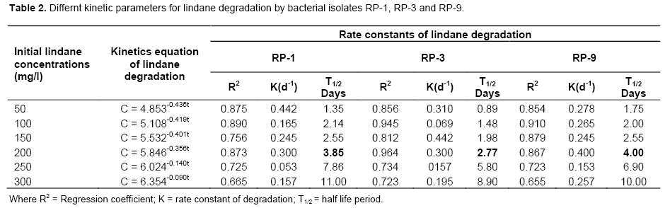
All the degradation experiments were carried out in triplicates. The isolates RP-1, RP-3 and RP-9 were inoculated into sterile basal mineral broth (50 mL) having different concentrations of lindane (50, 100, 150, 200, 250 and 300 ppm) and were incubated on a rotary shaker (120 rpm) at 30°C. Analysis of chloride ion release and residual lindane was carried out and uninoculated mineral medium was taken as the control. First order kinetic equation was used for calculating different parameters of lindane degradation by fitting experimental data:
Ct = C0e-kt
ln Ct = C0 – kt
Where, C0 = Initial lindane concentration; Ct = lindane concentration after reaction time t; K = degradation constant; T = reaction time.
Half- life for lindane biodegradation was calculated using the following formula:
T1/2 = ln 2/K
Physiological studies of lindane biodegradation
Substrate tolerance studies using different concentration of lindane (20, 40, 60, 80, 100 and 120 ppm) as a sole carbon source has been carried out for the said isolates. Different physiological parameters, that is, incubation time, temperature, pH, initial lindane concentration, shaking speed, on biodegradation rate has been optimized for the potent lindane degrading isolates.
Effect of different carbon sources
Effect of various carbon sources on lindane degradation viz. glucose, maltose, galactose, succinate, xylose, lactose as degra-dation activators and deactivators was also studied. For this experiment, mineral medium was supplemented with 1% of each carbon substrate, inoculated with 1 mL of particular bacterial inoculum and incubated at 30°C on shaker for 15 days.
GC- analysis
Residual lindane in the culture was determined qualitatively by GC-ECD method using Gas chromatograph (Shimadzu -2010 Plus model) equipped with ECD detector and DB-1701 (30 µm x 0.25 µm x 0.25 µm) column. For GC-ECD analysis, the residual lindane and the degradation product formed were extracted twice in 1 mL of hexane (HPLC Grade, Qualikam Chemicals, India). Elution were as follows: Helium as carrier gas, detector temperature; 350°C, oven temperature conditions; 90°C for 2 min, increase to 250°C at 5°C/min, increase to 250°C at 30°C/min and held for 5 min. Preliminary test with known standards showed the method capable of detecting about 1 µg/mL of lindane in the injected sample (2 µL). All the 9 isolates obtained after testing their dechlorinase activity were subjected to the same protocol along with S. japonicum as reference.
RESULTS AND DISCUSSION
Different physico-chemical characteristics of soil samples were analysed: pH; 7.4, organic carbon 61.1 ppm, available phosphate; 16.5 ppm, potassium; 49 ppm, ammonical nitrogen 20.8 ppm, nitrate nitrogen 10 ppm; sand 71%, silt 13% and clay 16%.
Isolation and screening of lindane degrading bacterial isolates
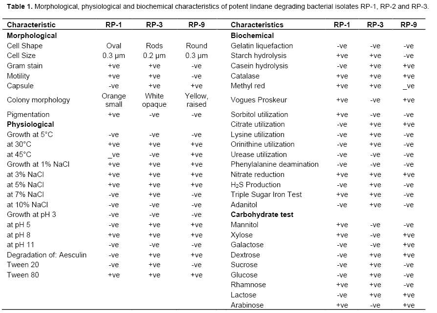
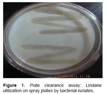
A total of 78 bacterial isolates were obtained which were tested for lindane utilization and dechlorinase enzyme activity. Plate clearance assay and dechlorinase enzyme assay were performed to confirm lindane degrading activity. Nine isolates were found positive for lindane utilization when screened on γ-HCH spray plates. They showed a prominent clearance zone after 9-12 days of incubation (Figure 1). Similar type of lindane degradation zones has been observed for other organisms e.g. bacterium Pesudomonas paucimobiliis (Senoo and Wada, 1989), fungus Conidiobolus 03-1-56 (Nagpal et al., 2008) and yeast Rhodotorula sp. VITJzNo3 (Salem et al., 2013). These isolates were further screened for enzyme activity using colorimetric assay. The phenol red indicator turned yellow when cell free extract of these nine isolates along with S. japonicum (as reference) were incubated in the presence of lindane (Figure 2). All these isolates were also found to be positive for dechlorination of dichloroethane (DCE). The colour change is due to decrease in pH. S. japonicum possess genes linA and linB which have been classified as haloalkane dehalogenases, responsible for the dechlorination of compounds like HCH and DCE. Dechlorination by linA results in accumulation of hydrochloric acid which in turn is responsible for lowering of pH (Nagata et al., 1997). When whole cells were inoculated into HEPES buffer containing lindane and incubated, no colour change was observed suggesting that enzymes having dechlorinase activity may be intracellular in nature (Thomas et al., 1996). It has already been reported in the case of S. paucimobilis UT26 that linA and linB genes are not excreted but are located in periplasmic space (Nagata et al., 1996a). However there is possibility that the same is not true for all lindane degrading micro-organisms, that is, some degraders may have dechlorinase enzymes present extracellularly.
Substrate tolerance
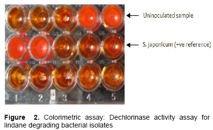
Both genetics and physiologies of microorganisms are involved in making them resistant/tolerant against any pesticide. It is observed that the tolerant microorganisms to lindane or any pesticide have biodegrading potential to break it down into smaller products which are later utilized by these organisms as carbon and nitrogen sources (Bellinaso et al., 2003). In this study, total ten isolates (along with the reference S. japonicum) were checked for their potential to withstand higher concen-trations of the substrate by inoculating into mineral broth having different concentration of γ-HCH. After one month, S. japonicum and three isolates RP-1, RP-3 and RP-9 were found to tolerate 100 ppm of lindane where as RP-2, RP-4 and RP-8 could tolerate up to 60 ppm. RP-6 and RP-7 were able to grow in medium having only 40 ppm of the active substrate and RP-5 showed growth only in very low concentration up to 20 ppm. Higher concentrations of lindane were found detrimental to growth of most of the bacterial isolates. Similarly, higher concentrations of toxic contaminants like cadmium have been reported to diminish bacterial growth (Kumar et al., 2010). After these observations, RP-1, RP-3 and RP-9 were selected to further biochemical, morphological and biodegradation studies (Figure 3 and Table 1).
Biodegradation studies
When the bacterial isolates were grown in mineral medium containing 100 mg/l of lindane, they were found to utilize lindane as a sole carbon source. There was a lag phase of two days before the bacterial growth started. Figure 4 shows that maximum OD600 was reached after 7 days of incubation for S. japonicum and RP-1 whereas in the case of RP-3 and RP-9, it was reported after 10 days of incubation. The substrate disappearance started after 3 days of incubation, that is, when the OD600 reached above 1.0 for all the three isolates along with reference. Extent of lindane mineralization was estimated by quantifying the release of inorganic chloride ions and analyzing the percent residual lindane in the medium. RP-1 and RP-3 showed 69.5 and 65% lindane degradation after 10 days of inoculation and their degradation rate was stable up to 15 days. RP-9 was able to degrade 62% of lindane after 15 days of inoculation and after this period the rate of degradation started decreasing for all the three bacteria (Table 3).
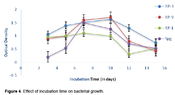
The concentration of Cl- in the medium increased during for first 15 days and after this period bacterial count as well as concentration of Cl- started diminishing which dissipated a linear relationship between growth and release of chloride ion or lindane mineralization.
Kinetics of lindane degradation
The degradation of lindane is dependent on the substrate concentration, which can be well explained by first order equation. The degradation kinetic of lindane was also studied in other organisms by different researchers, where the calculated half-life of lindane were as follows: 1.66 days for Rhodotorula sp. VITJzN03 (Salam et al., 2013); 4.78 days for Anabeana azotica (Zhang et al., 2012). In the present study, first order kinetic equation was used for calculating different parameters of degradation kinetics by fitting the triplicate values of experimental data. Degradation rate constant and half life period were calculated for different concentration of lindane by using first-order reaction model (Table 2). There was noticeable effect on lindane degradation with increasing initial concentration from 50 to 200 mg/L, however maximum degradation rate was observed for 100 mg/L of lindane. A drastic decrease in degradation rate was observed at concentrations higher than 200 mg/L, so this was taken as the optimum concentration for calculating half life period. The half-life periods calculated for RP-1, RP-3 and RP-9 were found to be 3.85, 2.77 and 4.00 days respectively. Previously, the degradation kinetic of lindane was also studied in other organisms isolated from different soils where the calculated half-life of lindane was described as follows: 3.66 days for Rhodotorula sp. VITJzN03 (Salam et al., 2013) and 4.78 days for Anabeana azotica (Zhang et al., 2012). Clearly, the calculated half-life of lindane, in the present study is shorter than in the earlier findings indicating the astonishing potency of the strain in lindane degradation.

Physiological studies for lindane biodegradation
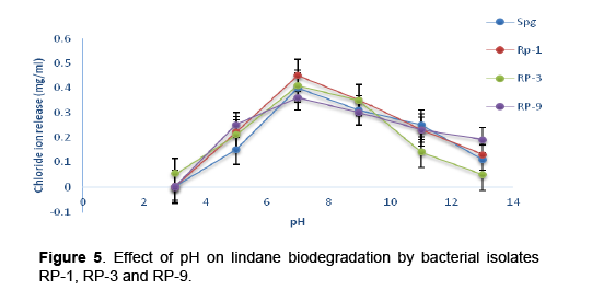
Cultural conditions such as growth period, incubation temperature, pH, revolution speed, substrate concentration have considerable effect on cell growth and degradation efficiency of micro-organisms (Kodama et al., 2001). For effective degradation study, the initial concentration of lindane was maintained at 50 mg/L in the optimization tests where pH, temperature, incubation time, shaking speed was also investigated. The effect of various parameters on lindane degradation by RP-1, RP-3 and RP-9 is depicted in Figures 4, 5 and 6. Strain RP-1, RP-3 and RP-9 showed better degradation at an initial pH of 7 which was considered as optimal pH. In the previous studies maximum lindane degradation was observed under a neutral pH of 7 (Siddique et al., 2002; Elcey and Kuhni, 2010). Salam et al (2014) also reported optimum pH as 7.0 for lindane degradation by Candida VITJzN04. In the case of temperature, maximum biomass and best course of degradation was observed at incubation temperature of 30°C.
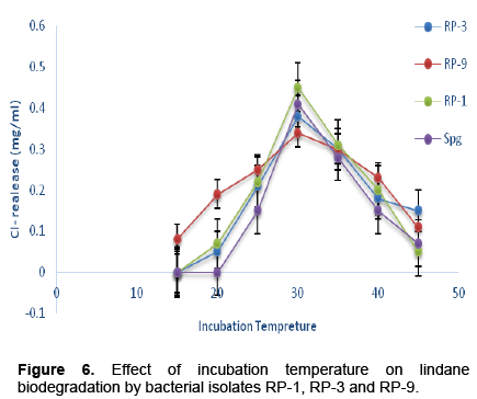
Synchronous observations at 30°C were reported earlier in fungal strain Rhodotorula sp. VITJzN03 by Salem et al. (2013) and in bacterial strain P. aeruginosa (Zhang et al., 2012). To demonstrate the effect of agitation on degradation of lindane, experimental flasks were incubated at different shaking speeds ranging from 80-120 rpm. The degradation was more prominent at 120 rpm and hence was considered optimum. In the case of inoculum size, the larger the inoculum size, the greater the efficiency of degradation of toxic compounds (Guillen-Jimeneza et al., 2012). In our study, different inoculum size (50-300ml/L) was used as the initial substrate concentration and 100 mg/L of lindane was found optimum for degradation activity. There is possibility that low concentration of substrate might not be able for induce the enzymes of degradation pathway. Similar results were reported by Kumar et al. (2006) in the case of other pollutants. Beyond 100 mg/L, the degradation rate significantly reduced, this limited growth and lindane degradation at higher concentrations could be attributed to the toxicity at higher concentrations of lindane. In the case of incubation period, a time course of 10-15 days was found to be optimum for lindane degradation.
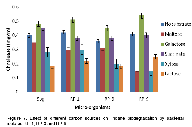
Effect of different carbon sources
Carbon sources, other than the target chemical, when present in medium may highly influence degradation rates. Accordingly, the effect of some of the common carbon sources was evaluated on the biodegradation of lindane.
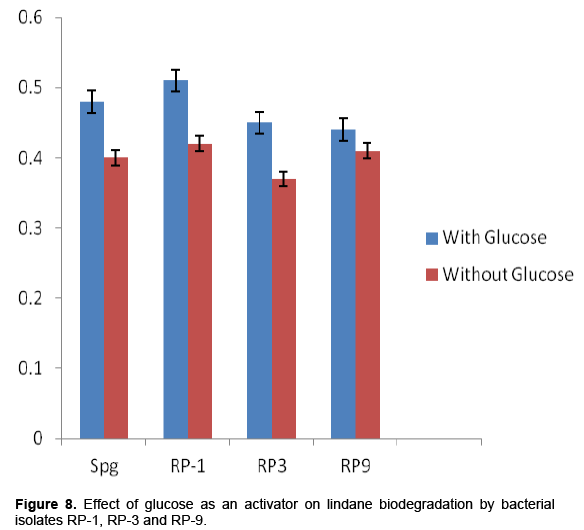
Addition of different carbon sources in the inoculated medium showed prominent effect on lindane degradation. It was found that galactose and succinate enhanced the degradation rate up to 10% whereas maltose, lactose and xylose decreased the degradation level up to 40% as compared to the sample having no additional carbon source (Figures 7 and 8). Addition of glucose as a co-substrate was found highly favorable for enhancement of lindane degradation. Also, in a previous research, succinate and glucose were found favorable for degradation of chlorinated pesticide like chlorpyrphos (Singh et al., 2004; Anuja Goerge, 2005). The enhanced rate of lindane degradation after addition of different C-sources might be due to the cometabolic process. However, many C-source are also reported to decrease the degradation rate, which might be due to the mechanism of catabolite repression or decrease in the rate of transcription either due to supercoiling of promoter DNA or by decreased binding of transcription factors.
GC-analysis
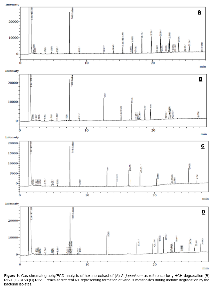
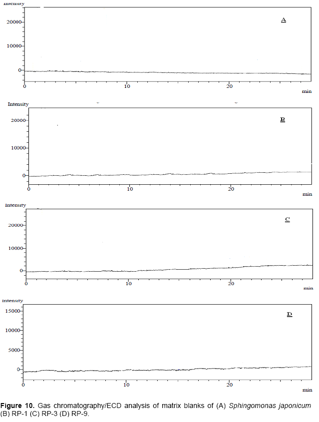
Gas chromatography analysis with electron capture detector was carried out to check lindane degradation by the bacterial isolates. Hexane extract of pure lindane was run as standard and respective peak was obtained at retention time (RT) 7.458. Sphingomonas japonicum was taken as the reference culture for comparison of peaks obtained for metabolites released during the degradation of lindane as a substrate. For S. japonicum as well as other isolates, no peaks were observed for the chromatographs obtained with blank matrix (Figure 10). Different peaks were obtained only in samples having media as well as lindane and inoculated with different bacterial isolates. The RT- values had also been compared with the literature available on lindane degradation. Bacterial strain RP-1 showed 69.5% degradation which is equivalent to degradation rate of S. japonicum whereas RP-3 and RP-9 degraded at rate slower than the reference culture. Various peaks at different RT values were obtained in common for all the three isolates which were comparative to peaks observed in case of S. japonicum during lindane degradation (Figure 9).
CONCLUSION
Bacterial isolates RP-1, RP-3 and RP-9 isolated from agricultural soil have been found to possess the ability to utilize and degrade higher concentrations of lindane. Once molecular characterization studies are completed, these can be considered as potential microbes for field trials regarding bioremediation of γ-HCH contaminated soils. Further research on metabolic pathway elucidation at molecular level is in progress.
CONFLICT OF INTERESTS
The authors declare no conflict of interest.
ACKNOWLEDGEMENTS
The authors are thankful to Department of Biotechnology, DCR University of Science and Technology Murthal, Sonepat, Haryana for providing all necessary research facilities and technical support.
REFERENCES
|
Anuja G (2005). Isolation, screening and selection of efficient chlorpyriphos degrading microorganisms. Ph.D Thesis, University of Agricultural Sciences, Dharwad. |
|
|
Bellinaso MDL, Greer CW, Perlba MC and Heriques JAP (2003). Biodegradation of herbicide trifluralin by bacteria isolated from soil. FEMS Microbial. Ecol. 43:191-194. |
|
|
Cappuccino J, Sherman N (2010). Microbiology: A Laboratory Manual. Ed. 9. Benjamin-Cummings Publishing Company, Subs of Addison Wesley Longman, Inc. |
|
|
Diez MC (2010). Biological Aspects Involved in the Degradation of Organic Pollutants. J Soil Sci. Plant Nutr.10(3):244-267. |
|
|
Elcey CD, Kunhi AAM (2010). Substantially enhanced degradation of hexachlorocyclohexane isomers by a microbial consortium on acclimation. J. Agric. Food Chem. 58:1046-1054. |
|
|
Greenberg AE, Clesceri LS, Eaton AD (1992). Standard Methods for the Examination of Water and Waste Water . 18 th ed. APHA, Washington. |
|
|
Guille’n-Jime’neza F, Cristiani-Urbinab E, Cancino-Dı’azc JC, Flores-Morenod JL, Barraga’n-Huertaa BE (2012). Lindane biodegradation by the Fusarium verticillioides AT-100 strain, isolated from Agave tequilana leaves: Kinetic study and identification of metabolites. Int Biodeterior. Biodegrad.74:36-47. |
|
|
Holloway P, Trevors JT, Lee H (1998). A colorimeteric assay for detecting haloalkane dehalogenase activity. J. Microbiol. Methods. 32:31-36. |
|
|
Kodama T, Ding L, Yoshida M, Yajima M (2001). Biodegradation of an s-triazine herbicide, simazine. J Mol. Catal. B Enzym. 11:1073-1078. |
|
|
Kumar A, Cameotra SS, Gupta S (2010). Screening and characterization of potential cadmium biosorbent Alcaligenes strain from industrial effluent. J. Basic Microbiol. 51:1-7. |
|
|
Kumar M, Gupta SK, Garg SK. Kumar A (2006). Biodegradation of hexachlorocyclohexane-isomers in contaminated soils. Soil Biol. Biochem. 38:2318-2327. |
|
|
Li YF (1999). Global technical hexachlorocyclohexane usage and its contamination consequences in the environment: from 1948 to 1997. Sci. Total Environ. 232:121-158. |
|
|
Nagata Y, Endo R, Ito M, Ohtsubo Y, Tsuda M (1997). Aerobic degradation of lindane (γ- hexachlorocyclohexane) in bacteria and its biochemical and molecular basis. Appl. Microbiol. Biotechnol. 76:741-752. |
|
|
Nagata Y, Hatta T, Imai R, Kimbara K, Fukuda M, Yano K, Takagi M (1996a). Purification and characterizacion of γ-hexachlorocyclohexane (γ-HCH) dehydrochlorinase (LinA) from Pseudomonas paucimobilis. Biosci. Biotechnol. Biochem. 57:1582-1583. |
|
|
Nagpal V, Srinivasan MC, Paknikar KM (2008). Biodegradation of chexachlorocyclohexane(lindane) by a non- white rot fungus Conidiobolus 03–1-56 isolated from litter. Indian J. Microbiol. 48:134-141. |
|
|
Rajendran BR, Venugopalan V, Ramesh R (1999). Pesticide residues in air from coastal environment, South India. Chemosphere 39:1699-1706. |
|
|
Sahu SK, Patnaik KK, Sharmila M, Sethunathan N (1990). Degradation of Alpha-, Beta-, and Gamma-Hexachlorocyclohexane by a soil bacterium under aerobic condition. Appl. Environ. Microbiol. 56(11): 3620-22. |
|
|
Salam JA, Das N (2014). Lindane degradation by Candida VITJzN04, a newly isolated yeast strain from contaminated soil: kinetic study, enzyme analysis and biodegradation pathway. World J. Microbiol. Biotechnol. 30:1301-1313. |
|
|
Salam JA, Lakshmi V, Das D, Das N (2013). Biodegradation of lindane using a novel yeast strain, Rhodotorula sp. VITJzN03 isolated from agricultural soil. World J. Microbiol. Biotechnol. 29:475-487. |
|
|
Senoo K, Wada H (1989). Isolation and identification of an aerobic γ-HCH- decomposing bacterium from soil. Soil Sci. Plant Nutr. 35:79-87. |
|
|
Siddique T, Okeke BC, Arshad M, Frankenberger WT (2002). Temperature and pH effects on biodegradation of hexachlorocyclohexane isomers in water and soil slurry. J. Agric. Food Chem. 50:5070-5076. |
|
|
Singh BK, Walker A, Alun J, Morgan W, Wright DJ (2004). Bioremediation of Chlorpyrifos by Enterobacter Strain B-14 and Its Use in Biodegradation of Contaminated Soils Appl. Environ. Microbiol. 70(8):4855. |
|
|
Thomas JC, Berger F, Jacquier M, Bernillon D, Baud-Gras-set F, Triffaut N, Normand P, Vogal TM, Simonet P (1996). Isolation and characterization of a novel γ-hexachlorocyclohexane degrading bacterium. J of Bacteriology. 178:6049-6055. |
|
|
Zhang H, Hu C, Jia X, Xu Y, Wu C, Chen L, Wang F (2012). Characteristics of c-hexachlorocyclohexane biodegradation by a nitrogen-fixing cyanobacterium. Anabaena azotica. J. Appl. Phycol. 24:221-225. |
|
Copyright © 2024 Author(s) retain the copyright of this article.
This article is published under the terms of the Creative Commons Attribution License 4.0


