African Journal of
Microbiology Research

  • Abbreviation: Afr. J. Microbiol. Res.
  • Language: English
  • ISSN: 1996-0808
  • DOI: 10.5897/AJMR
  • Start Year: 2007
  • Published Articles: 5233

Full Length Research Paper

Internal transcribed spacers (ITS) based identification of Trichoderma isolates and biocontrol activity against Macrophomina phaseolina, Aspergillus niger and Meloidogyne incognita

Khalid S. Abdel-lateif
  • Khalid S. Abdel-lateif
  • Department of Genetics, Faculty of Agriculture, Menoufia University, Egypt.
  • Google Scholar
Ramadan A. Bakr
  • Ramadan A. Bakr
  • Agricultural Botany Department, Faculty of Agriculture, Menoufia University, Egypt.
  • Google Scholar


  •  Received: 09 June 2018
  •  Accepted: 27 July 2018
  •  Published: 14 August 2018

 ABSTRACT

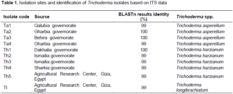
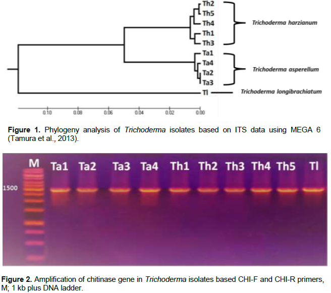
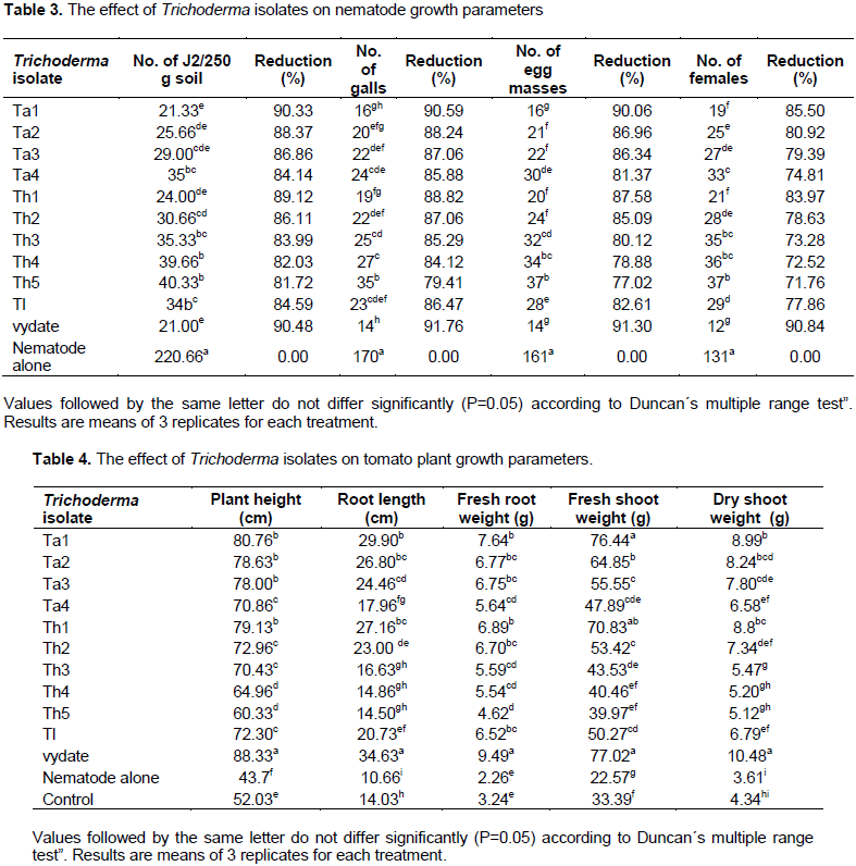
Ten Trichoderma isolates were isolated from different locations in Egypt. Amplification and sequencing of internal transcribed spacers (ITS) was employed to identify Trichoderma isolates that exhibited from 99 to 100% identity with three species of Trichoderma: Trichoderma harzianum, Trichoderma asperellum and Trichoderma longibrachiatum. The biocontrol activity of Trichoderma isolates against Macrophomina phaseolina, Aspergillus niger and Meloidogyne incognita was tested in vitro and under greenhouse conditions. The results show that the isolate Th2 (T. harzianum) gave the best antagonism against M. phaseolina and A. niger with inhibition rates of 72.85 and 64.28%, respectively. Moreover, the isolate Ta1 (T. asperellum) was the best efficient isolate in reduction of each second stage juveniles (J2), number of galls, egg masses and females per root system with 90.33, 90.59, 90.06 and 85.50%, respectively. Treatment with Trichoderma isolates improved tomato growth parameters (root length, plant height, roots and shoots fresh weight and shoots dry weight).


Key words: Trichoderma, internal transcribed spacers (ITS), antagonism, Macrophomina phaseolina, Aspergillus niger, Meloidogyne incognita.


 INTRODUCTION

The chemical pesticides are the first choice for farmers to control plant pathogens in order to obtain abundant crop yield (Junaid et al., 2013). It was estimated that 12% of crop loss is due to plant pathogens (Sharma et al., 2012). The long term uses of pesticides can cause severe environmental and health problems in addition to their expensive costs for developing countries. Trichoderma  is a famous saprophytic fungus that can be isolated from any soil and can be used as efficient biocontrol agent due to its high ability to reproduce and colonize (Pandya et al., 2011; Pal and Gardener, 2006; El-Hassan et al., 2013; Rao et al., 2015). Trichoderma fight against many fungal phytopathogens such as Phythium, Phytophthora, Macrophomina,  Aspergillus,  Rhizoctonia  and  Fusarium using many biocontrol weapons as mycoparasitism, production of antibiotics and competition of pathogens for food and space (Benítez et al., 2004; Rayatpanah et al., 2011; Krishnamurthy and Shashikala, 2006). Recently, the global attention rely on Trichoderma as safe alternative of pesticides and this led to increase in the number of  Trichoderma biocontrol products (Woo et al., 2014).
 
 
Macrophomina phaseolina and Aspergillus niger are considered destructive pathogens of many important and economically crops causing huge loses of each quantity and quality of crop yield (Khaledi and Taheri, 2016; Njoki et al., 2017; Bhale et al., 2013). M. phaseolina is a dangerous fungus that can infect many of important crops as corn, cotton, sesame, soy bean and sunflower causing diseases such as soy bean charcoal rot (Khaledi and Taheri, 2016; Aly et al., 2007). In addition, A. niger is a common saprophytic fungus that can cause dangerous diseases as black-rot and collar rot in crops like onion and peanut leading to yield loss from 5 to 40% (Khokhar et al., 2012; Gajera et al., 2011; Bhale et al., 2013). Moreover, root knot nematode (Meloidogyne incognita) is one of virulent parasites that attack many crops and can lead to destructive damages in crop yield range from 40 to 50% (Bakr et al., 2011; Katooli et al., 2010; Sikora and Fernandez, 2005; Karssen and Moens, 2006; Maqbool and Kerry, 1997). The previous studies indicated a vital role for Trichoderma species in biocontrol of M. phaseolina, Aspergillus species and M. incognita (Al-Hazmi and Javeed, 2016; Khaledi and Taheri, 2016;  Mendoza  et al., 2015; Shoaib et al., 2018; Gajera et al., 2011; Sharon et al., 2011). As the cell wall of nematodes and pathogenic fungi is mainly composed of chitin, Trichoderma have chitinase enzymes able to degrade the cell wall of these pathogens (Loc et al., 2011; Haran et al., 1995; Ike et al., 2006). Identification of Trichoderma spp. to be applied in the field of biological control is an important issue. It was reported that identification of Trichoderma based on morphological characters can give misleading results (Fahmi et al., 2016). Recently, molecular  identification   based   on  internal  transcribed spacers (ITS) amplification and sequencing is common and highly trusted (Savitha and Sriam, 2015; Fahmi et al., 2016; Oskiera et al., 2015; Jiang et al., 2016).
 
In this study, ten isolates of Trichoderma spp. were isolated from soil rhizosphere of different locations in Egypt, characterized on molecular level and screened for their antagonistic ability against M. phaseolina, A. niger and root knot nematode.


 MATERIALS AND METHODS

Isolation of Trichoderma isolates
 
Ten isolates of Trichoderma spp. were isolated from soil rhizosphere of different sites in Egypt (Table 1) cultivated with different crops as described by Fahmi et al. (2016). Several soil samples were obtained from a 15 cm depth, placed in sterile bags and transferred to the laboratory for isolation process. Serial dilutions were made and 250 μL of 103 dilution was distributed onto petri dishes containing Potato Dextrose Agar medium. The plates were then incubated at 28°C for one week for isolation of single colonies.
 
 
Soil borne pathogens
 
Isolates of M. phaseolina and A. niger were kindly provided by Faculty of Science, Zagazig University, Egypt.
 
DNA extraction from Trichoderma isolates
 
DNA isolation of Trichoderma was performed as described by Al-Samarrai and Schmid (2000). The clear sharp bands were indicator for the quality of DNA.
 
Molecular identification of Trichoderma isolates
 
Polymerase chain reaction (PCR) was utilized to amplify the internal transcribed spacer regions of Trichoderma using ITS1 (5' - TCC GTA GGT GAA CCT GCG G - 3') and ITS4 (5' - TCC TCC GCT TAT TGA TAT GC - 3') primers. PCR conditions were performed as described by Loc et al. (2011). PCR products were first purified using QIAquick PCR Purification Kit (QIAGEN Cat. No. 28104). Sequencing  was  performed  using  Big  Dye Terminator v3.1 Cycle Sequencing Kit in a total volume of 20 μL using 3500 Genetic Analyzer, Applied Biosystems (Daejeon, Korea).
 
PCR amplification of Chitinase gene
 
Amplification of chitinase gene was done using specific primers: CHI-F (5-ATG TTG GGC TTC CTC GGA-3) and CHI-R (5-TTC GGG ATG GTT GTC ATA CTG-3) as described by Loc et al. (2011).
 
Antagonism of Trichoderma isolates
 
The antagonism of Trichoderma isolates against M. phaseolina and A. niger was evaluated in dual culture plate technique by measuring the radial growth inhibition of pathogen(s) as described by Abdel-lateif et al. (2017).
 
Multiplication of M. incognita
 
Root-knot nematode, M. incognita was identified by observation of perineal patterns according to Hartman and Sasser (1985). Multiplication was done under greenhouse conditions at the experimental greenhouse, Department of Agricultural Botany, Faculty of Agriculture, Menoufia University, Egypt. Susceptible tomato seedlings Cv. GS (Solanum lycopersicum Mill) was transplanted in plastic pots 15 cm in diameter filled with sterilized clays and soil (1:2 v/v). Seedlings were then inoculated by one egg mass of M. incognita. Agricultural operations were carried as needed.
 
Preparation of M. incognita inoculum
 
Two months heavily galled tomato roots previously infested with M. incognita were used to prepare nematode inoculum. Massive infested roots removed from the pots and gently washed using tap water to separate the adherent soil particles. Then roots were cut into small pieces and macerated for two periods of 10 s each at high speed by using a blender. The macerated root solution then placed in a jar containing 0.5% of sodium hypochlorite (NaOCl) as described by Hussey and Barker (1973). The Jar was vigorously shaken for 3 min to release the eggs from the egg matrix with the help of NaOCl. The resulting solution then poured through serial sieves to separate the root tissue. Eggs were collected on the last sieve (20 μm) and washed several times with tap water to eliminate NaOCl residuals. The eggs were then transferred to a flask containing tap water. The number of eggs/ ml was estimated by counting 4 samples of 1 ml using a counting dish under a stereomicroscope.
 
Preparation of Trichoderma inoculum
 
The Trichoderma isolates were cultured on Potato Dextrose Agar (PDA) and incubated at 26°C for 10 days. The conidia were collected from the surfaces of plates by flooding with sterile distilled water and gently scraping the colony surface with a sterile scrapper (Jansson et al., 1985).
 
Greenhouse experiment
 
Greenhouse experiment was carried out to evaluate the effect of 10 Trichoderma isolates on M. incognita. Four weeks old tomato seedlings   (Cv. GS)   were   transplanted  into  plastic  pots  (15 cm diameter) containing sterilized sand-clay soil (2:1 v/v). Each plant is inoculated with 3000 eggs of M. incognita and Trichoderma spore suspension (108 g-1 of soil) around the young hairy roots and were mixed well within the pots. Control treatments included untreated seedlings and nematode treated seedlings without Trichoderma. Treatment with vydate (abroad spectrum nematicide) was used for comparison.
 
The treatments were replicated 3 times in a completely randomized block design under greenhouse conditions. Tomato plants were observed, watered and fertilized with a nutrient solution according to Epstein (1972).
 
Plant growth and nematode parameters
 
Two months after nematode inoculation, tomato plants were carefully uprooted. The roots were cautiously washed with running tap water. Plant growth parameters as root length (cm), plant height (cm), fresh weight of roots (g), fresh weight of shoots (g) and dry weight of shoots (g) were recorded. Number of galls/root system was counted directly, while number of second stage juveniles (J2S)/250 g soil was evaluated using serial sieves and modified Baermann technique as described by Goody (1957). Egg-masses were stained prior to counting by dipping the infected roots in 0.015% Phloxine-B solution for 20 min as described by Daykin and Hussey (1985). Number of females/root system was determined by cutting the root system to small pieces and submerging the roots in a beaker full of tap water for four days at room temperature until they became soft. Roots pieces were then washed through 500 and 250 μm sieves to separate the females from the root debris and counted under a stereomicroscope.
 
Data and cluster analysis
 
BLASTn was used to compare Trichoderma sequencing results with known sequences on NCBI site (http://www.ncbi.nlm.nih.gov/). The alignment and phylogenetic analyses were conducted using MEGA version 6 (Tamura et al., 2013). The obtained data of antagonism was analyzed using costat 6.3 version program. Analysis of variance and comparison of means were done at the 5% level of significance according to the Duncan's multiple range test (1955).


 RESULTS AND DISCUSSION

Isolation and molecular identification of Trichoderma isolates
 
Ten isolates of Trichoderma were isolated from rhizosphere soil samples collected from different sites in Egypt (Table 1). PCR based on ITS primers was used to amplify ITS region and gave one band about 600 bp. The PCR products were sequenced and the sequencing data was entered on NCBI site to search BLAST and compare these data with published ITS data. The amplified ITS regions of Trichoderma isolates were exhibited from 99 to 100% identity with three species of Trichoderma; five isolates were found to belong to Trichoderma harzianum (Th1, Th2, Th3 Th4 and Th5), four isolates classified as Trichoderma asperellum (Ta1, Ta2, Ta3 and Ta4), while one isolate (Tl) was identified as Trichoderma longibrachiatum (Table 1).
 
The phylogeny analysis supported the ITS identification and divided the isolates of Trichoderma into three clusters, the first cluster included the isolates of T. harzianum (Th1, Th2, Th3, Th4 and Th5). The second cluster included the isolates of T. asperellum (Ta1, Ta2, Ta3 and Ta4); finally, the third cluster contained the isolate Tl of T. longibrachiatum (Figure 1).
 
These results confirm the efficiency of barcode DNA and are compatible with previous studies that employed ITS to identify Trichoderma spp. (Wu et al., 2017; Fahmi et al., 2016; Savitha and Sriram, 2015; Oskiera et al., 2015).
 
Chitinase gene of Trichoderma
 
However, chitin is key component of pathogen cell walls, Trichoderma secrete highly active chitinases that can destroy these cell walls and feed on them (Seidl-Seiboth et al., 2014; Hassan et al., 2015; Prasetyawan et al., 2018). Chitinase 42 is one of Trichoderma endochitinases  that  can  degrade  the  β-1,  4-glycosidic bonds between the N-acetyl glucosamine residues of chitin (Hassan et al., 2015). In this study, PCR based specific primers (CHI-F and CHI-R) was utilized to amplify and detect the chitinase 42 gene in Trichoderma isolates as shown in Figure 2. The PCR gave one band about 1500 bp homolog to that obtained by Loc et al. (2011) and confirmed the presence of this gene in all tested isolates. The amplification of chitinase gene confirms that these isolates are Trichoderma since the primers are specific for Trichoderma chitinases.
 
Evaluation of Trichoderma antagonism against M. phaseolina and A. niger
 
The antagonism of Trichoderma isolates against M. phaseolina and A. niger was evaluated in vitro by measuring the radial growth inhibition of the two pathogens (Table 2 and Figure 3).


The isolates Th2, Ta2, Th3 and TI were the most efficient isolates  in suppression growth of M. phaseolina  with 72.85, 71.43, 67.14 and 65.69%, respectively. In addition, the isolate Th5 gave only 25.71% radial growth inhibition against M. phaseolina. For antagonism of A. niger, the isolates Th5 and Th2 were the most active isolates in suppression growth of A. niger with 71.43 and 64.28% respectively, while the isolate Tl exhibited the least radial growth inhibition against the same pathogen (31.43%). In general, the isolate Th2 of T. harzianum showed good antagonism against M. phaseolina and A. niger with inhibition rates 72.85 and 64.28%, respectively. Previous studies explained that T. harzianum is more vigorous in inhibition growth of pathogenic fungi than the other species of Trichoderma (Abdel-lateif et al., 2017; Shoaib et al., 2018; Al-Hazmi and Javeed, 2016). Mycoparasitism is one of Trichoderma biocontrol mechanisms that depend on secretion of lytic enzymes like chitinases and cellulases to destroy the cell walls of pathogens (Gajera et al., 2012). It was proved that chitin is major cell wall component of most pathogenic fungi and  Trichoderma   is   excellent   producer  of  chitinases which can degrade the chitin and therefore killing them (Benítez et al., 2004; Agrawal and Kotasthane, 2012; Gajera et al., 2012).
 
Trichoderma genus is proved to be an excellent control agent in inhibition growth of M. phaseolina, Aspergillus spp. and root knot nematode under in vitro and green house conditions (Al-Hazmi and Javeed, 2016; Khaledi and Taheri, 2016; Khalili  et al., 2015; Mendoza  et al., 2015; Shoaib  et al., 2018; Athira, 2017; Krishnamurthy and Shashikala, 2006). The variability in aggressiveness of the same Trichoderma isolates against M. phaseolina and A. niger may be due to the variation in pathogen cell wall structure and the defense ability of the pathogen.
 
The effect of Trichoderma isolates on nematode
 
The efficacy of Trichoderma isolates against M. incognita on tomato was evaluated in greenhouse experiments and the  results   were  compared  with  those  of  vydate  and control. The results show that treatment of tomato seedlings with Trichoderma decreased significantly root galling, egg masses, number of females and second stage juveniles (J2) per root system (Table 3), while improved tomato growth parameters (root length, plant height, roots and shoots fresh weight and shoots dry weight) as indicated in Table 4. The isolates of T. asperellum (Ta1, Ta2 and Ta3) and the isolates of T. harzianum (Th1 and Th2) exhibited high inhibition of nematode growth in levels comparable with those of vydate. The isolate of T. asperellum (Ta1) was the best efficient isolate in reduction of each second stage juveniles (J2), number of galls, egg masses  and  females per root system with 90.33, 90.59, 90.06 and 85.50%, respectively and effect of this isolate was similar to vydate. Wu et al. (2017) isolated new strain of T. asperellum with high antagonism against pathogens of cucumber wilt and corn stalk rot. 
 
 
On the contrast, the isolate T. harzianum (Th5) was the least isolate in reduction of each second stage juveniles (J2), number of galls, egg masses and females per root system with 81.72, 75.41, 77.02 and 71.76%, respectively, as compared to vydate. It was shown that the growth of nematode was increased in absence of Trichoderma. These results highlight the significance of Trichoderma as excellent control agent in inhibition growth of nematode (Spiegel et al., 2007; Chen and Dickson, 2004; Sharon et al., 2011). Interestingly, most of the isolates that exhibited high inhibition rate of nematode growth also improved each root length, plant height, roots fresh weight, shoots fresh, and dry weight of tomato plants (Table 4). These results are compatible with other studies showing the ability of Trichoderma in growth inhibition of nematode on various crops and enhancing the growth of host plants (Al-Hazmi and Javeed, 2016; Izuogu and Abiri, 2015; Sharon et al., 2011; Mascarin et al., 2012).
 
The variability among Trichoderma isolates in their aggressiveness against the tomato nematode  may be due to the difference in their genetic structure, origin of isolates, pathogen cell wall structure and quantity of lytic enzymes secreted by Trichoderma (Al-Hazmi and Javeed, 2016; Abdel-lateif et al., 2017).


 CONCLUSION

Identification of Trichoderma based on ITS markers exhibited high efficiency in discrimination among different Trichoderma spp. isolates. Trichoderma isolates showed variability in their aggressiveness against M. phaseolina, A. niger and M. incognita. In general, the isolate Th2 of T. harzianum showed the best antagonism against M. phaseolina and A. niger, while the isolate Ta1 of T. asperellum was the best efficient isolate in reduction of nematode growth.
 
In general, treatment of tomato with Trichoderma isolates improved their growth parameters (root length, plant height, roots and shoots fresh weight and shoots dry weight) as compared to untreated control. These results confirm the efficacy of Trichoderma as excellent biocontrol agent and also as plant growth promoting.


 CONFLICT OF INTERESTS

The authors have not declared any conflict of interests.


 REFERENCES

Abdel-lateif K, El-Zanaty AF, Helwa M (2017). Genetic diversity of Trichoderma isolates and their antagonism against Rhizoctonia Solani and Phythium aphnidermatum. Menoufia Journal of Agriculture Biotechnology 2:63-73.


Agrawal T, Kotasthane AS (2012). Chitinolytic assay of indigenous Trichoderma isolates collected from different geographical locations of Chhattisgarh in Central India. Springer Plus 1:73-82.
Crossref


Al-Hazmi AS, Javeed MT (2016). Effects of different inoculum densities of Trichoderma harzianum and Trichoderma viride against Meloidogyne javanicaon tomato. Saudi Journal of Biological Sciences 23:288-292
Crossref


Al-Samarrai TH, Schmid J (2000). A simple method for extraction of fungal genomic DNA. Letters in Applied Microbiology 30: 53-56.
Crossref


Aly AA, Abdel-Sattar MA, Omar MR, Abd-Elsalam KA (2007). Differential antagonism of Trichoderma sp. against Macrophomina phaseolina. Journal of Plant Protection Research 47(2).


Athira K (2017). Efficacy of fungicide and bio-control agents against root rot of black Gram (Vigna mungo L.) caused by Macrophomina phaseolina (Tassi) Goid. International Journal of Current Microbiology and Applied Sciences 6(10):2601-2607.
Crossref


Bakr RA, Mahdy ME, Mousa EM (2011). A survey of root-knot and citrus nematodes in some new reclaimed lands in Egypt. Pakistan Journal of Nematology 29:165-170.


Benítez T, Rincón AM, Limón MC, Codón AC (2004). Biocontrol mechanisms of Trichoderma strains. International Microbiology 7:249-260


Bhale UN, Wagh PM, Rajkonda JN (2013). Antagonistic confrontation of Trichoderma spp against fruit rot pathogens on Sapodilla (Manilka razapota L.). Journal of Yeast and Fungal Research 4(1):5-11


Chen S, Dickson DW (2004). Biological control of nematodes by fungal antagonists. In: Nematology Advances and Perspectives. Nematode Management and Utilization, vol. II. (Chen ZX, Chen SY, Dickson DW, eds).Wallingford: CAB International, UK pp. 977-1039.
Crossref


Daykin ME, Hussey RS (1985). Staining and histopathological techniques in nematology. pp. 39-48. In: K. R. Barker, C. C. Carter, J. N. Sasser, eds. An Advanced Treatise on Meloidogyne: Volume II. Raleigh, NC: North Carolina State University Graphics.


Duncan B (1955). Multiple range and Multiple F test. Biometrex 11:1-42.
Crossref


Elad Y, Hadar Y, Chet I, Henis Y (1981). Biological control of Rhizoctonia solani by Trichoderma harzianum in carnation. Plant Disease 65:675- 677.
Crossref


El-Hassan SA, Gowen SR, Pembroke B (2013). Use of Trichoderma hamatum for biocontrol of lentil vascular wilt disease: efficacy, mechanisms of interaction and future prospects. Journal of Plant Protection Research 53(1).
Crossref


Epstein E (1972). Mineral Nutrition of Plants: Principles and Perspectives. John Wiley, New York.


Fahmi AI, Eissa RA, El-Halfawi KA, Hamza HA, Helwa MS (2016). Identification of Trichoderma spp. by DNA Barcode and Screening for Cellulolytic Activity. Journal of Microbial and Biochemical Technology 8:202-209.
Crossref


Gajera H, Kalu K, Vakharia D (2011). Bioefficacy of Trichoderma isolates against Aspergillus niger Van Tieghem inciting collar rot in groundnut (Arachis Hypogaea L.). Journal of Plant Protection Research 51(3).
Crossref


Gajera HP, Bambharolia RP, Patel SV, Khatrani TJ, Goalkiya BA (2012). Antagonism of Trichoderma spp. against Macrophomina phaseolina: Evaluation of coiling and cell wall degrading enzymatic Activities. Journal of Plant Pathology and Microbiology 3:149.


Goody JB (1957). Laboratory method for work with plant and soil nematodes. Ministry of Agric., Fisheries and Food: Tech. Bull. 2 London, England 44 p.


Haran S, Schickler H, Oppenheim A, Chet I (1995). New components of the chitinolytic system of Trichoderma harzianum. Mycological Research 99:441-446.
Crossref


Hartman K, Sasser JN (1985). Identification of Meloidogyne species on the basis of differential hosts test and perineal pattern morphology. In: Barker KR, Carter CC, Sasser JN, editors. An advanced treatise on Meloidogyne. Volume II. North Carolina State University Graphics, Raleigh, North Carolina: Methodology. A cooperative publication of the Department of Plant Pathology and the United States Agency for International Development pp. 69-77.


Hassan M, EL-fiky ZA, Said MA, Ahmed NF (2015). Chitinase and cellulase genes sequencing for some Egyptian Trichoderma species isolated from rhizosphere and assay of their activity. Egyptian Journal of Genetics and Cytology 44:15-30.


Hussey RS, Barker KR (1973). Comparison of methods for collecting inocula of Meloidogyne spp., including a new technique. Plant Disease Report 57:1025-1028.


Ike M, Nagamatsu K, Shioya A, Nogawa M, Ogasawara W, Okada H, Morikawa Y (2006). Purification, characterization, and gene cloning of 46 kDa chitinase (Chi 46) from Trichoderma reesei PC-3-7 and its expression in Escherichia coli. Applied Microbiology and Biotechnology 71(3): 294-303.
Crossref


Izuogu NB, AbiriTO (2015). Efficacy of Trichoderma harzianum T22 as a biocontrol agent against root- knot nematode (Meloidogyne incognita) on some soybean varieties. Croatian Journal of Food Science and Technology 7(2):47-51.
Crossref


Jansson HB, Jeyaprakash A, Zuckerman BM (1985). Control of root-knot nematodes on tomato by the endoparasitic fungus Meriaconiospora. Journal of Nematology 17:327-329.


Jiang H, Zhang L, Zhang J, Ojaghian MR, Hyde KD (2016). Antagonistic interaction between Trichoderma asperellum and Phytophthora capsici in vitro. Journal of Zhejiang University-Science B 17(4): 271-281.
Crossref


Junaid JM, Dar NA, Bhat TA, Bhat AH, Bhat MA (2013). Commercial biocontrol agents and their mechanism of action in the management of plant pathogens. International Journal of Modern Plant and Animal Sciences 1(2): 39-57.


Karssen G, Moens M (2006). Root-knot nematodes. In: Perry, R.N. and Moens, M. (Eds). Plant Nematology. CABI publishing pp. 59-90
Crossref


Katooli N, Moghadam EM, Taheri A, Nasrollahnejad S (2010). Management of root knot nematode (Meloidogyne incognita) on cucumber with the extract and oily of nematicidal plants. International Journal of Agricultural Research 5:582-586.
Crossref


Khaledi N, Taheri P (2016). Biocontrol mechanisms of Trichoderma harzianum against soybean charcoal rot caused by Macrophomina phaseolina. Journal of Plant Protection Research 56(1).
Crossref


Khalili E, Javed MA, Huyop F, Rayatpanah S, Jamshidi S, Abdul Wahab R (2016). Evaluation of Trichoderma isolates as potential biological control agent against soybean charcoal rot disease caused by Macrophomina phaseolina. Biotechnology and Biotechnological Equipment 30(3):479-488
Crossref


Khokhar I, Haider MS, Mukhtar I, Mushtaq S (2012). Biological control of Aspergillus niger, the cause of Black-rot disease of Allium cepa L. (onion), by Penicillium species. Journal of Agriculture and Biology 29(1):23-28.
Crossref


Krishnamurthy YL, Shashikala J (2006) Inhibition of aflatoxin B1 production of Aspergillus flavus, isolated from soybean seeds by certain natural plant products. Letters in Applied Microbiology 43:469-474.
Crossref


Loc NH, Quang HT, Hung NB, Huy ND, Phuong TT, Ha TTT (2011). Trichoderma asperellum Chi42 genes encode chitinase. Mycobiology 39(3): 182-186
Crossref


Maqbool MA, Kerry B (1997). Plant Nematodes Problems and their Control in the Near East Region. Proc. Expert Consultation of Plant Nematode Problems and Their Control in the Near East Region Karachi, Pakistan 315 p.


Mascarin GM, Junior MFB, Filho JVdeA (2012). Trichoderma harzianum reduces population of Meloidogyne incognita in cucumber plants under greenhouse conditions. Journal of Entomology and Nematology 4:54-57.


Mendoza JLH, Pérez MIS, Prieto JMG, Velásquez JDQ, Olivares JGG, Langarica HRG (2015). Antibiosis of Trichoderma spp strains native to northeastern Mexico against the pathogenic fungus Macrophomina phaseolina. Brazilian Journal of Microbiology 46(4):1093-1101
Crossref


Njoki LM, Okoth SA, Wachira PM (2017). Effects of medicinal plant extracts and photosensitization on aflatoxin producing Aspergillus flavus (Raper and Fennell). International Journal of Microbiology 5:1-9.
Crossref


Oskiera M, Szczech M, Bartoszewski G (2015). Molecular identification of Trichoderma strains collected to develop plant growth-promoting and biocontrol agents. Journal of Horticulture Research 23(1): 75-86.
Crossref


Pal KK, Gardener BM (2006). Biological Control of Plant Pathogens. The Plant Health Instructor. 


Pandya JR, Sabalpara AN, Chawda SK (2011). Trichoderma: a particular weapon for biological control of Phytopathogens. Journal of Agricultural Technology 7(5): 1187-1191.


Prasetyawan S, Sulistyowati L, Aulanni'am (2018). Glucanase and chitinase from some isolates of endophytic fungus Trichoderma spp. IOP Conference Series: Materials Science and Engineering 299:012026
Crossref


Rao KLNM, Raju KS, Ravisankar H (2015). Cultural conditions on the production of extracellular enzymes by Trichoderma isolates from tobacco rhizosphere. Brazilian Journal of Microbiology 47(1):25-32


Rayatpanah S, Nanagulyan SG, Alav SV, Razavi M, Ghanbari-Malidarreh A (2011). Pathogenic and genetic diversity among Iranian isolates of Macrophomina phaseolina. Chilean Journal of Agricultural Research 72:40-44.
Crossref


Savitha MJ, Sriram S (2015). Morphological and molecular identification of Trichoderma isolates with biocontrol potential against Phytophthora blight in red pepper. Pest Managment in Horticulture Ecosystems 21(2):194-202.


Seidl-Seiboth V, Ihrmark K, Druzhinina I, Karlsson M (2014). Molecular evolution of Trichoderma chitinases. Biotechnology and Biology of Trichoderma 2014:67-78.
Crossref


Sharma RA (2012). Brief review on mechanism of Trichoderma fungus use as biological control agents. International Journal of Innovations in Biological Sciences 2:200-210.


Sharon E, Chet I, Spiegel Y (2011). Trichoderma as a biological control agent. In: Davies, K., Spiegel, Y. (Eds.), Biological Control of Plant-Parasitic Nematodes: Building Coherence between Microbial Ecology and Molecular Mechanisms, Progress in Biological Control. Springer, Netherlands pp. 183-201.
Crossref


Shoaib A, Munir M, Javaid A, Awan ZA, Rafiq M (2018). Anti-mycotic potential of Trichoderma spp. and leaf biomass of Azadira chtaindica against the charcoal rot pathogen, Macrophomina phaseolina (Tassi) Goid in cowpea. Egyptian Journal of Biological Pest Control 28:26.
Crossref


Sikora RA, Fernandez E (2005). Nematode parasites of vegetables, In: Luc, M., Sikora, R.A, Bridge, J. (Eds.). Plant Parasitic Nematodes in Subtropical and Tropical Agriculture, CABI Publishing, Wallingford, UK pp. 319-392.
Crossref


Spiegel Y, Sharon E, Bar-Eyal M (2007). Evaluation and mode of action of Trichoderma isolates as biocontrol agents against plant-parasitic nematodes. IOBC. WPRS. Bulletin 30:129-133.


Tamura K, Stecher G, Peterson D, Filipski A, Kumar S (2013). MEGA6: Molecular Evolutionary Genetics Analysis version 6.0. Molecular Biology and Evolution 30:2725-2729.
Crossref


Woo SL, Ruocco M, Vinale F, Nigro M, Marra R, Lombardi N, et al (2014). Trichoderma-based Products and their Widespread Use in Agriculture. The Open Mycology Journal 8(1).
Crossref


Wu Q, Sun R, Ni M, Yu J, Li Y, Yu C, Dou K, Ren J, Chen J (2017). Identification of a novel fungus, Trichoderma asperellum GDFS1009, and comprehensive evaluation of its biocontrol efficacy. PLOS ONE 12(6):e0179957.
Crossref


          */?>