African Journal of
Microbiology Research

  • Abbreviation: Afr. J. Microbiol. Res.
  • Language: English
  • ISSN: 1996-0808
  • DOI: 10.5897/AJMR
  • Start Year: 2007
  • Published Articles: 5233

Full Length Research Paper

Molecular diagnosis of multidrug-resistant tuberculosis from culture-positive isolates using line probe assay in North Karnataka

Reshma Bellad
  • Reshma Bellad
  • Department of Microbiology, JNMC, KAHER Nehru Nagar, Belgavai-590010, Karnataka, India.
  • Google Scholar
Mahantesh Nagamoti
  • Mahantesh Nagamoti
  • Department of Microbiology, JNMC, KAHER Nehru Nagar, Belgavai-590010, Karnataka, India.
  • Google Scholar


  •  Received: 23 July 2019
  •  Accepted: 13 July 2020
  •  Published: 31 July 2021

 ABSTRACT

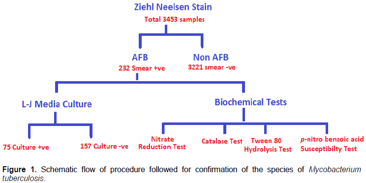
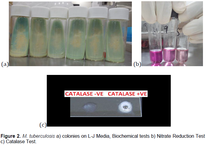
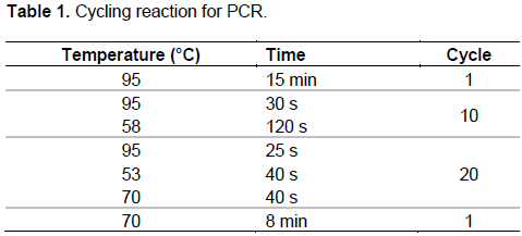
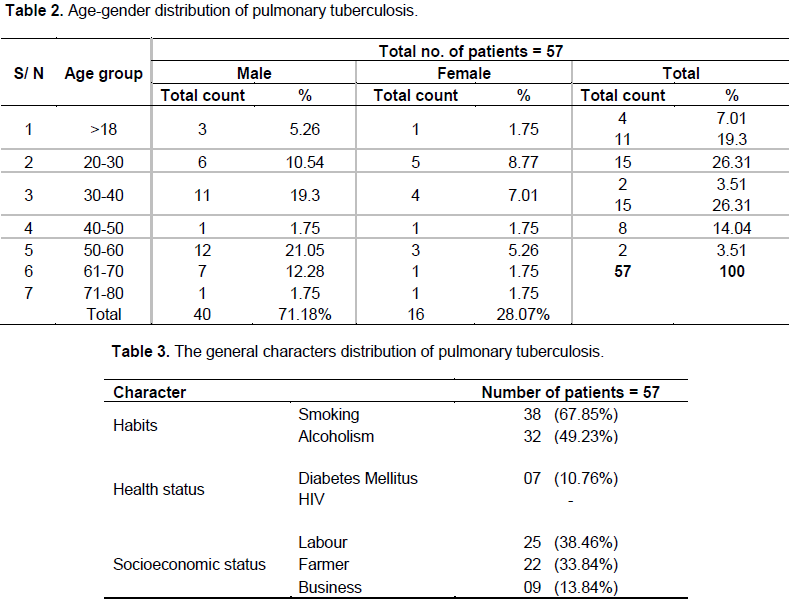
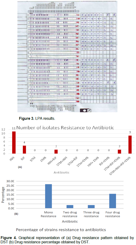
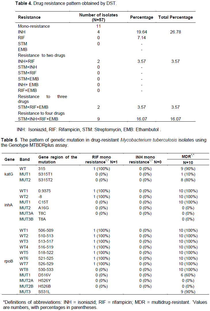
The control of tuberculosis (TB) has become a global health challenge due to the emergence of multidrug-resistant tuberculosis (MDR-TB) in Mycobacterium tuberculosis (MTB). This highlights the need for faster and more accurate detection of tuberculosis cases. The study aims to detect MDR-TB strains of pulmonary tuberculosis using resistance ratio method and to compare the diagnostic value of drug susceptibility testing (DST) with line probe assay (LPA) using Genotype MTBDRplus. All the sputum samples were tested for Acid Fast Bacilli (AFB) by Ziehl-Neelsen’s staining method and were cultured on Lowenstein-Jensen (L-J) media. The identification and confirmation of M. tuberculosis were done using various biochemical tests. DST was carried against the first line of anti-TB drugs. MTB positive samples were subjected to LPA. A total of 57 samples were subjected to DST and LPA for the detection of drug resistance of MTB to RIF and INH after conventional detection methods were applied to all the samples. Among these 57 MTB samples, 11 (19.29%) were resistant to INH, 4 (7.14%) were resistant to RIF; eleven (19.29%) isolates were identified as MDR-TB. LPA revealed 54 MTB positive among 57 MTB culture-positive samples and 3 showed invalid results. In LPA, MDR-TB was found in10 samples (17.54%) in which one was RIF-resistant. The study concludes risk factors that resulted in the development of TB are biomedical, socio-cultural, and behavioral interactions. LPA can be used as a rapid diagnostic technique for the detection of MDR-TB.

Key words: Tuberculosis (TB), multidrug resistance (MDR-TB), drug susceptibility testing (DST), line probe assay (LPA).


 INTRODUCTION

Tuberculosis (TB) caused by Mycobacterium tuberculosis (MTB) is one of the leading causes of deaths due to infectious diseases in developing nations, including India.  According to the World Health Organization (WHO), 1.8 million people died due to TB, and 10.4 million people were infected with TB in 2015. Overall, 95% of the total TB deaths occurred in low- and middle-income countries. Six countries had accounted for about 60% of the global total, with India leading the burden of tuberculosis in the world (WHO, 2016). Globally India has become a home for one-fourth of TB burden patients. In 2015, around 280, 00, 00 TB cases occurred in India, and 48, 0000 people had died due to this disease. The global emergence of Multidrug-resistant tuberculosis (MDR-TB) is a major public health problem as it causes a major challenge to control the disease and the mortality rates associated with it. India has a high burden of patients with MDR-TB. Annually, according to India reports, 130,000MDR-TB cases among which 79,000 MDR-TB cases are pulmonary TB (TB Statistics for India; WHO, 2008).
 
Early diagnosis of MDR-TB is necessary for the effective treatment and control of MDR-TB strains. It is known that resistance to isoniazid (INH) and rifampicin (RIF) is a key factor to determine the efficacy of the currently recommended standard treatment regimens. (Telenti et al., 1997; Cavusoglu et al., 2002; de Viedma et al., 2002; Morcillo et al., 2002; Saribas et al., 2003). Conventional techniques for MDR-TB detection are time-consuming and require sophisticated laboratory infrastructure, which causes delay in reporting the results. This in turn delays the proper treatment which increases the risk of transmission of the disease (Chauhan and Arora, 2004). Despite various measures available for the detection of MDR-TB, the prevalence of MDR-TB rate has remained unchanged in recent years (Havumaki et al., 2017). There is an urgent need for standardizing rapid molecular tests such as Line Probe assay (LPA) (Hillemann et al., 2005).
 
Identification of infectious cases is an important step for TB control programs worldwide. Detection of AFB in sputum by smear microscopy continues to be the mainstay diagnostic technique since its introduction in the late nineteenth century (Chakravorty and Tyagi, 2005). Drug susceptibility testing in Lowenstein-Jensen (L-J) culture media remains the cornerstone and gold standard for the diagnosis of resistance patterns in TB. There are different conventional methods (proportion, resistance ratio, and absolute concentration methods), the radiometric method, and other newer methods used for determining antimicrobial susceptibility patterns. The most extensively used is the proportion (PR) and the resistance ratio (RR) methods. The RR method compares the resistance of unknown strain of tubercle bacilli (test organism) with that of a standard laboratory strain of M. tuberculosis (H37Rv) (Acharya et al., 2010).
 
The drug resistance of Indian isolates varies from 52.2 to 2% (Iqbal et al., 2003). As there are variations in the resistance pattern to address this, the hospital-based study was undertaken to perform microscopy, biochemical analysis, and drug susceptibility testing using the RR method for the first line of anti-TB drugs. This method gives faster results than other DST methods.The revolution of TB diagnosis started in 2008 when WHO and Foundation of Innovative Diagnostics (FIND) endorsed the use of Line Probe Assay (LPA) which was developed by Genotype MTBDRplus (Hain  Life  Science, Nehren, Germany) for the detection of MDR-TB (Nathavitharana et al., 2017). The assay is based on the multiplex polymerase chain reaction (PCR) method and uses reverse hybridization to identify MDR-TB from MTB culture and smear-positive sputum specimens (Barnard et al., 2012). LPA detects the wild-type sequencing or specific mutations associated with the rpoB gene for RIF resistance, the katG gene for high-level INH resistance, and inhA regulatory region gene for low-level INH resistance (Chauhan and Arora, 2004). It targets a mutation in the 81 base pair “core region” of the rpoB gene which detects almost 95% of RIF resistant strains and ahpC-oxy R intergenic region which detects 5-10% mutation in INH resistant (Nathavitharana et al., 2017). The study aimed to compare the conventional drug susceptibility testing (DST) and LPA for the detection of RIF and INH resistance in MTB.


 MATERIALS AND METHODS

Sample size and patient recruitment
 
The study was approved by the Institutional Ethical Board, KLE Academy of Higher Education and Research, Belagavi (Karnataka, India). Patients attending Out Patient Department (OPD) and wards of the Karnataka Lingayat Education Society (KLES) Dr.Prabhakar Kore Hospital and Medical Research Centre with symptoms such as fever, night sweats, and cough for more than 2 weeks with sputum, chest pain from January 2013 to December 2014 were included in the study. Non-TB patients, extra-pulmonary TB, and culture-negative samples excluded. The sampling method for this study was inverse sampling. The estimated sample size according to this method should be 125 positive cultures. However, in this study 232 smear, positive samples were obtained out of which 75 samples were culture-positive. Hence to establish the validity of results, bootstrapping analysis was carried out (Haldane, 1945).
 
Collection of samples
 
Samples were collected after the Ethical clearance was obtained from the Institutional Ethical Board of KLE University Belagavi (Karnataka, India).The study was carried out at the Department of Microbiology, J.N Medical College, KLE University, and Belagavi, India between January 2013 and December 2014. This is a Hospital-based study. Sputum samples of all the 3453 patients (1 sputum sample per patient) were collected from Hi-Tech Laboratory, KLES Dr.Prabhakar Kore Hospital, Belagavi from January 2013 to December 2014. Patients attending Out Patient Department (OPD) and wards of the KLES Dr. Prabhakar Kore Hospital and Medical Research Centre, who have fever, night sweats, cough for more than 3 weeks with sputum and chest pain were included sputum sample was collected in a clean, dry, sterile wide-neck, leak-proof screw cap container (Figure 1).
 
Microscopic technique
 
From purulent part of the specimen, and subjected to smear microscopy by Ziehl-Neelsen (ZN) staining for acid-fast bacilli in laboratory level 3 biosafety level 2. The grading of smears was done according to the guidelines provided by the Revised National Tuberculosis Control Program (RNTCP) of India (WHO, 2009). The results of the microscopic examination were reported according to the Revised National Tuberculosis Control Programme (RNTCP). The processing of samples was carried out in a biosafety cabinet with standard procedures (Revised TB Programme, 2005; Central TB Division, 2005).
 
 
Culture technique
 
All sputum specimens were handled at the bio-safety level (BSL-3). Only smear-positive samples were cultured by Petroff's method (N-acetyl-L-cysteine and sodium hydroxide (NALC-NaOH) the sediments were suspended in distilled water. The supernatant was discarded after centrifugation and two slopes of Löwenstein–Jensen (LJ) media was taken for inoculation of each sample. The inoculated LJ media was incubated at 37°C. Reading and interpretation was done for the colony formation every week, preferably twice within the first week. The contaminated cultures and rapidly growing mycobacteria (colonies apparent in less than 7 days) were removed. M. tuberculosis colonies were developed within 3-4 weeks. Cultures were kept for up to 8 weeks before being reported as negative (Figure 2a) (Protocol for Processing, 1998).
 
 
Biochemical tests
 
The identification and confirmation of M. tuberculosis were done using various biochemical tests. Catalase Test- Into two screw-cap test-tubes 0.5 ml of phosphate buffer was dispensed (pH7.0). One tube was placed in the water-bath at 68°C for 20 min. Another tube was left at room temperature (Figure 2c).
 
Nitrate reduction test
 
Two loopful of bacterial growth was emulsified in 0.2 ml distilled water; then 2 ml of the substrate medium was added. Incubation was done at 37°C for 2 h. To each tube in sequence one drop of reagent 1 (HC1), two drops of reagent 2 (Sulfanilamide), and two drops of reagent 3 (n-naphthyl ethylenediamine dihydrochloride) was added. The development of a  red  colour  indicated  a  positive
reaction (Figure 2b).
 
Tween-80 hydrolysis
 
A volume of 0.5 ml freshly prepared Tween 80–peroxide substrate was added to each tube. The formation of bubbles was observed. The release of the oleic acid from Tween 80 results in the change of colour from the neutral indicator yellow to red within 5-10 days.
 
p-nitro benzoic acid (PNB) susceptibility
 
Single slope of LJ medium was inoculated containing Para-Nitro Benzoic acid (PNB Sodium Salt) 500 mcg/ml with bacterial suspension. It was incubated at 37°C and growth was recorded on the 28th day (Manual on isolation, 1998).
 
Drug susceptibility testing (DST)
 
Minimal Inhibitory Concentration (MIC) on solid LJ medium was used for DST which is expressed as a resistance ratio method according to the standard operating procedure of RNTCP. All the culture-positive MTB isolates (showing colonies more than 20) were further analyzed by DST resistant ratio method. The media containing two-fold dilutions of the primary anti-TB drugs were prepared as follows: INH, 0.5and 1.0 µg/ml; RIF, 32.0 and 64.0 µg/ml; streptomycin (SM), 16.0 and 32.0 µg/ml; and ethambutol (EMB), 4.0 and 8.0 µg/ml. One drop (100 µl) of 1 mg/ml bacillary suspension (McFarland No.1) from a Pasteur pipette was spread on the surface of each drug-containing slope of media of different concentrations. The same procedure was done for the H37RV strain, which was used as a positive control for the test. All the tubes were incubated at 37°C for 4 weeks and were observed every week. The growth was examined after 28 days and this was defined by the presence of 20 or more colonies in the drug-containing media (Figure 2a and b). The isolates were considered resistant when the growth appeared on the drug-containing media (Revised TB Programme 2014). MTB in all isolates were identified by the slow growth rate, colony morphology and biochemical tests such as the incapability to grow on PNB acid, niacin positive and catalase-negative test, nitrate negative test (Chauhan et al., 1998).
 
Line probe assay (LPA)
 
The bacterial colonies from cultures were used for DNA extraction (Bhawan, 2009). The bacterial DNA was extracted from the colonies of solid media according to the manufacturer’s instructions. Molecular grade water of 300 µl was added to and the DNA was by vortexing. This bacterial suspension was centrifuged for 15 min at 10,000 rpm. The supernatant was aspirated and the pellet was resuspended in 100 µl distilled water. The specimens were heat-killed at 95°C for 20 min, sonicated for 15 min and centrifuged at 13000 rpm for 5min. The supernatant containing DNA was transferred into a fresh tube. The extracted DNA was kept in4°C and was used within 1-7 days. The procedure of LPA was performed according to the manufacturer’s instructions (Hain Lifescience, Nehren, Germany, 2012). It consists of three steps: 1) multiplex PCR, 2) amplification and 3) reverse hybridization (Khanna et al., 2010). To avoid contamination, these steps were carried out in three separate rooms with restricted access and unidirectional flow.
 
A total of 5 µl DNA was added to 45µl of the master mix for each PCR reaction and the amplification procedure for cultured isolates were followed as per the directions given by the manufacturer (QIAGEN, Hilden, Germany). The cycling reaction is given in Table 1. Hybridization and detection is the final step in the assay, which was performed by using all the materials and reagents provided by the kit manufacturer (GmbH, Hain Life Science). A twelve well plastic tray was used for all three steps: denaturation, hybridization, and detection. In this tray, a denaturation buffer of 20 µl was dispensed to this 20 µl of DNA amplicons and mixed thoroughly. The solution was incubated at room temperature for 5 min. After denaturation, a 30 min hybridization step was done by adding 1ml of pre-warmed green hybridization buffer (HYB). After the aspiration of HYB 1ml of preheated red stringent wash buffer was added. To remove the excess STR buffer 1 ml of rinse (RIN) solution was added which was followed by the addition of previously prepared conjugate solution. Once again the strips were washed by 1ml of RIN solution and 1 ml of distilled water. In the final step, 1 ml of diluted substrate solution was added to each well and the tray was washed with 1 ml of distilled water. The bands were air-dried and the staining reaction was observed, showing whether the probes have been hybridized or not (GmbH, Hain Life Science).
 
The LPA strip has 27 reaction zones or bands for verification of the test procedures. The strips of LPA consist of two internal controls; conjugate control (CC) and amplification control (AC). The presence of positive MTB control tuberculosis-complex DNA band (TUB) reaction indicates the given specimen is positive for MTB complex. The test is considered valid only when wild type (WT) and mutation (MUT)  bands are present, if these two bands are absent the test is considered invalid (GmbH, Hain Life Science).


 RESULTS

All the 3453 sputum samples were subjected to Z-N staining. The smear examination yielded the following bacillary load: +1 AFB in 92 (39.65%), 2+ in 69 (29.75%) and 3+ in 71 (30.6%). Of these 232, 167 samples (71.18%) were males and 65 samples (28.07%) females (Table 2). Smoking (67.85%) and alcoholism (49.23%) were the commonest predisposing conditions and 72.3% were labour and farmer by profession (Table 3).
 
 
Out of 232 smear-positive samples, 75 were culture-positive, 70 were culture-negative, 34 were contaminated and 53 had less than20 colonies. Biochemical tests were performed for 65 samples (10 were excluded due to issues associated with culture handling). The samples were further tested by biochemical analysis using catalase, nitrate reduction test, tween-80 hydrolysis, and p-nitrobenzoic acid (PNB) susceptibility for the confirmation of MTB. Out of these, 60 (25.86%) isolates were MTB and 5 isolates (2.1%) were non-tuberculosis Mycobacteria (NTM).
 
Among 60, 3 yielded less than 20 colonies in a culture which were excluded from further tests. The remaining 57 samples were subjected to DST analysis (Figure 4). Among these 57 MTB samples, 11 (19.29%) were resistant to INH, 4 (7.14%) were resistant to RIF, and11 (19.29%) isolates were identified as MDR-TB through the resistance ratio method of DST (Table 4). LPA revealed 54 MTB positive among 57 MTB culture-positive samples and 3 showed invalid results (Figure 3). The TUB band was absent in all five NTM specimens. In LPA 10 samples (17.54%) were MDR-TB, in which one was RIF-resistant (Table 5).


 DISCUSSION

TB has existed for millennia and remains a major global health problem. In developing countries laboratory plays a crucial role in diagnosing TB. It is one of the top 10 causes of death worldwide, ranking above HIV/AIDS as one of the leading causes of death from an infectious disease (WHO, 2016). Thus, the present study was undertaken to study the drug resistance pattern, the risk factor associated with TB, and recommending the use of the resistance ratio method for DST. The risk factors which resulted in the development of TB were biomedical, socio-cultural, and behavioral interactions (Balaji et al., 2010).
 
One of the predisposing factors for the cause of Pulmonary TB was low economic status as these classes of people were more illiterate and have fewer health facilities which resulted in more mortality (Gupta et al., 2011). TB was more common in males (71.18%) when compared to females (28.07%). The accompanying risk factors (Smoking, Alcoholism, and DM), may have triggered the disease more in males as they are prone to such lifestyle (Acharya et al., 2010; Deodhar et al., 1999). Alcoholism is one of the most important morbid factors for TB infection (Fleming et al., 2006). This study predicts that Ziehl-Neelsen staining is rapid and inexpensive, but lacks sensitivity and specificity. It cannot be used to distinguish between the various members of the Mycobacterium and also requires a high amount of organisms in the specimen. Due to its low sensitivity, there are high chances of false negatives. Resistance testing is too expensive with modern techniques. In such cases as conventional L-J based approach, the DST method may be a suitable alternative. There are studies carried out in Bijapur and Pakistan where the isolation rate was 34.74 and 25.84% respectively, which can be compared to these results. There can be a variety of reasons for culture-negative; the organism may have lost their ability to grow on culture media or patients on treatment with regimens have negative results (Gaude et al., 2014).
 
Drug susceptibility testing was carried on all first-line anti-tubercular drugs-INH, RIF, EMB, and STM. It was observed that 11 specimens were sensitive to all 4 drugs.
 
INH mono-resistance was comparable, INH+RIF resistance is nearly 50% reduced in our case, resistance to STM+RIF+EMB combination is reduced to one third, and resistance to all four drugs is double in this study. The MDR-TB rate is highly variable between countries and in between the states of India i.e. Delhi (33.7%), Bihar (15%), Mumbai (51%), Gujarat (17.4%) and Tamil Nadu (20.3%) (Tripathy et al., 2015). The resistance of MTB in this study was (19.64%) which can be compared to Tamil Nadu and Gujarat. Since drug-resistant TB has increased in incidence and interfered with TB control programs, monitoring of drug resistance patterns is very much important to prevent MBR-TB outbreaks. So, all isolates of M. tuberculosis should be tested for their susceptibilities to the primary anti-tubercular drugs.
 
Of the conventional culture-based techniques for antimicrobial susceptibility testing, the Resistance Ratio (RR) and the Proportion (PR) methods are commonly used. The resistance ratio method is still in use in many countries especially the United Kingdom (Kent and Kubica, 1985). However, WHO has recommended the use of the proportion method to be used for determining drug susceptibility of M. tuberculosis. The RR method has been used in this study this compares the resistance of the unknown strain with that of the control strain on the same batch of the medium. Resistance can be expressed as the ratio of the MIC (Minimum Inhibitory Concentration) of the test strain to the MIC of the control strain in the same test. The RR method was convenient for inoculum preparation and it required a shorter time to perform. Interpretation of the result was rather simple when it can compare to other methods.
 
In MDR-TB detection, the conventional culture and DST on the solid LJ media is a time-consuming process. However, an early diagnosis of RIF and INH drug-resistant MTB is essential for effective treatment and control of MDR-TB. With the advent of molecular techniques and the development of commercial or in-house DNA hybridization or amplification methods, the results of MDR-TB can be obtained fast (Ahmed et al., 2017). With the introduction of LPA for the rapid diagnosis of drug-resistant TB, there has been a significant reduction in time to start the treatment in MDR suspected cases.
 
The present study was conducted to determine the pattern of mutations in MTB using LPA and to compare with DST for the detection of RIF and INH resistance in culture-positive isolates. The findings of this study showed that many mutations, which occurred in the rpoB and katG genes, are comparable to those reported in other countries (Taniguchi et al., 1996; Siddiqi et al., 2002). The existence of common mutations in the rpoB gene, at codons 526 and 531 isolates from India and other countries, supports the assumption that these mutations are common for many drug-resistant strains around the globe (Telenti et al., 1993; Sun et al., 2008). In this study, resistance to RIF was higher than that of INH in LPA. This similarity was also observed in Ethiopia (Meaza et al., 2017). The common mutation associated with RIF was similar to the present study.
 
The LPA test failed to detect mono- INH resistant strains in 11 specimens, which were detected by conventional DST. One MDR-TB sample detected by DST showed the false result in LPA as MTB which could be due to the presence of PCR inhibitors during the process of DNA extraction. Similar results were also observed in another study conducted in central India (Desikan et al., 2017). Among 57 isolates, 10 were RIF resistant strains and one was RIF mono-resistant of MDR-TB which had a mutation on rpoB S531L diagnosed by the presence of MUT3 band (Table 3). The most frequent mutation found in INH was a katG mutation, which occurred more commonly in MDR-TB strains than in INH mono-resistant strains. RIF resistance is associated with the mutations in 81 base pairs region (codon 527 to 533) of the rpoB gene (Yue et al., 2003). The finding of dominant mutations for RIF resistance in rpoB S531L in the present study is similar to a previously published report (Miotto et al., 2006).
 
In this study, no mutations were observed in the inhA gene region. Similarly, a low frequency of inhA gene mutations was reported in Ethiopia (Omer et al., 2016). A study from north India also has reported a low frequency of INH resistance mutation in the inhA gene. This could suggest that there are possibilities of mutation in other codons of the katG and inhA gene (Omer et al., 2016). The high prevalence of mutations within the rpoB core region in the MDR-TB strains isolated from India indicates the potential of a rapid diagnostic test for the detection of drug-resistant MTB. As stated, the detection of mutations in the rpoB gene is very effective for the diagnosis of drug resistance to RIF in MTB complex since the mutations in the hot spot region are prevailing. The invalid results observed in the present study were due to the lower bacillary load in sputum specimens or culture-negative samples, which emphasizes not using the LPA test, directly for smear-negative clinical specimens (Chauhan and Arora, 2004).
 
The limitation of LPA by Genotype MTBDR plus assay is that it requires sophisticated infrastructure, well-trained and skilled laboratory personnel. The test does not provide convenient results with sputum specimens, which have a lower bacillary load (Yadav et al., 2013). However, the use of LPA can lead to the earlier initiation of appropriate drug therapy which will thereby prevent further transmission of the drug-resistant strains. Applying the LPA method to detect drug resistance in the MTB isolates in clinical laboratories, require further research and method validation, aiming to enable the patients receive appropriate standardized MDR-TB treatment regime at an early stage of the illness.


 CONFLICT OF INTERESTS

The authors have not declared any conflict of interests.


 ACKNOWLEDGMENTS

The authors appreciate the Head of Department of Microbiology and Dean, J.N. Medical College, KAHER, Belagavi, and National JALMA Institute for Leprosy and other Mycobacterial Diseases, Agra (UP) for their help in providing the conventional and molecular technical platform for the study.


 REFERENCES

Acharya S, Ghimire P, Khadka D, Nepali S (2010). Comparison of Proportion and Resistance Ratio Methods for Drug Susceptibility Testing of Mycobacterium tuberculosis isolated from Patients Attending National Tuberculosis Centre, Nepal. SAARC Journal of Tuberculosis, Lung Diseases and HIV/AIDS 5(1):13-20.
Crossref


Ahmed S, Shukla I, Fatima N, Varshney SK, Shameem M (2017). Evaluation of genotype MTBDRplus line probe assay in detection of rifampicin and isoniazid resistance in comparison to solid culture drug susceptibility testing in a tertiary care centre of western Uttar Pradesh. Indian Journal of Medical Microbiology 35:568-574.
Crossref


Balaji V, Daley P, Anand AA, Sudarsanam T, Michael JS, Sahni DR, Chordia P, George IA, Thomas K, Ganesh A, John KR (2010). Risk Factors for MDR and XDR-TB in a Tertiary Referral Hospital in India. PLoS ONE 53:e9527.
Crossref


Barnard M, Parsons L, Miotto P, Cirillo D, Feldmann K, Gutierrez C, Somoskovi A (2012). Molecular detection of drug resistant tuberculosis by line probe assay-Laboratory manual for resource-limited settings. Geneva: FIND.


Bhawan N (2009). Training Manual for Mycobacterium tuberculosis Culture and Drug susceptibility testing. National Tuberculosis Institute, Bangalore.


Cavusoglu C, Hilmioglu S, Guneri S, Bilgic A (2002).Characterization of rpoB mutations in rifampin-resistant clinical isolates of Mycobacterium tuberculosis from Turkey by DNA sequencing and line probe assay. Journal of Clinical Microbiology 40(12):4435-4438.
Crossref


Central TB Division (2005). Directorate General of Health Services, Ministry of Health & family Welfare, Government of India.


Chakravorty S, Tyagi J (2005). Novel Multipurpose Methodology for Detection of Mycobacteria in Pulmonary and Extrapulmonary Specimens by Smear Microscopy, Culture, and PCR. Journal of Clinical Microbiology 43:2697-2702.
Crossref


Chauhan LS, Arora VK (2004). Central TB Division, Directorate General of Health Services, Ministry of Health and Family Welfare; Indian Academy of Paediatrics, Management of Paediatric Tuberculosis under the Revised National Tuberculosis Control Program (RNTCP). Indian Paediatrics 41:901-905.


Chauhan MM, Mahadev B, Balasangameshwara, VH (1998). Manual on isolation, identification and sensitivity testing of Mycobacterium tuberculosis. National Tuberculosis Institute, Bangalore, India.


de Viedma DG, Infantes MDSD, Lasala F, Chaves F, AlcaláL, Bouza E (2002). New real-time PCR able to detect in a single tube multiple rifampin resistance mutations and high-level isoniazid resistance mutations in Mycobacterium tuberculosis. Journal of Clinical Microbiology 40(3): 988-995.
Crossref


Deodhar LL, Miskeen P, Chomal S (1999). Drug resistance in tuberculosis. Bombay Hospital Journal 41:253.


Desikan P, Panwalkar N, Mirza SB, Chaturvedi A, Ansari K, Varathe R, Chourey M, Kumar P, Pandey M (2017). Line probe assay for detection of Mycobacterium tuberculosis complex: An experience from Central India. The Indian Journal of Medical Research 145(1):70.
Crossref


Fleming MF, Krupitsky E, Tsoy M, Zvartau E, Brazhenko N, Jakubowiak, W, McCaul ME (2006). Alcohol and drug use disorders, HIV status and drug resistance in a sample of Russian TB patients. International Journal of Tuberculosis Lung Diseases 10:565-570.


Gaude GS, Hattiholli J, Kumar P (2014). Risk factors and drug-resistance patterns among pulmonary tuberculosis patients in the northern Karnataka region, India. Nigerian Medical Journal 55:327-333.
Crossref


HainLifesciences GmbH Germany (2012). GenoType MTBDR Plus VER 2.0. Instructions for Use IFU-304A-02

View  


Gupta S, Shenoy VP, Mukhopadhyay C, Bairy I, Muralidharan S (2011). Role of risk factors and socioeconomic status in pulmonary tuberculosis: A search for the root cause in patients in a tertiary care hospital, South India. Tropical Medicine and International Health 16:74-78.
Crossref


Haldane JBS (1945). On a method of estimating frequencies. Biometrika 33(3):222-225.
Crossref


Havumaki J, Hillemann D, Ismail N, Omar SV, Georghiou SB, Schumacher SG, Boehme C, Denkinger CM (2017). Comparative accuracy of the REBA MTB MDR and HainMTBDRplus line probe assays for the detection of multidrug-resistant tuberculosis: A multicenter, non-inferiority study. PloS ONE 12(3):0173804.
Crossref


Hillemann D, Weizenegger M, KubicaT, Richter E, Niemann S (2005). Use of the genotype MTBDR assay for rapid detection of rifampin and isoniazid resistance in Mycobacterium tuberculosis complex isolates. Journal of Clinical Microbiology 43(8):3699-3703.
Crossref


Iqbal S, Iqbal, R, Khan MM, Hussain I, Akhtar A, Shabbir I (2003). Comparison of Two Conventional Techniques used for the Diagnosis of Tuberculosis. International Journal of Agriculture and Biology 5:545-547.


Kent PT, Kubica GP (1985). Public health Mycobacteriology: A guide for the level III laboratory. US Department of Health and Human Services, Public health service, CDC, Georgia.


Khanna A, Raj VS, Tarai B, Sood R, Pareek PK, Upadhyay DJ, Sharma P, Rattan A, Saini, KS, Singh H (2010). Emergence and molecular characterization of extensively drug-resistant Mycobacterium tuberculosis clinical isolates from the Delhi Region in India Antimicrobial agents and chemotherapy 54(11):4789-4793.
Crossref


Manual on Isolation, Identification and Sensitivity (1998). Testing of Mycobacterium tuberculosisntiindia.kar.nic.in/cdc district level.


Meaza A, Kebede A, Yaregal Z, Dagne Z, Moga S, Yenew B, Diriba G, Molalign H, Tadesse M, Adisse D, Getahun M (2017). Evaluation of genotype MTBDR plus VER 2.0 line probe assay for the detection of MDR-TB in smear-positive and negative sputum samples. BMC Infectious Diseases 17(1):280.
Crossref


Miotto P, Piana F, Penati V, Canducci F, Migliori GB, Cirillo DM (2006). Use of genotype MTBDR assay for molecular detection of rifampin and isoniazid resistance in Mycobacterium tuberculosis clinical strains isolated in Italy. Journal of Clinical Microbiology 44(7):2485-2491.
Crossref


Morcillo N, Zumarraga M, Alito A, Dolmann A, Schouls L, Cataldi A, Kremer K, Van Soolingen D (2002). A low-cost, home-made, reverse- line blot hybridization assay for rapid detection of rifampicin resistance in Mycobacterium tuberculosis. International Journal of Tuberculosis and Lung Disease 6:959-965.


Nathavitharana RR, Cudahy PGT, Schumacher SG, Steingart KR, Pai M, Denkinger CM (2017). Accuracy of line probe assays for the diagnosis of pulmonary and multidrug-resistant tuberculosis: A systematic review and meta-analysis. European Respiratory Journal 18:49-51.
Crossref


Omer ZB, Mekonnen Y, Worku A, Zewde A, Medhin G, Mohammed T, Pieper R, Ameni G (2016). Evaluation of the GenoTypeMTBDRplus assay for detection of rifampicin-and isoniazid-resistant Mycobacterium tuberculosis isolates in central Ethiopia. International Journal of Mycobacteriology 5(4):475-481.
Crossref


Protocol for Processing Specimens for Culture (1998).


Revised National TB Control Programme (2014). Training Manual for Mycobacterium tuberculosis Culture & Drug susceptibility testing.

View


Revised National Tuberculosis Control Programme Laboratory Network (2005) Guidelines for quality assurance of smear microscopy for diagnosing tuberculosis. TBC India.


Saribas Z, Kocagoz T, Alp A, Gunalp A (2003). Rapid Detection of Rifampin Resistance in Mycobacterium tuberculosis Isolates by Heteroduplex Analysis and Determination of Rifamycin Cross-Resistance in Rifampin-Resistant Isolates. Journal of Clinical Microbiology 41(2):816-818.
Crossref


Siddiqi N, Shamim M, Hussain S, Choudhary RK, Ahmed N, Prachee, Banerjee S, Savithri GR, Alam M, Pathak N, Amin A, Hanief M, Katoch VM, Sharma SK, Hasnain SE (2002). Molecular Characterization of Multidrug-Resistant Isolates of Mycobacterium tuberculosis from patients in North India. Antimicrobial Agents and Chemotherapy 46 (2):443-450.
Crossref


Sun Z, Chao Y, Zhang X, Zhang J, Li Y, Qiu Y, Liu Y, Nie L, Guo A, Li C (2008). Characterization of Extensively Drug-Resistant Mycobacterium tuberculosis Clinical Isolates in China. Journal of Clinical Microbiology 46(12):4075-4077.
Crossref


Taniguchi H, Aramaki H, Nikaido Y, Mizuguchi Y, Nakamura M, Koga T, Yoshida S (1996). Rifampicin resistance and mutations of the rpoB gene in Mycobacterium tuberculosis.FEMS Microbiology Letters 144:103-108.
Crossref


TB Statistics for India (National and State Statistics)

View


Telenti A, Honoré N, Bernasconi C, March J, Ortega A, Heym B, Takiff HE, Cole ST (1997). Genotypic assessment of isoniazid and rifampin resistance in Mycobacterium tuberculosis: A blind study at reference laboratory level. Journal of Clinical Microbiology 35(3):719-723.
Crossref


Telenti A, Imboden P, Marchesi F, Lowrie D, Cole S, Colston MJ, Matter L, Schopfer K, Bodmer T (1993). Detection of rifampicin-resistance mutations in Mycobacterium tuberculosis. Lancet 341:647-650.
Crossref


Tripathy S, Kumar R, Singh SD (2015). Prevalence of Multidrug Resistant Pulmonary Tuberculosis in North Bihar. Journal of Clinical and Diagnostic Research 9:09-12.
Crossref


World Health Organization (2008). Molecular line probe assays for rapid screening of patients at risk of multi-drug resistant tuberculosis (MDR-TB). Policy statement WHO, Geneva, Switzerland TBC.

View


World Health Organization (2009). Guidelines for surveillance of drug resistance in tuberculosis (No. WHO/HTM/TB/2009.422). Geneva: World Health Organization.


World Health Organization (2016). Global Tuberculosis Report, WHO, Geneva, Switzerland.

View


Yadav RN, Singh BK, Sharma SK, Sharma R, Soneja M, Sreenivas V, Myneedu VP, Hanif M, Kumar A, Sachdeva KS, Paramasivan CN, Vollepore B, Thakur R, Raizada N, Arora SK, Sinha S (2013). Comparative Evaluation of GenoTypeMTBDRplus Line Probe Assay with Solid Culture Method in Early Diagnosis of Multidrug-Resistant Tuberculosis (MDR-TB) at a Tertiary Care Centre in India. PLoS ONE 8(9):72036.
Crossref


Yue J, Shi W, Xie J, Li Y, Zeng E, Wang H (2003). Mutations in the rpoB Gene of Multidrug- Resistant Mycobacterium tuberculosis isolates from China. Journal of Clinical Microbiology 41(5):2209-2212.
Crossref


          */?>