ABSTRACT
Molecular identification of four endophytic fungi (Penicillium singorense, Curvularia geniculata, Aspergillus neoflavipes and Alternaria alternata) from two important medicinal plants, Calotropis procera (L.) R.Br. and Catharanthus roseus (L.) G.Don. was carried out by 18SrRNA sequencing. Genetic diversity using two RAPD primer viz. OPB-07 and OPC-06 showed good result with high polymorphism between the four strains. The sample DRC1 Accession No. MG322179 indicated strong homology with Penicillium singorense, strain DI16-118 (Accession No. LT558940), sample DRC2 Accession No. MG322180 with Curvularia geniculata strain F1 (Accession No. KX022497), sample DRC3 Accession No. MG322181 with Aspergillus neoflavipes strain AJR1 ribosomal gene (Accession No. KX218387), sample DRC4 Accession No. MH021686 with A. alternata strain AE1 18SrRNA gene, partial sequence, ITS1, 5.8S rRNA gene and ITS 2 complete sequence (Accession No. KY676196). Phylogenetic tree showing relatedness of sample DRC1 Accession No. MG322179 with 19 strains of Penicillium sp, DRC2 Accession No. MG322180 with 34 strains of Curvularia sp, DRC3 Accession No. MG322181 with 24 strains of Aspergillus sp and DRC4 Accession No. MH021686 with 16 strains of Alternaria sp of diverse origin. The present investigation thus gave an insight for the detection and genetic diversity study among the fungal endophytes isolated from two important Indian medicinal plants (C. procera and C. roseus) and it would probably the first report of its own kind from West Bengal, India.
Key words: Calotropis procera, Catharanthus roseus, National Center for Biotechnology Information (NCBI), homology search, endophytic fungi, DNA marker, Random amplification of polymorphic DNA (RAPD) analysis, 18SrRNA sequencing.
Medicinal plants are the reservoir of biopharmaceutical compound traditionally used for the treatment of human ailment. Deforestation or constant use of medicinal plants makes them threatened or endangered (
Chinedu, 2017). Conservation of medicinally important plant through sustainable practices is one of the methods for their availability. Another alternative approach for the production of secondary metabolites or biopharmaceutical is the search for fungal endophytes having medicinal importance. The long association of fungi with plants in mutuality’s mode probably makes fungi rich sources for bioactive compounds (Pimentel, 2011).
The living organism is made up of DNA, RNA and protein, similar organism has the similar type of these molecules. Molecular phylogeny generally made with such data to infer a "relationship tree" that shows the probable
evolution of various organisms. After the discovery of
Sanger sequencing in 1977 it became possible to isolate and identify these molecular structures (Sanger and Coulson, 1975; Sanger et al., 1977). Studies on the diversity of fungal endophytes are expected to yield potential sources of natural products and bioactive compounds for medicinal, agricultural and industrial uses, such as new antibiotics as well as novel biocontrol agents (Molina et al., 2012). Molecular identification and phylogeny of an organism generally accomplished by the help of hereditary molecular differences, mainly in DNA sequences. DNA sequencing is therefore specific identification of an individual than conventional culture-based methods: identification is refined by including internal transcribed spacer (ITS) sequence analyses with phenotypic characterization. Nuclear-encoded ribosomal DNA (rDNA) gene has been the gene of interest to analyze phylogenetic relationships and resolve taxonomic problems in different taxonomic levels (Cai et al., 2005; Jeewon et al., 2004). The internal transcribed spacer (ITS) regions of the ribosomal DNA (rDNA) have become a more popular marker for systematic and phylogenetic studies of closely related species of animals, plants and fungi (Von der Schulenburg et al., 2001), recently phylogenetic and taxonomic analysis in 100 endophytic fungi from
Chenopodium quinoa has been carried out (Gonzalez-Teuber et al., 2017). Endophytic fungi often possess 'cryptic' morphological characters (Ganley et al., 2004). The cryptic morphological identity exhibited by fungal endophytes can be resolved with the help of molecular genetic marker-based identification (Bhagobaty and Joshi, 2011).
Two common medicinal plant, namely Calotropis procera and Catharanthus roseus commonly available in tropical area, mainly West Bengal has potential to provide various active ingredients of pharmaceutical importance. In the natural product research, secondary compounds leads to search an alternative way to obtain already existing active ingredients and new active phyoto-chemical from the mutulaistic symbionts mainly fungi, has gained momentum rapidly (Carvalho et al., 2016).
In the present investigation, an attempt has been made to identify endophytic fungi from C. procera (L.) R.Br. and C. roseus (L.) G.Don., the two important medicinal plant and phylogenetic classification with the collective patterns generated by partial 18S rDNA sequencing and random amplification of polymorphic DNA (RAPD).
Genomic DNA isolation
Genomic DNA extraction from pure culture isolated from C. procera and C. roseus of Alternaria sp (Sample No.1), Penicillium sp (Sample No. 3), Curvularia sp ( Sample No. 4) and Aspergillus sp (Sample No. 5) was performed following the standard protocol (Gontia- Mishra et al., 2013) with little modification. In brief fungal mycelium (800 mg of each) were taken from pure and fresh fungal culture, crushed with (3.2 ml) extraction buffer (0.1 M Tris HCl pH 8,10 mm EDTA pH 8, (2.5M) NaCl, 3.5% CTAB,150 μl of 20 mg/ ml of proteinase K) with sterilized fine sand in preatuoclaved mortar pestle. The mixture was transferred to 10 ml centrifuge tube (Tarsons) vortexes vigorously, then incubated in a water bath at 65°C for 30 min. The samples were then centrifuged at 10,000 rpm for 10 min at room temperature. Supernatant was collected and equal volume of phenol-chloroform- isoamyl alcohol (25:24:1) was added mix thoroughly, followed by centrifugation at 10.000 rpm for 10 min at room temperature. Supernatant was collected and equal volume of chloroform-isoamyl alcohol (24:1) was added, mix thoroughly followed by centrifugation at previous condition. Supernatant was then collected, equal volumes of chilled isopropanol added, allowed to precipitate at - 20°C freezers for overnight. Samples were the again centrifuge at 13.000 rpm for 15 min to form the DNA pellet. Supernatant was discarded; DNA pellet washed thoroughly with 1 ml of absolute ethanol by centrifugation at 500 rpm for 5 min. DNA pellet was dried under Laminar Air Flow, dissolved in 200 μl of sterile high performance liquid chromatography (HPLC) grade water. The genomic DNA was treated with RNase A according to manufacturer’s instruction to remove RNA and the purified DNA was quantified spectrophotometrically (NanoDrop 8000 spectrophotometer) to know their concentration and quality.
Amplification of 18S rRNA gene with ITS 1 and ITS 4 primer
Polymerase chain reaction (PCR) amplification of the 18S rRNA gene of four fungal isolates was performed using the respective genomic DNA with the primers ITS1: 5’- TCCGTAGGTGAACCTGCGG-3’ and ITS4: 5’- TCCGCTTATTGATATGC-3’ (White et al., 1990). PCR was performed in 25 μl reaction volume containing 2.5μl of 10X Taq DNA polymerase, 2.0 μl of 2.0 mM dNTP mix, 15 pmol of each primer ITS1 and ITS4, 50 ng template DNA and 1.5U of Taq polymerase (Promega) using the following program: initial denaturation at 94°C for 1 min followed by 35 cycles of denaturation at 94°C for 30 s, annealing at 53.5°C for 45 s, extension at 72°C for 1 min and a final extension at 72°C for 5 min on a thermal cycler (ABI Veriti Thermal Cycler).
18 SrRNA detection and sequencing of amplicon
The amplicon generated were resolved in 1.5% agarose gel with ethidium bromide containing a (100 bp) DNA ladder. The applicon was the enzymaticaly purified followed by EDTA-Ethanol purification method. The bi-directional DNA sequencing reaction of PCR amplicon was carried out with 18S sequencing primers (1F CTGGTGCCAGCAGCCGCGGYAA, 4R CKRAGGGCATYACWGACCTGTTAT) using BDT v3.1 Cycle sequencing kit on ABI 3730xl Genetic Analyzer. The PCR condition was 94°C 3 min, 94°C 30 s, 48°C 30 s, 72°C 1 min, 72°C 7 min, 4°C forever, the total cycles were 30.
Submission of 18Sr RNA gene sequence in the NCBI database and construction of phylogenetic tree
The partial 18S rRNA gene sequence of selected fungal antagonists was submitted to NCBI GenBank for obtaining Accessions. Parsimony Methods were conducted in MEGA 5 software. The evolutionary history was inferred by using the Maximum Likelihood (Tamura et al., 2004). Evolutionary Genetics Analysis was performed using Maximum Likelihood, Evolutionary Distance (Tamura et al., 2011). The bootstrap value was calculated and shown in the dendrogram.
RAPD polymerase chain reaction
PCR was carried out in a final reaction volume of 25 µl in ABI Veriti Thermal Cycler with three RAPD primers: OPB-07 5’- GGTGACGCAG -3’ (10 nts.) OPC-06 5’- GAACGGACTC-3’ (10 nts.) and OPC-07 5’- GTCCCGACGA-3’ (Gonita-Mishra et al., 2013). Composition of reaction mixture for PCR is : Nuclease free water to make-up volume for 25 μl, DNA 50 ng, Primer (10 pmole) 1.0 μl, 2X PCR Master Mix 10 μl, total Volume 25 μl. PCR tubes containing the mixture were tapped gently and spin briefly. The PCR tubes with all the components were transferred to thermal cycler. The PCR protocol designed for 40 cycles for the primers used is: initial denaturation 94°C, 4 min; denaturation 94°C, 1 min; annealing 37°C, 1 min; extention 72°C 2 min and final extension 72°C, 5 min.
Visualization of PCR product
To confirm the targeted PCR amplification, 10 μl of PCR product from each tube was mixed with 2 μl of 6X gel loading dye and electrophoreses on 2.0% agarose gel containing ethidium bromide and co run with 100 bp DNA ladders, at the constant 5 V/cm for 45 min in 1X TAE buffer. The amplified products were visualized under UV light and documented by a gel documentation system (BIORAD).
Data analysis and construction of dendrograms from RAPD profile
Clear and distinct bands amplified by RAPD primers were scored for the presence (1) and absence (0) of the corresponding band through OPC-06 and OPB-07 primers, in the gDNA of the sample. The data were entered into the binary matrix and subsequently analyzed. Using FreeTree Software, cluster analysis was performed by agglomerative technique using the UPGMA (Unweighted Pair Group Method with Arithmetic Mean, replication no. 3) method. Relationships between the samples were graphically represented in the form of dendrograms.
Qualitative and quantitative estimation of genomic DNA
Nanodrop reading of genomic DNA of four samples under study viz: 1,3,4 and 5 showed high yield and good quality 847.4 ng/ μl (Alternaria sp), 469.8 ng/ μl (Penicillium sp), 811.3 ng/ μl Curvularia sp and 240.3 mg/μl (Aspergillus sp) respectively.
Amplification of 18S rRNA using ITS1 and ITS 4 primer
Amplification of the rDNA regions of four sample under study with ITS1 and ITS 4 oligonucleotide primers yielded amplicon of approximately 900 bp as estimated by 1.5% agarose gel electrophoresis using known standard DNA ladder.
Sequencing of 18SrRNA gene
The four 18SrRNA amplicon subjected to sequencing namely Sample No.1 (Alternaria alternata DRC 4), Sample No. 3 (Penicillium sp DRC1), Sample No.4 (Curvularia sp DRC2), Sample No. 5 (Aspergillus sp DRC 3) gave a good sequence reading of standard fragment size. The sequences used for the final homology search, phylogenetic analysis were done after manual counting trimming and corresponded to the ITS 1 and 2 complete regions, 5’ portion of the 18S gene, 5.8 S complete sequences and the 3’ end of the 28S gene.
GenBank accession followed by homology searching
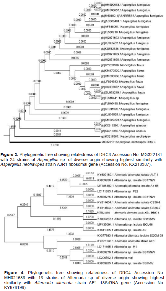
The FASTA format of four (sample DRC1, DRC2, DRC3, DRC4) 18 SrRNA sequence after submitting in NCBI database, the GenBank Accession were obtained (DRC1 MG322179, DRC2 MG322180, DRC3 KX218387, DRC4 MH021686) respectively. Sample 3 that is, DRC1 Accession No. MG322179 showed strong homology with Penicillium singorense, strain DI16-118, sample 4 that is, DRC2 Accession No. MG322180 showed strong homology with Curvularia geniculata strain F1, sample 5 that is, DRC3 Accession No. MG322181 showed strong homology with Aspergillus neoflavipes strain AJR1 ribosomal gene, sample 1 that is, DRC4 Accession No. MH021686 showed strong homology with Alternaria alternata strain AE1 18SrRNA gene, partial sequence, ITS1, 5.8S rRNA gene and ITS 2 complete sequence Accession No. KY676196 (Table 1). The phylogenetic analysis of these four isolated and identified strain reveals their relatedness with respective species (Figures 1, 2, 3 and 4).
RAPD analysis
Among the three RAPD primer (OPB- 07, OPC-06 and OPC-07) tested to identify genetic diversity good amplification was responded by two primers namely OPB-07 and OPC-06. The RAPD profile of four samples showed polymorphism and genetic diversity among them with fragment size of approximately 150 to 1.5 kb (Figure 5). The highest amplification was recorded with OPB-07 primer. RAPD marker has also been used (Gontia-Mishra et al., 2013) to amplify the genomic DNA of several species of Aspergillus and showed similar result
Dendrograms from RAPD profile
Dendrogram constructed from two RAPD profile depicted in Figures 6 and 7 reveals their relationship. In both the tree there are two groups showing the close relationship with sample number 1 (Alternaria sp.) and three (Penicillium sp.) and other with sample number 4 (Curvularia sp.) and 5 (Aspergillus sp.). Both the groups in other hand distantly related in the case of both primers (Figures 6 and 7).
Total homology searching between the four fungal isolates namely Penicillium sp, Curvularia sp, Aspergillus sp. and Alternaria sp. revealed that Alternaria sp., Penicillium sp. and Curvularia sp., Aspergillus sp. are also closely related to each other. The molecular analyses proved that these four fungal isolates to the diverse genera.
After thorough investigation for the presence of fungal endophytes from two important Indian medicinal plants (
C. procera and
C. roseus), four pure fungal isolates of
Penicillium sp.,
Curvularia sp.,
Aspergillus sp. and
Alternaria sp. has been identified by using molecular tools and techniques. From the perusal of literature it has been revealed that the four isaolated fungi have great medicinal value, as they produce several bioactive compounds of pharmaceutical importance.
Penicillium singorense is a species of the genus
Penicillium which was isolated from house dust (Visagie et al., 2014).
Curvularia is a hyphomycete fungus which is a facultative saprophyte of many plant species and common in soil. Most
Curvularia are found in tropical to temperate regions. However literature reveals that
Curvularia affinis,
Curvularia fallax, and
Curvularia senegalensis are synonymous to C.
geniculata. The presence of the mycotoxins, fumifungin, fumiquinazoline A/B and D, fumitremorgin B, gliotoxin, sphingofungins, subroutines, and verruculogen in
Aspergillus fumigatus a close relative of A. n
eoflavipes has been reported (Tamiya et al., 2015). The presence of isochromenone, from an endophytic fungus
Aspergillus fumigatus isolated from
Bacopa monnieri plant and proof its antioxidant and antitubercular activity has been investigated (Thakur et al., 2015). The methanol extract of
A. fumigatus inhibits the growth of the virulent strain of
Mycobacterium tuberculosis HRV with minimum inhibitory concentration 500 µg/ml.
Fungal endophytes are reported to secrete a diverse group of biomolecules extracellularly which are capable of reducing metal salts at a rapid scale under optimized conditions. One such endophyte is isolated from healthy leaves of Andrographis paniculata and employed for rapid synthesis of silver nanoparticles. The synthesized nanoparticles were evaluated for bactericidal activity against significant human pathogens (Azmath et al., 2016). The presence of paclitaxel from newly described endophytic fungus Pestalotiopsis microspora, isolated from the pine needles of T. wallichiana has been reported (Strobel et al., 1996). This study indicates that a large scale production of the natural product paclitaxel can be made at a lower cost just by mass production of the fungus in culture. Paclitaxel can be obtained from Yew (Taxus sp.), but such obtaining requires destruction of trees. Chemical synthesis of paclitaxel by cell and tissue culture from the Taxus sp are cost effective and production is not up to the mark (Guenard et al., 1993). Recently, paclitaxel as well as other toxins have been detected in shells and leaves of Corylus avellana and also in leaves of Ocimum basilicum (Ottaggio et al., 2008; Gangadevi and Muthumary 2017). It is well known that the tissues of living plant residue the endophytic fungi which posses analogical ability to perform a biosynthesis of secondary metabolites as the host plant. The ability of the endophytic fungus of Hazel plant Penicillium aurantiogriseum NRRL 62431 to independently synthesize paclitaxel was detected by liquid chromatography-mass spectrometry and proton nuclear magnetic resonance. The presence of Taxol from Phoma sp isolated from Calotropis gigantea has been detected (Hemamalini et al., 2015). The genome of Penicillium aurantiogriseum NRRL 62431 was sequenced and gene candidates that may be involved in paclitaxel biosynthesis were identified by comparison with the 13 known paclitaxel biosynthetic genes in Taxus (Yang et al., 2014). Two new metabolites, from a soil fungus, Curvularia affinis strain HS-FG-196. pyrenocine J (1) and pyrenochaetic acid D (2), together with two known metabolites, pyrenocine A (3) and pyrenochaetic acid A (4) has reported (Zhang et al., 2012). Pyrenocine J showed cytotoxic activity against the human hepatic cancer cell line HepG2 with an IC (50) value of 28.5 μg/ml. The presence of different mycotoxins, gliotoxin, sphingofungins, subroutines, and verruculogen in Aspergillus fumigatus a close relative of A. neoflavipes has also been reported (Tamiya et al., 2015) which are of great commercial value. The capacity of producing Vinblastin, an anticancerous drug from Alternaria sp isolated from C. roseus was reported (Guo et al., 1998). The paclitaxel a highly valued anticancerous compound was also produced by A. alternata TPF6 isolated from Taxus chinensis var. mairei (Tian et al., 2006).
The present study highlights the detection and genetic diversity among the fungal endophytes isolated from two important Indian medicinal plants. The endophytic fungi are expected to be a potential source of many natural bioactive products including paclitaxel and other taxanes. The search for a bioactive compound from fungal endophytes gaining momentum, thus the identification of fungal endophytes would be of great importance to the discovery of the newly active compound. The present study would probably the first report of its own kind from West Bengal, India.
The authors have not declared any conflict of interests.
Authors are thankful to the Secretary, Oriental Institute of Science and Technology for extending the facilities to conduct this research work. Authors highly appreciate the technical assistance of Dr. Anindya Sundar Panja for Bioinformatics works.
REFERENCES
|
Azmath P, Baker S, Rakshith D (2016). Mycosynthesis of silver nanoparticles bearing antibacterial activity. Saudi Pharmaceutical Journal 24(2):140-146.
Crossref
|
|
|
|
Bhagobaty RK, Joshi SR (2011). Multi-loci Molecular Characterisation of Endophytic Fungi Isolated from Five Medicinal Plants of Meghalaya, India. Mycobiology 39(2):71-78.
Crossref
|
|
|
|
|
Cai L, Jeewon R, Hyde KD (2005). Phylogenetic evaluation and taxonomic revision of Schizothecium based on ribosomal DNA and protein coding genes. Fungal Diversity 19:1-21.
|
|
|
|
|
Chinedu E (2017). Deforestation and the future of herbal medicine practice. Journal of HerbMed Pharmacology 6(3):94.
|
|
|
|
|
Carvalho PLN, Oliveira Silva E, Chagas-Paula DA, Luiz JHH, Ikegak M (2016). Importance and implications of the production of phenolic secondary metabolites by Endophytic Fungi: A Mini-Review. Mini-Reviews in Medicinal Chemistry 16(4):259-271.
Crossref
|
|
|
|
|
Gangadevi V, Muthumary J (2007). Endophytic fungal diversity from young, mature and senescent leaves of Ocimum basilicum L. with special reference to taxol production. Indian Journal of Science and Technology 1(1):1-15.
|
|
|
|
|
Ganley RJ, Brunsfeld SJ, Newcombe G (2004). A community of unknown, endophytic fungi in western white pine. Proceedings of National Academy of Science USA 101(27):10107-10112.
Crossref
|
|
|
|
|
González-Teuber A, Vilo V, Bascu-án-Godoy L (2017). Molecular characterization of endophytic fungi associated with the roots of Chenopodium quinoa inhabiting the Atacama Desert, Chile. Genomics Data 11:109-112.
Crossref
|
|
|
|
|
Gontia-Mishra I, Tripathi N, Tiwari S (2013). A simple and rapid DNA extraction protocol for filamentous fungi efficient for molecular studies. Indian Journal of Biotechnology 13:536-539.
|
|
|
|
|
Guenard D, Gueritte-Voegelein F, Potier P (1993). Taxol and taxotere: discovery, chemistry and structure activity relationship. Accounts of Chemical Research 26:160-167.
Crossref
|
|
|
|
|
Guo B, Li H, Zhang L (1998). Isolation of the fungus producing vinblastine. Journal of Yunnan University (Natural Science Edition) 20:214-215.
|
|
|
|
|
Hemamalini V, Kumar DM, Rebecca AI, Srimathi S, Muthumary J, Kalaichelvan P (2015). Isolation and Characterisation of Taxol producing endophytic fungi Phoma sp. from Calotropis gigantea and its anti-proliferative studies. Journal of Academia and Industrial Research 3(12):645-649.
|
|
|
|
|
Jeewon R, Liew ECY, Hyde KD (2004). Phylogenetic evaluation of species nomenclature of Pestalotiopsis in relation to host association. Fungal Diversity 17:39-55.
|
|
|
|
|
Molina G, Pimentel MR, Bertucci TCP, Pastore GM (2012). Application of fungal endophytes in biotechnological processes. Chemical Engineering Transaction 27:289-294.
|
|
|
|
|
Ottaggio L, Bestoso F, Armirotti A, Balbi A, Damonte G, Mazzei M et al., (2008). Taxanes from Shells and Leaves of Corylus avellana. Journal of Natural Product 71(1):58-60.
Crossref
|
|
|
|
|
Pimentel MR, Molina G, Dionísio AP, Junior MRM, Pastore GM (2011). The use of endophytes to obtain bioactive compounds and their application in biotransformation Process. Biotechnology Research International pp. 1-11.
Crossref
|
|
|
|
|
Sanger F, Coulson AR (1975). "A rapid method for determining sequences in DNA by primed synthesis with DNA polymerase". Journal of Molecular Biology 94(3):441-448.
Crossref
|
|
|
|
|
Sanger F, Nicklen S, Coulson AR (1977). "DNA sequencing with chain-terminating inhibitors". Proceedings of National Academy of Science USA 74(12):5463-5467.
Crossref
|
|
|
|
|
Strobel G, Yang X, Sears J, Kramer R, Sidhu RS, Hess WM (1996). Taxol from Pestalotiopsis microspora, an endophytic fungus of Taxus wallachiana. Microbiology 142:435-440.
Crossref
|
|
|
|
|
Tamiya H, Ochiai E, Kikuchi K, Yahiro M, Toyotome T, Watanabe A, Yaguchi T, Kamei K (2015). Secondary metabolite profiles and antifungal drug susceptibility of Aspergillus fumigatus and closely related species, Aspergillus lentulus, Aspergillus udagawae, and Aspergillus viridinutans. Journal of Infection and Chemotherapy 21(5):385-391.
Crossref
|
|
|
|
|
Tamura K, Nei M, Kumar S (2004). Prospects for inferring very large phylogenies by using the neighbor-joining method. Proceedings of National Academy of Science USA 101:11030-11035.
Crossref
|
|
|
|
|
Tamura K, Peterson D, Peterson N, Stecher G, Nei M, Kumar S (2011). MEGA5: Molecular Evolutionary Genetics Analysis using Maximum Likelihood, Evolutionary Distance, and Maximum Parsimony Methods. Molecular Biology and Evolution 28(10):2731-2739.
Crossref
|
|
|
|
|
Thakur JP, Haider R, Singh DK, Kumar BS, Vasudev PG, Luqman S, Kalra A, Saikia D, Negi AS (2015). Bioactive Isochromenone isolated from Aspergillus Fumigatus, Endophytic Fungus from Bacopa monnieri. Microbiological Research 6(1):14-18.
|
|
|
|
|
Tian R, Yang Q, Zhou G, Tan J, Zhang L, Fang C (2006). Taxonomic study on a taxol producing fungus isolated from bark of Taxus chinensis var. mairei. Journal of Wuhan Botanical Research 24:541-545.
|
|
|
|
|
Visagie CM, Houbraken J, Frisvad JC, Hong SB, Klaassen CH, Perrone G, Seifert KA, Varga J, Yaguchi T, Samson RA (2014) . "Identification and nomenclature of the genus Penicillium". Studies in Mycology 78:343-371.
Crossref
|
|
|
|
|
Von der Schulenburg JHG, Hancock JM, Pagnamenta A, Sloggett JJ, Majerus MEN, Hurst GDD (2001). Extreme length and length variation in the first ribosomal internal transcribed spacer of ladybird beetles (Coleoptera; Coccinellidae). Molecular Biology and Evolution 18: 648-660.
Crossref
|
|
|
|
|
White TJ, Bruns T, Lee S, Taylor J (1990). Amplification and direct sequencing of fungal ribosomal RNA genes for phylogenetics. In: Innis MA, Gelfand DH, Sninsky JJ, White TJ, (Eds), PCR Protocols: a guide to methods and applications. Academic Press, San Diego pp. 315-322.
|
|
|
|
|
Yang Y, Zhao H, Barrero RA, Zhang B, Sun G, Wilson IW, Xie F, Walker KD, Parks JW, Bruce R, Guo G, Chen L, Zhang Y, Huang X, Tang Q, Liu H, Bellgard MI, Qiu D1, Lai J, Hoffman A (2014). Genome sequencing and analysis of the paclitaxel-producing endophytic fungus Penicillium aurantiogriseum NRRL 62431. BMC Genomics 15(1):69.
Crossref
|
|
|
|
|
Zhang H, Mao LL, Qian PT, Shan WG, Wang JD, Bai H (2012). Two new metabolites from a soil fungus Curvularia affinis strain HS-FG-196. Journal of Asian Natural Product Research 14(11):1078-1083.
Crossref
|
|