Full Length Research Paper

ABSTRACT
Attachment of bacteria to food processing equipment surfaces can lead to product contamination, spoilage, and surface destruction. Most of bacteria in their habitat live in communities of different degrees of complexity, in general composing biofilms. Once installed the biofilm, the resistance of these sessile organisms increases, making it difficult thus the action of sanitizers. This multidisciplinary study led to the description regarding the effect of antimicrobial solutions of essential oils of Syzygium aromaticum and Thymus vulgaris and their combination on biofilm formed by Staphylococcus aureus ATCC 25923 on AISI 304 stainless steel and polypropylene surfaces and evaluate the possible effects of these oils on Staphylococcus aureus cells after treatment with sanitizing solutions based on these natural compounds. All sanitizing solutions showed antibacterial potential, being effective in reducing bacterial biofilms on these surfaces. The solution containing the combination of essential oils was the most efficient by reducing 7.38 and 6.58 Log CFU.cm-2 of cells adhered on the surfaces of AISI 304 stainless steel and polypropylene, respectively, after 5 min of contact.
Key words: Antimicrobial effect, microorganism, Syzygium aromaticum, Thymus vulgaris.
INTRODUCTION
MATERIALS AND METHODS
Microbiological analyzes were performed in the Food Microbiology Laboratory, Food Science Department, Universidade Federal de Lavras (UFLA), MG. The electron micrographs of the structure of mature biofilms and sanitized were obtained from scanning electron microscopy, from the Laboratory of Electron Microscopy and Ultrastructural Analysis, Plant Pathology Department, Universidade Federal de Lavras (UFLA), MG.
Microorganism used, standardization, inoculum preparation and storage
The microorganism used in the development of this study was S. aureus ATCC 25923. The culture of S. aureus was maintained at -18°C in microcentrifuge tubes with freezing medium [glycerol (150 mL), peptone (5 g), yeast extract (3 g), NaCl (5 g), H2O (1.000 mL), pH 7.2 ± 7.4]. During the experiment, subcultures were made for the maintenance of cultures. Aliquots were transferred to the microcentrifuge tubes containing tryptic soy broth (TSB) and incubated at 37°C/24 h. After culturing, 1 mL of the culture was dispensed into sterile microcentrifuge tubes and centrifuged at 6,000 x g for 8 min in a microcentrifuge. After removing the supernatant, the content was again coated with freezing medium and stored at -18°C.
For reactivation and use of the strain, 10 µL of the culture was inoculated in tubes containing 3 mL of TSB and incubated at 37°C/24 h. After incubation, 20 µL of the inoculum was removed and transferred to 200 mL of TSB. The number of cells per millilitre was quantified using standard curve and the growth monitored by spectrophotometry at 600 nm and then counting on plates. The bacterial culture was standardized to a concentration of 108 CFU/mL.
Experimental model of biofilm formation
Preparation and cleaning of coupons
The bacterial adhesion was conducted on AISI 304 stainless steel and polypropylene coupons with 1 mm thickness and dimensions of 10x20 mm.
The AISI 304 stainless steel coupons were cleaned individually with 100% acetone, submerged in detergent, rinsed with sterile distilled water, dried and cleaned with 70% ethanol (v/v). After cleaning, the coupons were again washed with sterile distilled water, dried for 2 h in oven at 60°C and autoclaved at 121°C/15 min (Rossoni and Gaylarde, 2000). As for the polypropylene coupons, they were initially immersed in a solution of commercial 0.3% peracetic acid for 30 min under stirring at 50 rpm at 50°C. They were then soaked in sterile distilled water at 80°C for 5 min and at room temperature for 1 minunder agitation of 50 rpm. The coupons were dried at 40°C for 2 h and autoclaved for 15 min at 115°C (Oulahal et al., 2008).
Adhesion of bacterial cells to surfaces
In two Petri dishes (140 mm diameter) 45 AISI 304 stainless steel coupons and 80 mL TSB were added and inoculated with 108 CFU/mL of culture. In two other Petri dishes the same procedures were employed, however, with 45 polypropylene coupons, with the aim of promoting the formation of biofilms on these surfaces. The plates were incubated at 37°C with orbital agitation of 50 rpm. At 48-hintervals, the coupons were collected, washed with peptone water (0.1% w/v) five times and immersed in TSB contained in sterile plates. This procedure was performed five times in order to complete the formation of the biofilm after 10 days of incubation (Joseph et al., 2001).
Enumeration of adhered cells
For enumerating the adherent cells, one AISI 304 stainless steel and one polypropylene coupon was removed from each Petri dish every two days of incubation. These were washed with peptone water (0.1% w/v) five times to remove planktonic cells and the sessile cells were collected using a standard sterile cotton swab. The swabs were transferred to tubes containing peptone water (0.1% w/v) was then agitated in a vortex for 2 min. After this procedure, serial dilution was carried out in which 0.1 mL aliquots were plated and the number of viable cells quantified in Triptic Soy Agar (TSA), using the surface seeding technique. The plates were incubated at 37°C/24 h, conducting a standard plate count at the end of this period, results expressed in CFU/cm2 (Joseph et al., 2001).
Obtention of essential oils
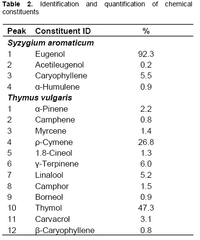
The essential oils of S. aromaticum and T. vulgaris were purchased through the Ferquima Ind. e Com. Ltda Company (Vargem Grande Paulista, São Paulo, Brazil); their physical and chemical parameters being described by the supplier, which produces and sells essential oils on an industrial scale.
Preparation of sanitizing solutions
To perform the sessile cell sensitivity test, four sanitizing solutions were formulated containing saline (NaCl 0.85% w/v), ethanol (p.a.95% v/v) and essential oil as shown in Table 1.
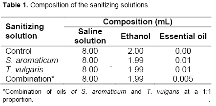
All sanitizing solutions contained a total volume of 10 mL and the amount of essential oils used in the formulation of the sanitizing solutions was defined from the Minimum Inhibitory Concentration (MIC) test results previously performed by disk diffusion technique (NCCLS, 2000 with modifications).
Treatment of biofilms with sanitizing solutions containing essential oils at different contact times
On the tenth day of cultivation, polypropylene and steel coupons were taken from each Petri dish, rinsed in 0.1% peptone water (v/v) five times to eliminate non-adherent cells, and immersed in the above sanitizing solutions for durations of 5 and 10 min at room temperature. After the sanitizing, the coupons were rubbed with standardized sterile swabs. The swabs were transferred to tubes containing 0.1% peptone water (v/v) and then agitated in a vortex for 2 min. After this procedure, serial dilution was conducted, 0.1 mL aliquots were plated and the number of viable cells determined in TSA medium, using the surface seeding technique. The plates were incubated at 37°C for approximately 24 h, conducting the standard plate count at the end of this period with, results expressed in CFU/cm2 (Joseph et al., 2001; with adaptations).
View the surfaces of coupons by scanning electron microscopy
The coupons of stainless steel and polypropylene containing biofilm and sanitizers were immersed in fixative solution (modified Karnovsky), pH 7.2, for 48 h. The coupons were cleaned by soaking for 10 mins in cacodylate buffer, and this procedure was repeated three times and posteriomente were fixed in 1% osmium tetroxide in water for three hours. After this process, were washed three times in distilled water and dehydrated in ethanol gradient (25, 50, 70, 90, 95%, 10 min and 100% three times for 10 min). Next was the drying of this material in a critical point apparatus (Bal-Tec CPD 030), mounting stubs and covered with a thin gold layer (Bal-Tec SCD sputter 050). Electron micrographs of the bacteria adhered to the surfaces of stainless steel and polypropylene were obtained before and after treatment with sanitizing solutions, and a scanning electron microscope Leo 040 Evo (Alves, 2004).
Experimental design and statistical analysis
A completely randomized design (CRD) was used in a 2 x 5 factorial outline with 3 replicates, the surface factor having 2 levels: stainless steel and polypropylene, the time factor with five quantitative levels: 48, 96, 144, 192 and 240 h. The enumeration of adhered cells on the stainless steel and polypropylene coupons after treatment with these sanitizing solutions at different contact times, used the CRD in a factorial scheme (4 x 2 x 2) with three replicates with the factor agents at four qualitative levels: control, S. aromaticum, T. vulgaris and combination, the factor surfaces with two qualitative levels: stainless steel and polypropylene, and the time factor with two quantitative levels: 5 and 10 min. Statistical analyzes were performed using the SISVAR (Ferreira, 2003) and R Development Core Team programs (R Development Core Team, 2004).
RESULTS AND DISCUSSION
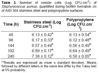
Table 2 shows the counts of sessile cells adhered to the surfaces of the AISI 304 stainless steel and polypropylene coupons during the biofilm formation process.
The adhesion of bacterial cells depends on factors such as physiology and cell morphology and physico-chemical properties of the contact surface. Gram negative microorganisms have greater ease of adhesion on surfaces when compared to Gram positive, as they have pili, flagella and fimbriae, as well as an outer membrane. Microorganisms electrically charged with negative charges have more difficulty to link directly to surfaces. The participation of a conditioning film formed by various compounds and molecules from the aqueous phase, will be decisive.
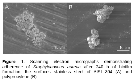
It can be observed that the microbial cells adhered similarly to both surfaces up to 192 h and differed significantly only at the end of the biofilm formation process that is at 240 h. The electron micrographs in Figure 1 illustrate cell adhesion of S. aureus on surfaces stainless steel of AISI 304 and polypropylene.
The adhesion of bacterial cells depends on factors such as physiology and cell morphology and physico-chemical properties of the contact surface.
The ability of S. aureus to adhere to solid surfaces pro-ducing compounds by multilayered cells embedded in a exopolysaccharide matrix is considered one of the relevant aspects of the epidemiology of this bacterium (Cucarella et al., 2001; Flach and Karnopp, 2005). This organization is extremely beneficial to all species of microorganisms, because it provides protection against adversity such as dehydration, colonization by bacterio-phages and antimicrobial resistance (Gilbert et al., 2003).
By the phenomenon known as passivation, chromium, due to its high affinity for oxygen, tends to combine with it, forming a thin layer of chromium oxide with an approximate thickness of 40Å. This passive layer is responsible for the corrosion resistance and the hydrophobicity of stainless steel.
In this context, in the case of initial adhesion, the more hydrophobic the bacterial cell, the greater its ability to bind directly to this surface. Similar considerations were observed by Meylheuc et al. (2006) and Sheng et al. (2007).
Thus, surfaces considered hydrophobic, such as stainless steel, allow the adhesion to occur more easily than less hydrophobic or hydrophilic surfaces, which is evidenced by counts and electron micrographs, which show improved adhesion of the cells on the surface of stainless steel at the end of 240 h incubation compared to the polypropylene surface.
In studies conducted by Boari et al. (2009), that consisted of evaluating S. aureus biofilm formation on stainless steel using milk as substrate and different growing conditions, biofilm formation by S. aureus was observed by scanning electron micrographs in all conditions tested, revealing the adhesion ability of this bacterium mainly to the stainless steel surface, which was also observed in electron micrographs of the present study.
In a review by Chmielewski and Frank (2003) it is shown that a layer of organic matter on the surface can promote and facilitate bacterial adhesion. Moreover, these authors state that the time of contact between cells and surfaces also influence the bacterial adhesion. The irreversible cell adhesion to surfaces occurs between 20 min and a maximum of 4 h of contact. After this period the removal of these cells requires the application of physical force, chemicals or heat. In this present study it is possible to observe that the bacterial cells have obtained adhesion to the stainless steel and polypropylene surfaces from 48 hand increasing, to a small extent, up to 240 h.
The probability of cells remaining irreversibly attached after sanitation procedures is high and corresponds to one of the main reasons for the formation of biofilms on surfaces that come into contact with food, becoming a constant source of contamination.
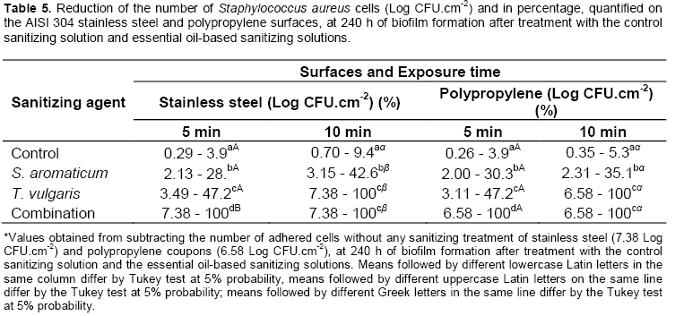
Table 3 presents the counts of sessile cells adhered to the surfaces of the AISI 304 stainless steel and polypropylene coupons after treatment with the control sanitizing solution and the essential oil-based sanitizing solutions. Table 4 shows the reduction percentage of the number of sessile cells after treatment with the sanitizing solutions.
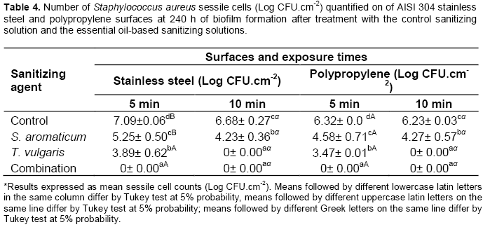
The effectiveness of the sanitizing solutions containing essential oils can be observed by the counts obtained after treatment of coupons on both surfaces and the reduction percentage of these cells. A significant difference in the counts and reduction percentage of adhered sessile cells can be noted among the different treatments (Table 5). All sanitizing solutions based on essential oils showed more superior antimicrobial activity than the control sanitizing solution.
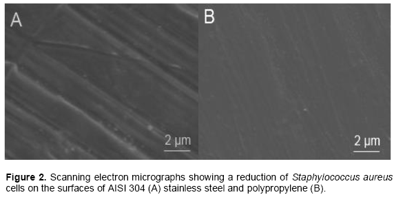
The effectiveness of the sanitizing solutions based on S. aromaticum, T. vulgaris, and their combination differ significantly from each other, their combination being the most effective to reduce the number of sessile cells adhered to the surfaces. It can be observed that the 5 min exposure of the coupons containing the biofilm to the sanitizing solution based on the combination of oils was effective to promote non-recovery of viable cells adhered to both surfaces. Scanning electron micrographs of Figure 2 show a reduction of S. aureus cells on surfaces stainless steel of AISI 304 and polypropylene.
The sanitizing solution based on T. vulgaris was more effective compared to S. aromaticum. This solution allowed the non-recovery of viable cells after exposure for 10 min to both surfaces. The sanitizing solution based on the essential oil of S. aromaticum was less effective, presenting a reduction in the number of sessile cells, but after 10 min of stainless steel and polypropylene coupon exposure to this solution, viable cells were still recovered.
As S. aureus is Gram positive, it is concluded that the cell wall does not serve as a barrier to the entrance of such antibacterial compounds through the cytoplasmic membrane. Since the cell wall of these bacteria is permeable, usually it does not restrict the penetration of these sanitizing agents (Schaffer and Messner, 2005).
The difference between the performance of the sani-tizing solutions within each phase of biofilm formation analyzed can be attributed to environmental and growth factors that are related to the concentration and nature of the chemical constituents, such as composition, func-tional groups and the structural configuration of the essential oil components (Chang et al., 2001).
Marques et al. (2007) show in their work that S. aureus, when in the form of biofilm, becomes more resistant to sanitizing agents.
The effects of colonization of surfaces where food is processed can result in various problems, been of an economic or public health nature. On the economic front, spoilage bacteria can contaminate food by changing its characteristics and resulting in economic losses. The risk to public health is the most serious problem, because the biofilm can transport pathogenic microorganisms and be a source of chronic contamination (Ribeiro-Furtini, 2005).
This multidisciplinary study led to the description of the sanitizing solutions essential oils of S. aromaticum and T. vulgaris and their combination on biofilm formed by S. aureus (ATCC 25923) on AISI 304 stainless steel and polypropylene surfaces. All solutions showed potential antibacterial sanitizers, being effective in reducing bacterial biofilms on these surfaces. The solution containing the combination of essential oils was more efficient by reducing 7.38 and 6.58 Log CFU.cm-2 cells adhered on the surfaces of AISI 304 stainless steel and polypropylene respectively, after 5 min of contact.
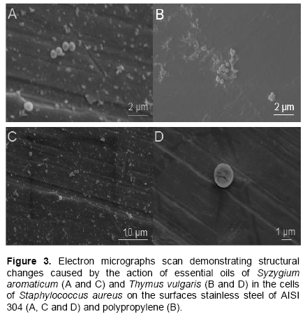
For the analysis of scanning electron micrographs obtained, were observed structural changes on the cell surface. Probably, these changes were caused by the action of essential oils in the cytoplasmic membrane, resulting in increased permeability and extravasation of intracellular constituents.
Studies by Oliveira et al. (2010) revealed, by means of electron micrographs of transmission electron microsco-py, alterations in the cell wall of Clostridium perfringens, when it was treated with essential oil of Satureja montana. The electron micrographs of cells not exposed to the culture that oil had continuous, smooth and thin cell walls and cell structures were defined. Already electron micrographs of cells that were exposed to treatment with the oil showed morphological changes in the wall as roughness and irregularities, leading to rupture of the wall and subsequent cell lysis.
Furthermore, binding and clustering of intracellular material in the cytoplasm of the cells were observed because the electron micrographs revealed that these cells lacked cytoplasm in certain regions due to functional loss of the membrane caused by the action of the chemical components of S. montana.
Damage in cells of C. perfringens were also observed by Si et al. (2009), evaluated the effect of different essential oils in the morphology of bacterial cells by scanning electron microscopy, where occurred formation of cracks in the cell walls of the cultures treated with essential oils, which can also be observed in this study (Figure 3D).
The scanning electron micrographs below (Figure 3) reveal structural alterations probably caused by the action of essential oils S. aromaticum and T. vulgaris cells of S. aureus on surfaces stainless steel of AISI 304 and polypropylene.
CONFLICT OF INTERESTS
The author(s) have not declared any conflict of interests.
ACKNOWLEDGMENTS
We acknowledge the financial support for this project by FAPEMIG, CNPq and CAPES.
REFERENCES
|
Alhede M, Bjarnsholt T, Jensen PO, Phipps RK, Moser C, Hristophersen L, Christensen LD, Van Gennip M, Parsek M, Hoiby N et al. (2009). Pseudomonas aeruginosa recognizes and responds aggressively to the presence of polymorphonuclear eukocytes. Microbiology. 155:3500-3508. |
|
|
Alves, E (2004). Introdução à microscopia eletrônica de varredura e de transmissão. Lavras: FAEPE-UFLA. 54. |
|
|
Bakkali F, Averbeck D, Idaomar M (2008). Biological effects of essential oils: a review. Food Chem. Toxicol. 46:446-475. |
|
|
Boari CA, Alves MP, Tebaldi VMR, Savian, TV, Piccoli RH (2009). Formação de biofilme em aço inoxidável por Aeromonas hydrophila e Staphylococcus aureus usando leite e diferentes condições de cultivo. Ciênc Tecnol. Aliment. 29:886-895. |
|
|
Burt S (2004). Essential oils: their antibacterial properties and potential applications in foods. Int. J. Food Microbiol. 94:223-253. |
|
|
Chang ST, Chen PF, Chang SC (2001). Antibacterial activity of leaf essential oils and their constituents from Cinnamomum osmophloeum. J. Ethnopharmacol. 77:123-127. |
|
|
Chmielewski RAN, Frank JF (2003). Biofilm formation and control in food processing facilities. Comp Rev Food Sci Food Safety. 2:22-32. |
|
|
Cucarella C, Solano C, Valle J, Amorena B, Lasa I, Penades JR (2001). Bap a Staphylococcus aureus surface involved in biofilm formation. J Bacteriol. 183:2888-2896. |
|
|
Di Pasqua R, Hoskins N, Betts G, Edwards M, Ercoline D, Mauriello G (2007). Membrane toxicity of antimicrobial coumpounds from essential oils. J. Agric. Food Chem. 55:4863-4870. |
|
|
Ferreira DF (2003). SISVAR - Sistema de análise de variância para dados balanceados: Programa de análises estatísticas e planejamento de experimentos. Versão 4.6. Software. Lavras: DEX/UFLA. |
|
|
Flach J, Karnopp C (2005). Biofilmes formados em matéria prima em contato com leite: fatores de virulência envolvidos. Acta Scientiae Vet. 33:291-296. |
|
|
Fuqua WC, Winans SC, Greenberg EP (1994). Quorum sensing in bacteria: the LuxR-LuxI family of cell density-responsive transcriptional regulators. J. Bacteriol. 176:269-275. |
|
|
Gilbert P, Mcbain AJ, Rickard AH (2003). Formation of microbial biofilm in hygienic situations: a problem of control. Int. Biodeterior. Biodegrad. 51:245-248. |
|
|
Gill AO, Holley RA (2006). Disruption of Escherichia coli, Listeria monocytogenes and Lactobacillus sakei cellular membranes by plant oil aromatics. Int. J. Food Microbiol. 108:1-9. |
|
|
Joseph B, Ottas SK, Karunasagar I (2001). Biofilm formation by Salmonella spp. On food contact surfaces and their sensitivity to sanitizers. Int. J. Food Microbiol. 64:367-372. |
|
|
Kalemba D, Kunicka A (2003). Antibacterial and antifungal proprieties of essential oils. Curr. Med. Chem. 10:813-829. |
|
|
Lambert RJW, Skandamis, PN, Coote PJ, Nychas GJE (2001). A study of minimum inhibitory concentration and mode of action of oregano essential oil, thymol and carvacrol. J. Appl. Microbiol. 91:453-462. |
|
|
Marques SC, Rezende JGOS, Alves LAFA, Silva BCS, Abreu LR, Piccoli RH (2007). Formation of biofilms by Staphylococcus aureus on surfaces of stainless steel and glass and its resistance to some selected chemical sanitizers. Brazilian J. Microbiol. 38:538-543. |
|
|
Meylheuc T, Methivier C, Renault M, Herry JM, Pradier CM, Bellon-Fontaine MN (2006). Adsorption on stainless steel surfaces of biosurfactants produced by gram-negative and gram-positive bacteria: consequence on the bioadhesive behavior of Listeria monocytogenes. Colloids Surf B. 52:128-137. |
|
|
NCCLS, National Committee for Clinical Laboratory Standards (2000). Methods for dilution antimicrobial susceptibility tests for bacteria that grow aerobically. Approved standard M7-A6, Wayne, Pa, USA. |
|
|
Oliveira MMMD, Brugnera DF, Cardoso MDG, Alves E, Piccoli RH (2010). Disinfectant action of Cymbopogon sp. essential oil in different phases of biofilm formation by Listeria monocytogenes on stainless steel surface. Food Control, Guildford, 21(4):549-553. |
|
|
Oulahal N, Brice W, Martial A, Degraeve P (2008). Quantitative analysis of survival of Spaphylococcus aureus or Listeria innocua on two types of surfaces: Polypropylene e strainless steel in contact with three different dairy products. Food Control 19:178-185. |
|
|
R Development Core Team (2004). R: a language and environment for statistical computing. Viena: R Foundation for Statistical Computing, 2004. http://www.R-project.org. Accessed 10 Jul 2013. |
|
|
Ribeiro-Furtini LL (2005). Caracterização e isolamento de microrganismos aderidos em tubulações de laticínios e seu comportamento frente a detergência. 80 p. Tese (Doutorado em Ciência dos Alimentos) - Universidade Federal de Lavras, Lavras, Brasil. |
|
|
Rossoni EMM, Gaylarde CC (2000). Comparison of sodium hypochlorite and peracet acid as sanitising agents for stainless steel food processing surfaces using epifluorescence microscopy. Int. J. Food Microbiol. 61:81-85. |
|
|
Schaffer C, Messner P (2005). The structure of secondary cell wall polymers: how gram-positive bacteria stick their cell walls together. Microbiol. 151:643-651. |
|
|
Shafahi M, Vafai K (2009). Bioï¬lm affected characteristics of porous structures. Int. J. Heat Mass Transfer. 52:574-581. |
|
|
Sheng X, Ting YP, Pehkonem SO (2007). Force measurements of bacterial adhesion on metals using a cell probe atomic force microscope. J. Colloid Interface Sci. 310:661-669. |
|
|
Shi X, Zhu X (2009). Biofilm formation and food safety in food industries. Trends Food Sci. Technol. 20:407-413. |
|
|
Si W, Ni X, Gong J, Yu H, Tsao R, Han Y, Chambers JR (2009). Antimicrobial activity of essential oils and structurally related synthetic food additives towards Clostridium perfringens. J. Appl. Microbiol. 106(1):213-220. |
|
|
Skandamis PN, Nychas GJ (2012). Quorum sensing in the context of food microbiology. Appl. Environ. Microbiol. 78:5473-5482. |
|
|
Tan SYE, Chew SC, Tan SYY, Givskov M, Yang L (2014). Emerging frontiers in detection and control of bacterial biofilms. Curr. Opin. Biotechnol. 26:1-6. |
|
Copyright © 2024 Author(s) retain the copyright of this article.
This article is published under the terms of the Creative Commons Attribution License 4.0


