African Journal of
Microbiology Research

  • Abbreviation: Afr. J. Microbiol. Res.
  • Language: English
  • ISSN: 1996-0808
  • DOI: 10.5897/AJMR
  • Start Year: 2007
  • Published Articles: 5233

Full Length Research Paper

Isolation of Salmonella species and some other gram negative bacteria from rats cohabitating with poultry in Ibadan, Oyo State, Nigeria

Jemilehin F. O.
  • Jemilehin F. O.
  • Department of Veterinary Microbiology and Parasitology, University of Ibadan, Nigeria.
  • Google Scholar
Ogunleye A.O.*
  • Ogunleye A.O.*
  • Department of Veterinary Microbiology and Parasitology, University of Ibadan, Nigeria.
  • Google Scholar
Okunlade A. O.
  • Okunlade A. O.
  • Department of Veterinary Microbiology and Parasitology, University of Ibadan, Nigeria.
  • Google Scholar
Ajuwape A. T. P.
  • Ajuwape A. T. P.
  • Department of Veterinary Microbiology and Parasitology, University of Ibadan, Nigeria.
  • Google Scholar


  •  Received: 24 September 2015
  •  Accepted: 06 November 2015
  •  Published: 07 August 2016

 ABSTRACT

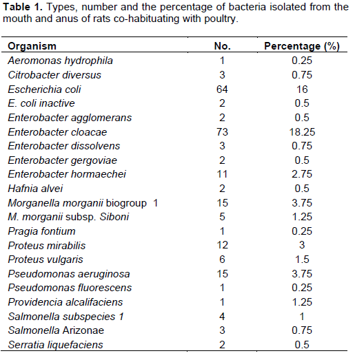
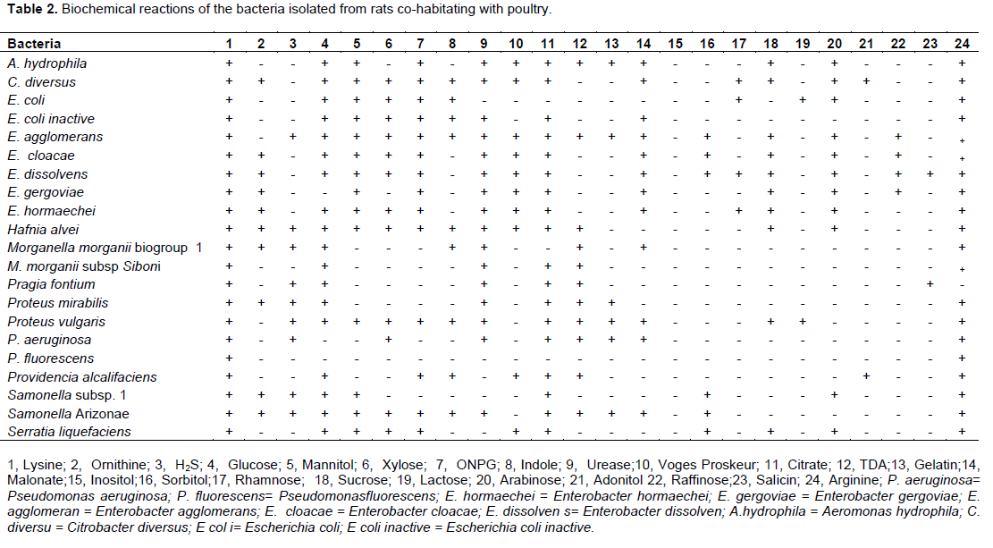
The aim of this study was to isolate Salmonella species and other enteric bacteria in rats cohabiting with poultry; in order to consider the potential role of rats in their transmissions to poultry and humans. Four hundred samples comprising 200 oral swabs and 200 anal swabs were collected from rats cohabiting with poultry from 5 local government areas in Ibadan. The samples were subjected to standard bacteriological analysis. A total of 228 Gram negative bacteria including 21 different species of both Lactose fermenters and non-Lactose fermenters were isolated. The identified organisms include: Salmonella subspecies 1, Salmonella Arizonae, Escherichia coli, Escherichia coli inactive, Pseudomonas aeruginosa, Pseudomonas fluorescens, Enterobacter cloacae, Enterobacter hormaechei, Enterobacter agglomerans, Enterobacter dissolvens, Enterobacter gergoviae, and Aeromonas hydrophila, Proteus mirabilis, Proteus vulgaris, Hafnia alvei, Morganella morganii biogp 1,Morganella morganii subspecies Siboni 1, Citrobacter diversus, Serratia liquefaciens, Pragia fontium and Providencia alcalifaciens. The organisms were identified using Oxoid Microbact GNB 24E® (MB24E) and accompanying computer software package (Oxoid Microbact®) 2000 version 2.03.  Some of the isolated bacteria from rats’ cohabitating with poultry have been associated with diseases in poultry and humans. The findings therefore serves to create fresh awareness among poultry farmers and other stakeholders in the industry from the studied area, that rats do not only constitute physical threat in terms of destruction of infrastructures and feeding on poultry feeds, they also pose a great risk in terms of transmission of bacterial infection to poultry and men associated with poultry production. Possible measures to control rats’ infestations within poultry houses are highlighted.

Key words: Rats, poultry, co-habitating, Gram negative Bacteria.


 INTRODUCTION

Rodents are known to be so numerous and very diverse in nature; and they are well acknowledged as a major contributors to human disease, right from the middle age (Meerburg et al., 2009;  Webster,  1996).   Black  rats  for example were associated with the transmission of plague long time ago; in the same way, more than 24 different infectious agents have been directly or indirectly reported as been transmitted by rodents  to  humans  (Meerburg et al., 2009; Webster,1996). Wild rodents serve as reservoirs and vectors for some pathogenic organisms like: Leptospira spp., Salmonella spp., Campylobacter spp., Trichinella spp., Toxoplasma spp. which are well known disease causing agents of food animals and humans (Gratz, 1994, Hiett et al., 2002). For instance, it had been noted that, rat population at a poultry farms can be a major reservoir of pathogenic bacteria and they are capable of transmitting the pathogens to the environment, food and animals (Rose et al., 2000). Henzler et al. (1998) demonstrated the direct association between the presence of rats and poultry diseases because they documented that effective control measure of rodents in the poultry house resulted in reduced bacterial infection of the associated poultry. It has also been shown that rodent control measures can effectively decrease Salmonella species in poultry houses (Rodenburg et al., 2004). Rats are common commensal pest (Meerburg et al., 2009), which  can  damage  the  food  products,  buildings, stored products, and also serve as potential vector of pathogenic diseases in human and animals. Rats may transmit the bacteria through feces, urine, and hair remnants (Meerburg et al., 2009).
 
Rodents are capable of perpetrating long existence of pathogens in the animal house and population. They can acquire infection through contact with faeces of infected livestock on the farm (Oosterom, 1991), from other wild animals such as wild birds or from their own family members as rodents. And since they tend to live close to each other, it encourages the infections to remain resident in the population for a long time (Singh et al., 1980; Hilton et al., 2002). Notably among the group of bacteria organisms carried and transmitted by rodents are enteric bacteria belonging to the Family enterobacteriaceae, of which Salmonella species; an important animal and human pathogen belongs.
 
Salmonella species are Gram-negative, non-spore forming rod-shaped bacteria, a member of the family Enterobacteriaceae (Jay et al., 2003). They are generally known to be actively motile with the exception of Salmonella Pullorum and Gallinarum; they grow at optimum temperature of 35 to 37°C and they are facultative anaerobic organisms (Jay et al., 2003). They are non-capsulated except Salmonella typhi (Cheesbrough, 2002). Persistence of Salmonella on farms is about two times higher for a farm where rodents are resident (Rose et al., 2000). According to Mehmood et al. (2011), bandicoot rat served as reservoir of bacterial infection of Escherichia coli, Klebsiella  species, Proteus  species and Salmonella species at poultry farms at Rawalpindi/Islamabad, Pakistan.
 
The Family Enterobacteriacea  to which Salmonella belong are a large family of Gram-negative bacteria that includes many of the more familiar pathogens, such as, E. coli, Yersinia pestis, Klebsiella and Shigella, Proteus, Enterobacter, Serratia and Citrobacter (Engelkirk, 2007). The family belongs to the order Enterobacteriales, the class Gammaproteobacteria and phylum Proteobacteria (Brenner et al., 2005). Members of the Enterobacteriaceae are rod-shaped, typically 0.6 to 6 μm in length and appear as small grey colonies on blood agar (Engelkirk, 2007). They are facultative anaerobes, they ferments sugars to produce lactic acid and various other end product (Ewing, 1986), most also reduce nitrate to nitrite (Mac Faddin, 2000). Bacteriological screening of rodents in close association with poultry houses for their carriage of pathogenic bacteria could be helpful to determine their role in bacterial disease transmission in poultry. The role of rodents, rat inclusive in destruction of poultry infrastructures is well appreciated among poultry farmers in Ibadan, Nigeria. There is however limited information on the types of bacteria carried by the rats, despite their often close interactions with poultry / poultry feeds and water sources. We therefore screened the oral and anal region of rats cohabitating with poultry from some poultry farms in Ibadan, Oyo State Nigeria to isolate and identify the kind of pathogen they carry as a mean of accessing their possible roles in disease transmission to poultry and human.


 MATERIALS AND METHODS

Sample location
 
Purposive samples were collected from poultry farms(comprising of both commercial and breeder flocks) in Suburbs of Ibadan, Oyo State, South-Western Nigeria (Latitude 7° 23' N and Longitude 3° 56' E) with known problems of rat infestation. The south-western States including; Oyo, Osun, Ogun, Ondo and Lagos are the main centers of poultry industry in Nigeria and Ibadan is a major and largest central city in the south-western States playing strategic roles in the national poultry production. Samples were collected from 5/11 local government areas located in Ibadan suburbs namely: Akinyele, Ido, Ona ara, Lagelu and Egbeda, where most of the poultry farms are located. One farm each was chosen from each LGA for sample collection in rats cohabiting with chicken flocks.
 
Sample collection
 
Two hundred rats were captured alive using adhesive glue boards. They were subsequently retrained with hands fortified with hand gloves for oral and anal sample collections. The oral and anal regions were sterilized with methylated spirit for aseptic collection of samples and to reduce contamination. A sterile swab stick was then inserted into the anus and mouth and rolled gently for sample collection. A total of four hundred samples were collected including; two hundred oral swab and two hundred anal swabs from rats cohabiting with poultry. The samples were transported to the laboratory and bacteriologically processed under 24 h of collection to reduce contamination and to ensure viability of organisms.
 
Bacteriological analysis
 
The samples were processed for bacteria isolation and identification using standard morphological, biochemical screening and serological procedures (Barrow and Felthams, 1993; Garcia and Isenberg, 2007; Cheesbrough, 2002). The oral and anal samples were streaked on MacConkey agar plates and inoculated into Selenite F Broth and subsequently incubated at 37°C for 24 h for primary isolation. Lactose fermenters were sub-cultured to Eosine Methylene Blue agar and incubated at 37°C for 24 h. Organisms that produced metallic sheen colonies on EMB were stored for further biochemical identification. Discrete colonies of Lactose and non- Lactose fermenters were also sub cultured from MacConkey agar on to Tryptose agar slope for further identification. Isolates that showed brick red colouration on Selenite F Broth were sub-cultured onto Deoxycholate Citrate Agar and incubated at 37°C for 24 h. All the Gram-negative isolates were identified with Oxoid Microbact GNB 24E® (MB24E) and accompanying computer software package (Oxoid Microbact®) 2000 version 2.03 according to the manufacturers procedures. The list of the reagents included in the kits are: Lysine, Ornithine, H2S, Glucose, Mannitol, Xylose, ONPG, Indole, Urease, Voges Proskeur, Citrate, Gelatine, TDA (Tryptophan deaminase), Malonate, Inositol, Sorbitol, Rhamnose,  Sucrose, Lactose, Arabinose, Adonitol, Raffinose, Salicin and Arginine.
 
Serology
 
All the suspected Salmonella species were subjected to slide agglutination test by standard method using Polyvalent Salmonella Antisera (Difcom Salmonella O Antiserum Poly A-I and V1) (Cheesbrough, 2002; Andrews et al., 2005).The Salmonella polyvalent antisera was reconstituted with 3mls of sterile 0.85% NaCl solution according to the manufacturer’s instructions. From test culture on a non selective media (Mueller Hilton), a discrete colony of the suspected Salmonella species organism was emulsified in  a drop of sterile saline on a glass slide. A drop  of the antiserum was then mixed with the emulsified culture on the slide,  rotated for one minute to observe for agglutination. Aggutination indicated a positive result while absence of agglutination indicated a negative result.


 RESULTS

Colonial morphology
 
One hundred and nineteen lactose fermenters were isolated on MacConkey agar; colonies were circular, 0.5 to 5 mm, pink coloured, with sweet odour and glistening surface. Forty-one non-lactose fermenting organisms were isolated from primary isolation; the colonies were light yellow, circular, translucent with entire edges (some had serrated edges), they were low convex, 0.5 to 2 mm with bad odour and with glistening surface. Sixty four isolates had characteristic metallic sheen on EMB. One hundred and thirty three isolates exhibited brick red coloration in selenite F Broth, 68 isolates grew on Deoxycholate Citrate Agar, out of which 32 isolates appeared as cream transparent to translucent circular, convex colonies with black centers, whereas 36 isolates appeared as white opaque convex colonies without black centers.
 
Biochemical test
 
A total of 10 genera of Enterobacteriaceae family namely: Citrobacter, Escherichia, Enterobacter, Hafnia, Morganella, Pragia, Proteus, Providencia, Salmonella and Serratia were identified to species level based on MicrobactR identification procedure. The other non-lactose fermenters identified include Pseudomonas species and Aeromonas hydrophilia (Table 1). Table 2 shows the various biochemical reactions of each of the isolated organism to the 24 biochemical tests included in the MicrobactR kit. Table 1 shows the details of the numbers and respective percentages of the identified organisms. Enterobacter species had the highest percentage (22.7%) while A. hydrophila had the lowest (0.25%). Only 7(1.75%) Salmonella organisms belonging to two serovars: namely, Salmonella subspecies 1, 4(1%) and Salmonella Arizonae 3(0.75) were isolated and identified. 


Serology
 
The identities of the 7 suspected Salmonella isolates were confirmed by slide agglutination with the Polyvalent Salmonella Antisera (Difcom Salmonella O Antiserum Poly A-I and V1),


 DISCUSSION

Members of the family Enterobacteriacea have been associated with diseases in poultry and humans (Yusop et al., 1991; Tsolis et al., 2008; Velge et al., 2012). In this current study only 2 groups of Salmonella was identified namely Salmonella subspecies 1 with an isolation rate of 1% and Salmonella Arizonae with an isolation rate of 0.75% giving a total isolation rate of 1.75%. These two subspecies of Salmonella have been implicated in poultry diseases and human infection. Brenner, 1998 stated that Salmonella enterica subspecies I are the predominant pathogens associated with birds and mammals; Velge  et al. (2012) also reported that strains of S. enterica subspecies 1 causes 99% of Salmonella infections in humans and other warm blooded animals. It produces cytotoxin and causes inflammatory diarrhea due to their ability to invade colonic epithelium, and they produce fever in humans (Tsolis et al., 2008; Guerrant and Steiner, 2005). On the other hand, Salmonella Arizonae subspecies have been associated with diseases in Turkey and Sheep (Grimont and Weill, 2007), although they are mostly isolated from reptiles and rarely isolated from man (Weiss et al., 1986). It was believed to be the cause of septiceamia, pneumonia, otitis media, brain abscess, meningitis, osteomyelitis, gastroenteritis and hepatic abscess in humans in many studies (Mclntyre et al., 1982, Petru and Richman, 1981 and Yusop et al., 1991).
 
The other 9 organisms belonging to the Enterobacterecea family were isolated and identified. Five  Enterobacter  species  with  highest isolation rate of 22.75% included, Enterobacter agglomerans (0.5%), Enterobacter cloacae (18.25%), Enterobacter dissolvens (0.75%), Enterobacter gergoviae (0.5%) and Enterobacter hormaechei (2.75%). This was followed by E. coli with an isolation rate of 16.5%. Morganella species isolation rate was 5% comprising: Morganella morganii biogroup 1(3.75%) and M. morganii subspecies Siboni (1.25%). Proteus species (4.5%) identified are; Proteus mirabilis (3%) and Proteus vulgaris (1.5%). The other bacteria isolates are Citrobacter diversus with isolation rate of 0.75%, Serratia liquefaciens and Hafnia alvei had the same isolation rate of 0.5% while Providentia alcalifaciens and Pragia fontium had the lowest isolation rate of 0.25% respectively.
 
Most of these organisms have been reported to be pathogenic to poultry and humans. For example, E. coli  according  to  Todar  (2007), can cause urinary tract infection (UTI), neonatal meningitis and intestinal disease (gastroenteritis) in humans. It is also the aetiology of avian colibacillosis which is considered as one of the principal causes of morbidity and mortality, associated with heavy economic losses to the poultry industry (Calnek et al., 1997). A group of E coli called E coli inactive was also isolated and identified, this was discovered to be non- lactose fermenters and also non-motile. This is in agreement with Le Minor and Richard (1993) and Holt et al. (1994) who discovered that a lactose-negative, non- motile organism thought to be Shigella species were inactive E. coli when identified serologically. M. morganii had been implicated in outbreaks of septicemia and bacteremia in humans and animals (Barragan Casas et al., 1999; Heard et al., 1988; Rowen and Lopez, 1998).  Serratia   liquefaciens   had   been implicated in the contamination of chicken carcasses (Lahellec et al., 1975) and it is also occasionally isolated from clinical specimens, although its pathogenic role has not been established (Grimont and Grimont, 1984). The presence of Proteus mirabilis in poultry meat had also been documented (Kim et al., 2005, Wong et al., 2013), and the presence of Proteus species in chicken droppings had been acknowledged as one of the route of its transmission as a zoonotic pathogens to vulnerable workers while handing infected chicken directly (Tonkic et al., 2010) or through fecal-contaminated poultry products as other Enteric bacteria (Lima-Filho et al., 2013). A more current report also associated poultry-borne Proteus species with zoonotic urinary tract infection (Armbruster et al., 2014).It was also associated with rheumatoid arthritis that is common in developing  countries  (Ebringer  and  Rashid, 2014).
 
Hafnia species has been recovered from mammals as well as from poultry where it has been identified as infectious agent (Farmer, 2003; Proietti et al., 2004). Additionally, it has been reported as agent of nosocomial infections and community acquired infections in humans (Günthard and Pennekamp, 1996), of urinary tract infection and sepsis in infants (Laupland et al., 2006). Likewise, Enterobacter species have been reported to cause disease in poultry (Nandi et al., 2013), it has also been isolated form egg shell of poultry together with other pathogenic Enterobacterecea (Musgrove et al., 2008). Some species of Enterobacter have also been isolated from patients' cerebrospinal fluid (Parodi et al., 2003).
 
Psedomonas species and Aeromonas hydrophila which do not belong to the Enterobacteriacea family were also isolated. Two species of Pseudomonas (4%) namely: Pseudomonas aeruginosa (3.75%) and Pseudomonas fluorescens (0.25%) were isolated and identified. The percentage isolation rate of 0.25% was obtained for Aeromonas hydrophila. These two organisms have been implicated in poultry diseases and are also of zoonotic importance. It has been reported that Pseudomonas  can  be  highly  virulent  causing  50 to 100%  mortality  in  experimentally  inoculated  4-week-old chickens (Saif et al., 2003). Fekadu (2010) also reported high mortality in newly  hatched  chickens  and  death  at  later  stage  of embryos  in  poultry  farms as a result of Pseudomonas infection. On the other hand, A. hydrophila has been isolated from organs of clinically sick commercial chickens in Jos, Nigeria (Dashe et al., 2014). According to Okpokwasili and Ogbulie (2001), disease caused by A. hydrophila is endemic in Nigeria and is the most important zoonotic disease of fish. It can be deduced from this study that rats cohabiting with poultry habour pathogens of poultry and humans, these rats should be put into consideration whenever the sources of  bacterial diseases  aetiologic agents in poultry and humans is been traced. From this study, rat cohabitating with poultry poses public health threat to poultry and humans as possible agents of disease transmission.
 
Preventive measures to reduce rats’ population in contact with poultry are highly recommended. This becomes imperative, since rat do not just constitute physical threat through their activities in destruction of poultry house infrastructures, they are also of great risk in the transmission of bacterial infection to poultry and man. Therefore adequate measures should be taken to prevent and eradicate rats from poultry houses and human abodes. Some of the recommended steps that could be taken to control rats population in and around poultry facilities include: Good sanitation; all feed bins must be kept in good repairs, and outside debris such as old equipments must be eliminated and grain of feed spillage which occurs during grain delivery must be cleaned immediately. Bushes around poultry houses should also be cleared regularly, as this  act as breeding  sites  for rodents.
 
Rodent-proof construction should be encouraged: A lasting form of rodent control is to keep them out of poultry houses by eliminating all openings through which rodents can enter a poultry houses, this is often cost prohibitive but it is an effective measure. Likewise, grain storage houses should be made rodent proof.
 
Farmers should be educated and enlightened on the role of rodents in the transmission of bacterial disease to poultry, this will enable them pay more attention to rodent control.


 CONFLICT OF INTERESTS

The authors have not declare any conflict of interest.


 REFERENCES

Andrews GP, Hromockyi AE, Coker C Maurelli AT (2005). Two novel virulence loci, MxiA and MxiB, in Shigella flexneri facilitate excretion of invasion plasmid antigens. Infect. Immun. 59:19-25.


Armbruster CE, Smith SN, Yep A, Mobley HL (2014). Increased Incidence of Urolithiasis and Bacteremia during Proteus mirabilis and Providencia stuartii co - infection due to synergistic induction of urease activity. J. Infect. Dis. 209:1524-1532.
Crossref


Barragan Casas JM, Hernandez MA, Garcinuno Jimenez MA, Gonzalo Molina P, Diaz C, Ibanez R, Serrano-Herranz R (1999). Bacteremia caused by digestive system endoscopy. Rev. Esp. Enferm. Dig. 91:105-116.


Barrow GH, Feltham RKA (1993). Cowan and Steel's manual for identification of medical bacteria, 3rd edition. Cambridge. UK. Cambridge University Press. p. 331.
Crossref


Brenner DJ, Krieg NR, Staley JT (2005). The Gammaproteobacteria. In: Bergey's Manual of Systematic Bacteriology 2B (2nd ed.). [1984 (Williams & Wilkins)]. George MGarrity, ed. New York: Springer. P, 1108. ISBN 978-0-387-24144-9. British Library No. GBA561951.


Brenner FW (1998). Modified Kauffmann-White scheme. Centers for Disease Control and Prevention, Atlanta, GA.


Calnek BW, Barnes HJ, Beard CW, McDougald LR, Saif YM (1997). Diseases of poultry 10th ed. Iowa State University Press; Ames, IA, USA. p. 1080.


Cheesbrough M (2002). District laboratory practice in tropical countries. E.C.B.S edition, Cambridge University Press 2:97-182.


Dashe YG, Raji MA, Abdu PA, Oladele BS, Olarinmoye D (2014). Isolation of Aeromonas hydrophila from Commercial Chickens in Jos Metropolis, Nigeria. Int. J. Poult. Sci. 13(1):26-30.
Crossref


Ebringer A, Rashid T (2014). Rheumatoid arthritis is caused by a Proteus urinary tract infection. APMIS 122:363-368.
Crossref


Engelkirk JD (2007). Laboratory Diagnostic of Infectious Disease: Essentials of Diagnostic Microbiology. 1st ed, Lippincolt Williams and Wilkins. ISBN-13: 9780781797016,ISBN-10:078179 7012


Ewing WH (1986). Edwards and Ewing's identification of Enterobacteria 4th ed. Elsevier Science, NewYork, NY, USA. 536pp.


Fekadu K (2010). Pseudomonas infection in chickens. J. Vet. Med. Anim. Health 2(4):55-85.


Garcia LS, Isenberg HD (2007). Clinical Microbiology Procedures Handbook Vol. 1, Second edn. update ASM Press American Society for Microbiology 1752 N St., N.W. Washington, DC 20036-290.


Gratz NG (1994). Rodents as carriers of disease, In: Rodent Pests and Their Control, ed. By Buckle AP and Smith RH. CAB International, Oxford, pp. 85-108.


Grimont PAD, Grimont F (1984). Genus Serratia Bizio 1823, 288AL, In: Krieg, N. R., and Holt, J. G.(ed.), Bergey's manual of systematic bacteriology, vol. 2.Williams and Wilkins. Baltimore pp. 477-484.


Grimont PAD, Weill FX (2007). Antigenic Formulae of Salmonella Serovars, Ninth Edition, World Health Organization Collaborating Center for Reference and Research on Salmonella. Institut Pasteur, Paris, France.


Guerrant RL, Steiner TS (2005). Principles and syndromes of enteric infections. In: Mandell GL, Bennett JE, Dolin R (ed), Principles and practice of infectious diseases, 6th ed, Elsevier Churchill. 1:1215-1230.


Günthard H, Pennekamp A (1996). Clinical significance of extraintestinal Hafnia alvei isolates from 61 patients and review of the literature. Clin. Infect. Dis. 22:1040-1045.
Crossref


Heard DJ, Jacobson ER, Clemmons RE, Campbell GA (1988). Bacteremia and septic arthritis in a West African dwarf crocodile. J. Am. Vet. Med. Assoc.192:1453-1454.


Henzler DJ, Kradel DC, Sischo WM (1998). Management and environmental risk factors for Salmonella enteritidis contamination of eggs. Am. J. Vet. Res. 59:824-829.


Hiett KL, Stern NJ, Fedorka-Cray P, Cox NA, Musgrove MT, Ladely S. (2002).Molecular subtype analyses of Campylobacter spp.from Arkansas and California poultry operations. Appl. Environ. Microbiol. 68:6220-6236.
Crossref


Hilton AC, Willis RJ, Hickie SJ (2002). Isolation of Salmonella from urban wild brown rats (Rattus norvegicus) in the West Midlands, UK. Int. J. Environ. Health Res. 12:163-168.
Crossref


Holt JG, Krieg NR, Sneath PHA, Staley JT, Williams ST (1994). Bergey's Manual of Determinative Bacteriology, ninth edition, Williams and Wilkins, Baltimore, pp. 175-222.


Jay LS, Davos D, Dundas M, Frankish E, Lightfoot D (2003). Salmonella. Ch 8 In: Hocking A.D. (ed) Foodborne microorganisms of Public health significance. 6th ed. Australian Institute of Food Science and Technology (NSW Branch), Sydney, pp. 207-266.


Kim SH, Wei CI, An H (2005). Molecular characterization of multidrug-resistant Proteus mirabilis isolates from retail meat products. J Food Prot. 68: 1408-1413.


Lahellec C, Meurier C, Bennejean, G, Catsaras M. (1975). A study of 5920 strains of psychrotrophic bacteria isolated from chickens. J. Appl. Microbiol. 38:89-97.
Crossref


Laupland KB, Church DL, Ross T (2006). Population-based laboratory surveillance of Hafnia alvei isolation in a Canadian health region. Ann. Clin. Microbiol. Antimicrob.18:12.
Crossref


Le Minor L, Richard C (1993). Methodes de laboratoire pour lidentification des enterobacteries. Institute Pasteur, Paris pp. 55-72.


Lima-Filho JV, Martins LV, Nascimento DC, Ventura RF, Batista JEC, Silva AFB, Ralph MT, Vaz RV, Boa-ViagemRabello C, Mendes daSilva I, Evêncio-Neto J(2013). Zoonotic potential of multidrug-resistant extraintestinal pathogenic Escherichia coli obtained from healthy poultry carcasses in Salvador, Brazil. Braz. J. Infect. Dis.17: 54-61.
Crossref


Mac Faddin, JF (2000). Biochemical Tests for Identification of Medical Bacteria, 3rd ed. Lippincolt Williams and Wilkins, Philadelphia, PA.


Mclntyre KE, Malone JM, Richards E, Axline SG (1982). Mycotic aortic pseudoaneurysm with aorticenteric fistula caused by Arizona hinshawii. Surgery 91:173-177.


Meerburg BG, Singleton GR, Kijlstra A (2009). Rodent-borne diseases and their risks for public health. Crit. Rev. Microbiol. 35:221-270.
Crossref


Mehmood A, Ansari MS, Hussain T, Akhter S, Khan SA, Hassan S, Khan AA, Rakha BA (2011). Bandicoot Rat (Ban dicota bengalen sis):A Novel Reservoir of Pathogenic Bacteria at Poultry Farms, Rawalpindi/Islamabad, Pakistan. Pak. J. Zool. 43(1):201-202.


Musgrove MT, Northcutt JK, Jones DR, Cox NA, Harrison MA (2008). Enterobacteriaceae and related organisms isolated from shell eggs collected during commercial processing. Int. J. Poult. Sci. 87:1211-1218
Crossref


Nandi SP, Sultana M, Hossain MA (2013). Prevalence and characterization of multidrug resistant zoonotic Enterobacter spp. in poultry of Bangladesh. Foodborne Pathog. Dis. 10:420-427.
Crossref


Okpokwasili GC, Ogbulie JN (2001). The biology andseasonality of Tilapia (Oreochromisniloticus) brown-patch syndrome. J. Trop. Aquacul. 16:88-100.


Oosterom J (1991). Epidemiological studies and proposed preventive measures in the fight against human salmonellosis. Int J Food Microbiol. 12:41-51.
Crossref


Parodi S, Lechner A, Osih R (2003). Nososomial Enterobacter meningitis: Risk factors, management and treatment outcomes. Clin. Infect. Dis. 37:159-66.
Crossref


Petru MA, Richman DD (1981). Arizona hinshawii infection of an artherosclerotic abdominal aorta. Arch. Int. Med. 141:537-538.
Crossref


Proietti PC, Passamonti F, Franciosini MP (2004). Hafnia alvei infection in pullets in Italy. Avian Pathol. 33:200-204.
Crossref


Rodenburg TB, Van der Hulst-Van Arkel MC, Kwakkel RP (2004). Campylobacter and Salmonella infections on organic broiler farms. NJAS-Wageningen J. Life Sci. 52:101-108.
Crossref


Rose N, Beaudeau F, Drouin P, Toux J, Rose V, Colin P (2000). Risk factors for Salmonella persistence after cleansing and disinfection in French broiler-chicken houses. Prev. Vet. Med. 39:9-20.
Crossref


Rowen JL, Lopez SM (1998). Morganella morganii early onset sepsis. Pediatr. Infect. Dis. J. 17:1176-1177.
Crossref


Saif YM, Barnes HJ, Glisson JR, Fadly AM, McDougald LR, David SE(2003). Diseases of Poultry 11th edition. Iowa State.


Singh SP, Sethi MS and Sharma VD (1980).The occurrence of Salmonellae in rodent, shrew, cockroach and ant. Int. J. Zoonoses 7:58-61.


Todar K (2007). Pathogenic E. coli, Online textbook of bacteriology. University of Wisconsin Madison Department of Bacteriology.


Tonkic M, Mohar B, Sisko-Kraljevic K, Mesko-Meglic K, Goic-Barisic I, Novak A, Kovacˇic´ A, Punda-Polic´ V(2010). High prevalence and molecular characterization of extended-spectrum beta-lactamase producing Proteus mirabilis strains in southern Croatia. J. Med. Microbiol. 59:1185-1190.
Crossref


Tsolis RM, Young GM, Solnick JV, Baumler AJ (2008). From bench to bedside: stealth of enteroinvasive pathogens. Nat. Rev. Microbiol. 6:883-892.
Crossref


Velge P, Wiedemann A, Rosselin M, Abed N, Boumart Z, Chausse AM, Grepinet O, Namdari F, Roche SM, Rossignol A, Virlogeux-Payant I (2012). Multiplicity of Salmonella entry mechanisms, a new paradigm for Salmonella pathogenesis. Open Microbiol. 1:243-258.
Crossref


Webster JP (1996).Wild brown rats(Rattus norvegicus)as a zoonotic risk on farms in England and Wales. Commun. Dis. Rep. CDR Rev. 6:R46-49.


Weiss SH, Blaser MJ, Paleologo FP, Black RE, Mcwhorter AC, Asbury MA, Carter GP, Feldman RA Brener DJ (1986). Occurrence and distribution of serotypes of the Arizona subspecies of Samonella strains in the United States from 1967-1976. J. Clin. Microbiol. 23: 1056-1064.


Wong MH, Wan HY, Chen S (2013). Characterization of multidrug-resistant Proteus mirabilis isolated from chicken carcasses. Foodborne Pathog. Dis. 10:177-181.
Crossref


Yusop C, Oh Hun k, Samuel YL, Ki Sup C, Toshio S (1991). Salmonella enterica subspecies diarizonae bacteria in an infant with enterits. Yonsei Med. J. 32(3):275-278.
Crossref


          */?>