African Journal of
Microbiology Research

  • Abbreviation: Afr. J. Microbiol. Res.
  • Language: English
  • ISSN: 1996-0808
  • DOI: 10.5897/AJMR
  • Start Year: 2007
  • Published Articles: 5233

Full Length Research Paper

Characterization of bacterial pathogens associated with milk microbiota in Egypt

Hager Yehia Shalaby
  • Hager Yehia Shalaby
  • Department of Microbiology and Immunology, Faculty of Pharmacy, Damanhour University, Damanhour, Egypt.
  • Google Scholar
Kholoud Baraka
  • Kholoud Baraka
  • Department of Microbiology and Immunology, Faculty of Pharmacy, Damanhour University, Damanhour, Egypt.
  • Google Scholar
Madiha Salah Ibrahim
  • Madiha Salah Ibrahim
  • Department of Microbiology, Faculty of Veterinary Medicine, Damanhour University, Damanhour Egypt.
  • Google Scholar
Eman Mohammad Khalaf
  • Eman Mohammad Khalaf
  • Department of Microbiology and Immunology, Faculty of Pharmacy, Damanhour University, Damanhour, Egypt.
  • Google Scholar


  •  Received: 31 August 2019
  •  Accepted: 07 October 2019
  •  Published: 31 October 2019

 ABSTRACT

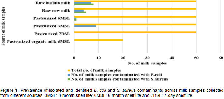
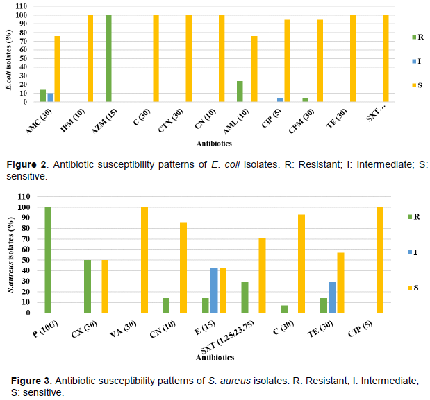
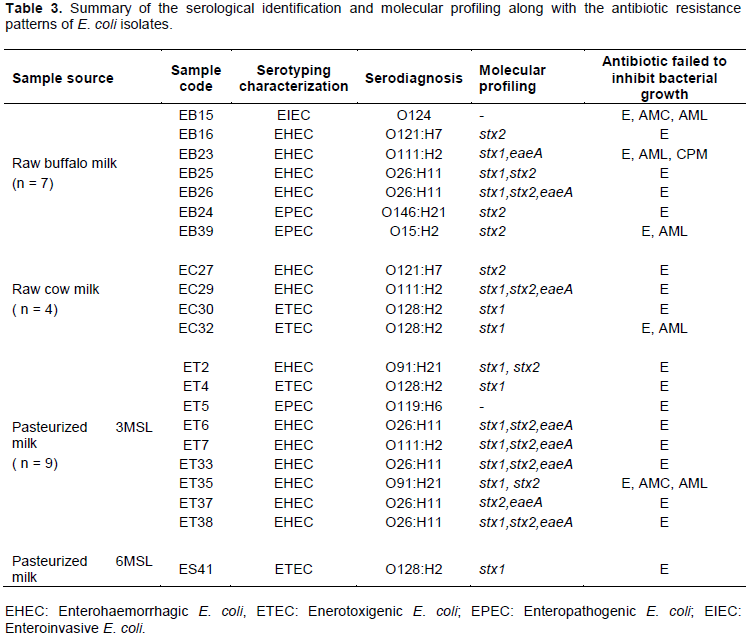
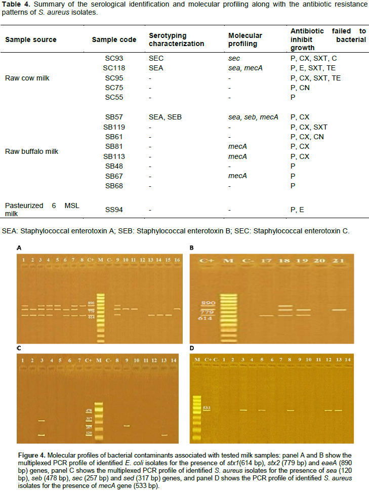
Milk is a substantial source of nutrients needed by all humans across lifespan development. Given its nutritional composition, milk is considered a vehicle for various microbes including beneficial and pathogenic bacteria. In this study, 270 milk samples comprising raw cow and buffalo milk and pasteurized milk with different shelf-life durations were tested along with pasteurized organic milk for the presence of Staphylococcus aureus and Escherichia coli. Collectively, 21 E. coli and 14 S. aureus isolates were cultivated and identified from total milk samples. All E. coli and S. aureus isolates exhibited resistance to erythromycin and penicillin, respectively. Serogroups O26, O128, and O111 were the most frequently identified amongst E. coli isolates, whereas staphylococcal enterotoxins (SEs) were inconsistently produced across S. aureus isolates. The molecular profile showed clustering of 6 isolates of E. coli by harboring stx1, stx2, eaeA genes, and 5 isolates of S. aureus by mecA gene. Findings revealed the bacteriological quality of popularly consumed milk in Egypt, including raw and pasteurized milk with preference to pasteurized organic milk and 7-day shelf life (7DSL) pasteurized milk. However, raw milk and 3MSL pasteurized milk were the major sources of E. coli and S. aureus, posing a serious public health issue.


Key words: Raw milk, pasteurization, Staphylococcus aureus and Escherichia coli, shelf-life.


 INTRODUCTION

Milk and dairy products are substantial sources of macro- and micronutrients needed by humans that make them prone   to    contamination    with    microbial   pathogens. Simultaneously, milk nutrients support the growth of specific beneficial microbes (e.g. lactobacilli and bifidobacteria)  that  promote  human  health  and  fitness
 
(Fernandes, 2009). Though the ingestion of contaminated milk either raw or pasteurized is the major cause of serious food-poisoning outbreaks, potentially result from microbial toxins production (Dhanashekar et al., 2012). Contaminated milk may harbor harmful microbes that lead to either milk spoilage (e.g. Pseudomonas and thermoduric microbes such as Clostridium and Bacillus) or the emergence of public health issues (e.g. Listeria, Salmonella, E. coli, and S. aureus) (Bennett et al., 2013; Quigley et al., 2013b). Milk is sterile at secretion in udder but is contaminated with extraneous microbes before leaving the animal udder (Elgadi et al., 2008). In developing countries especially rural areas, raw milk is directly used for either consumption or local dairy production (FAO, 2011; Zeinhom and Abdel-Latef, 2014).  Raw milk has a short shelf life that could be extended by heating. However, in the dairy industry, the shelf life of pasteurized milk is greatly influenced by the microbiological quality of the used raw milk (Murphy et al., 2016). In general, spoilage of commercialized milk and dairy products is attributable to various contamination sources including; pre-pasteurization psychrotophic growth, the degradable activity of heat-resistant microbial enzymes, and contamination after pasteurization process which is the most probable source (Sarkar, 2015). Gram-negative rods are the major psychrotrophic bacteria inhabiting raw milk (e.g., Enterobacteriaceae family including coliform bacteria that encompasses 5 to 33% of milk psychrotrophic bacteria) and proliferate during storage with the production of thermoresistant degradative enzymes (De Oliveira et al., 2015; Barbano et al., 2006;Mallet et al., 2012; Lewis and Gilmour, 1987). In addition, some Gram-positive bacteria contaminate raw milk with less frequent existence compared to Gram-negative psychrotrophs such as Staphylococcus species (Vithanage et al., 2016).
 
In general, pasteurization and ultra-high temperature (UHT) sterilization are the most commonly used techniques in the dairy industry for proper preservation and prolonged usability periods (Rais et al., 2013). Pasteurization meant to destroy common pathogens inhabiting raw milk microbiotas, especially those responsible for milk spoilage and influencing the shelf-life duration. Furthermore, pasteurization inactivates microbial enzymes that catalyze the breakage of milk macro-nutrients (e.g., lipids and proteins) and result in spoilage and invalidity of dairy products for consumption (Sarkar, 2015). In UHT treatment, heating is applied in the range of 135 to 150°C for up to 4 s for safe commercial dairy products combined with prolongation of the milk shelf-life duration (up to 12 months) (Vranješ et al., 2015). Though, aseptic packaging is crucial in both techniques that assure safety and extended usability of final dairy products (Deeth, 2017).
 
There is a  considerable  number  of  published  studies that have been conducted on the prevalence of E. coli and S. aureus in milk (Kandil et al., 2018;Vahedi et al., 2013). Milk and dairy products are one of the major causes of the transmission of pathogenic E. coli strains into the human  (Ombarak et al., 2019; Momtaz et al., 2012). With the advent of the high throughput sequencing technology, Escherichia coli was reported as a dominant inhabitant of the healthy human gut microbiome (Desmarchelier and Fegan, 2016). However, some E. coli strains exhibited virulence traits that enabled them to infect different body organs and cause illness (Awadallah et al., 2016; Zeinhom and Abdel-Latef, 2014).  Noteworthy, diarrheagenic E. coli strains increasingly become the leading cause of pediatric diarrhea. The most important diarrheagenic E. coli that threaten human health worldwide are enteropathogenic E. coli (EPEC) (the etiological agent of watery diarrhea in infants), enterohemorrhagic E. coli (EHEC) (leads to hemorrhagic colitis and hemolytic-uremic syndrome), entero-aggregative E. coli (EAEC) (causes persistent diarrhea), and enterotoxigenic E. coli (ETEC) (known to cause traveler's diarrhea) (Nataro and Kaper, 1998). The pathogenicity of diarrheagenic E. coli is attributed to possessing genetically encoded virulence traits. For instance, enterohemorrhagic E. coli  (EHEC) causes illness through the expression of intimin outer membrane protein encoded by eae gene and required for tissue colonization along with the production of Shiga toxins (ST) (e.g., Stx1, Stx2 or Stx2 variants) (Kaper et al., 2004). However, Enteropathogenic E. coli (EPEC) lacks ST genes, but exhibits its pathogenicity through the formation of A/E lesions on the intestinal cells, and is identified as eae-harboring diarrheagenic E. coli (Aidar-Ugrinovich et al., 2007).
S. aureus is a facultative anaerobic Gram-positive coccus and one of the world top pathogens that causes food-poisoning (Tirado and Schmidt, 2001; Hennekinne et al., 2012). Globally, enterotoxigenic S. aureus is implicated in udder infection of dairy cows combined with improper handling and poor storage conditions that result in frequent contamination of milk and dairy products. S. aureus produces several toxins including classical staphylococcal enterotoxins (SE) (SEA to SEE), in addition to other new types (SEG to SElU2) (Argudín et al., 2010). S. aureus could be inactivated by pasteurization however, thermostable SEs were found to retain their biological activity after the thermal treatment (Asao et al., 2003). Furthermore, more recent evidence suggests that SEA is the leading cause of staphylococcal food poisoning worldwide (Argudín et al., 2010). In order to verify the prevalence of genes encoding SE in S. aureus isolated from milk and dairy products, the phenotypic/serotypic assays of SE production should be conducted (Morandi et al., 2007). Of the classical techniques  used  for  SE  serotyping   analysis,   the  gel- diffusion test,  agglutination test, and reverse passive latex agglutination (RPLA) test kits (Wu et al., 2016). When compared to molecular techniques, the serological tests have limited sensitivity and specificity for SEs detection and cannot be used for total quantification of  SE (Wu et al., 2016).
 
So far, culture-dependent methods are still used as a routine protocol for the microbial assessment of raw and pasteurized milk. However, the detection of bacterial species that exist at subdominant levels is needed since the conventional laboratory methods are not enough to support the in vitro growth of milk-associated microbiota (Quigley et al., 2013a). Nowadays, culture-based foodborne pathogen detection methods have been developed to reduce the inspection time and improve product quality. One of the most informative and cost-effective molecular-based detection techniques is the multiplex PCR, which enables the screening of multiple target genes within a single reaction (Postollec et al., 2011).
 
In developing countries, consumption of raw milk is not prohibited and the advanced pasteurization techniques are still neither regulated nor implemented. Given the nutritional importance of milk and its widespread consumption particularly, among women and children, the study aimed to investigate the bacteriological quality of popularly consumed milk in the Delta area, Egypt for the presence of E. coli and S. aureus as major milk contaminants. The identified isolates were subjected to further testing for their potential pathogenicity through serotypic characterization and molecular profiling along with their antibiotic susceptibility profile.


 MATERIALS AND METHODS

Samples collection
 
Two hundred and seventy milk samples (10 ml each) were randomly collected (from January to June 2017) from local grocery stores and farmer vendors in El-Beheira governorate that represents Delta area in Egypt as street vendors are coming from different villages of neighbor Delta governorates. The milk samples included 100 samples of raw milk (50 samples of cow milk and 50 samples of buffalo milk), and 170 samples of pasteurized milk (50 samples of 6-month shelf life (6MSL), 50 samples of 3-month shelf life (3MSL), 50 samples of 7-day shelf life (7DSL) and 20 samples of pasteurized organic milk (6MSL)) (Figure 1 and Table S1A). All milk samples were collected in an icebox and brought to the laboratory to assess them for the presence of E. coli and S. aureus contaminants.
 
 
Isolation and identification of E. coli
 
Under aseptic conditions, 1 ml of each milk sample was drawn, homogenized with 10 ml of nutrient broth and incubated overnight at 37°C. Next day, 100 µl of the cultivated broth were streaked on MacConkey agar plate and incubated overnight for selection of enteric Gram-negative (Gm-ve) bacteria. Every lactose-fermenting (LF) colony was picked up using sterile toothpicks and streaked on Eosin methylene blue (EMB) agar plate, then incubated overnight at 37°C for further purification. Colonies exhibited blue-black color with a metallic green sheen were isolated and examined under a light microscope for gram stain. E. coli candidates were biochemically confirmed using indole, methyl Red, Voges Proskauer, citrate, triple sugar iron, and urease tests (Table S1B) according to Kreig and Holt (1984) and Miller (1992).
 
Isolation and identification of S. aureus
 
For   isolation   of   S.  aureus,  100 µl  of  overnight  cultivated  milk samples were streaked on Mannitol salt agar (MSA) plate and incubated overnight for bacterial growth. A yellow colony grown on a red/pink (MSA) medium was picked up and then streaked on a Baired parker (BP) agar plate for further purification. Every unique single colony was gram stained and visualized under the light microscope. The identification of S. aureus isolates was confirmed by performing a specific scheme of biochemical tests including coagulase, oxidase and DNase tests (Table S1C) according to MacFaddin (2000)and Lachica et al. (1971). 50% glycerol stocks of all identified bacterial isolates under this study were prepared and stored at -80°C freezer for further experiments.
 
Antibiotic susceptibility testing
 
The susceptibility of E. coli and S. aureus isolates to antibiotics were tested using the agar disk diffusion method. 11 antibiotics including ampicillin 10 µg (AML), amoxicillin/clavulanic 30 µg (AMC), imipenem 10 µg (IPM), cefipime 30 µg (CPM), cefotaxime 30 µg (CTX), gentamicin 10 µg (CN), azithromycin 15 µg (AZM), chloramphenicol 30 µg (C), tetracycline 30 µg (TE), sulphamethoxazole/trimethoprim 1.25/23.75 (SXT) and ciprofloxacin 5 µg (CIP) were used for the screening of E. coli isolates. With respect to testing S. aureus isolates, 9 antibiotics including penicillin 10U (P), cefoxitin 30 µg (CX), vancomycin 30 µg (VA), gentamicin 10 µg (CN), erythromycin 15 µg (E), chloramphenicol 30 µg (C), tetracycline 30 µg (TE), sulphamethoxazole/trimethoprim 1.25/23.75 (SXT) and ciprofloxacin 5 µg (CIP) were used. Following 16 to 18 h of aerobic incubation at 37°C, the plates were examined for bacterial growth and the diameter of inhibition zones surrounding antibiotic disks were scored in millimeter (mm). The zone diameters were interpreted as resistant (R), intermediate (I) or susceptible (S) according to  (CLSI, 2017).
 
Serotyping of E. coli isolates
 
Serotyping of E. coli isolates was performed using rapid diagnostic E. coli antisera sets (Denka Seiken Co, Japan) for lab diagnosis of Enteropathogenic serotypes according to the manufacturer's instructions. All antisera were obtained and absorbed with the corresponding cross-reacting antigens to remove the non-specific agglutinins.
 
Staphylococcal enterotoxins (SE) production test using SET-RPLA assay
 
S. aureus isolates were tested for enterotoxin production (SEA to SED) using SET-RPLA assay (SET-RPLA; Denka Seiken Co. Ltd., Tokyo, Japan) (Park and Szabo, 1986). The serotypic assay was performed according to the manufacturer’s instruction
 
Genomic DNA purification
 
DNA was purified from E. coli and S. aureus isolates along with used reference strains using a genomic DNA purification QIAamp kit (Qiagen, Germany) according to the manufacturer’s recommendations. The used reference strains for E. coli were: E. coli O157:H7 Sakai (positive for stx1, stx2, eaeA, and hylA genes) and E. coli K12 DH5α (a non-pathogenic negative control strain). Whereas enterotoxigenic S. aureus strains ATCC 13565 (positive for sea gene), ATCC 14458 (positive for  seb  gene),  ATCC  19095 (positive for sec gene), ATCC 23235 (positive for sed gene), 95-S-739 (positive for mecA gene) were used as positive controls for S. aureus molecular profiling, and S. xylosus ATCC 29971 was used as a negative control.
 
Molecular shiga toxin profiling and eaeA gene in E. coli isolates
 
The multiplexed-PCR technique was used for molecular profiling of E. coli isolates through amplification of shiga toxin-encoding genes; stx1, stx2 along with intimin-encoding gene (eaeA). The PCR reaction was performed using primers listed in (Table 1) in a Thermal Cycler (Master Cycler, Eppendorf, Hamburg, Germany). Approximately 50 ng of bacterial DNA was added to 12.5 μl DreamTaq Green PCR Master Mix (2X) (Thermo), 0.5 µl (5 pmol) of each primer and the final volume was adjusted to 25 μl by adding sterile ultrapure water. The amplification conditions started by initial denaturation for 3 min at 95°C followed by 35 cycles of 95°C for 20 s, 58°C for 40 s, and 72°C for 90 s. The final cycle was followed by 72°C final extension for 5 min. The amplified DNA fragments were separated by 1.5% of agarose gel electrophoresis (Applichem, Germany, GmbH) in 1x TBE buffer and captured as well as visualized on a UV transilluminator. A 100 bp plus DNA Ladder (Qiagen, Germany, GmbH) was used to determine each amplicon size and strains; E. coli O157:H7 Sakai and E. coli K12 DH5-α were used as a positive and negative control, respectively.
 
Molecular enterotoxin profiling and mecA gene in S. aureus
 
The genotypic profile of S. aureus isolates was generated based on the presence of sea, seb, sec and sed SE-encoding genes using multiplexed PCR along with conventional PCR for mecA gene amplification. PCR conditions used in E. coli molecular profiling were adapted by changing the annealing temperature to 50°C for 1 min and 56°C for 30 s for multiplexed and conventional PCR, respectively. S. aureus strains ATCC 13565, ATCC 14458, ATCC 19095, ATCC 23235 and 95-S-739 were used as positive controls for sea, seb, sec, sed and mecA genes, respectively and S. xylosus ATCC 29971 was used as a negative control. Sequences of the used primers are listed in (Table 2).


 RESULTS

Prevalence of E. coli and S. aureus contaminants across milk samples
 
In the current study, a total of 21 (7.8%) E. coli isolates were identified in particular, from raw and pasteurized 3MSL milk samples (Figure 1 and Table S1B). At the other side, raw and pasteurized 6MSL milk samples were the main sources of S. aureus isolates (14 isolates, accounting for 5.2% of the total milk samples) (Table S1C). Interestingly, pasteurized 7DSL and organic 6MSL samples exhibited negative bacterial growth (Figure 1).
 
Antibiotic susceptibility testing
 
Findings  revealed  the  resistance of all E. coli isolates to  erythromycin (100%) whereas 24 and 14% of the total E. coli isolates exhibited resistance to amoxicillin and amoxicillin/clavulanic acid, respectively. Of note, all E. coli isolates were susceptible to imipenem, chloramphenicol,  gentamicin,   cefotaxime,   tetracycline, and sulfamethoxazole (Figure 2). Similarly, all S. aureus isolates showed resistance to penicillin followed by far behind cefoxitin (50%) and sulfamethoxazole (29%). Meanwhile, vancomycin and ciprofloxacin inhibited the growth of all S. aureus isolates (Figure 3).
 
 
Serotyping of E. coli and S. aureus isolates
 
The serological typing of E. coli isolates showed that EHEC was the most dominant pathotype accounting for 62% (13 out of 21 isolates), followed by far behind ETEC (19%, 4 isolates), EPEC (14%, 3 isolates), and EIEC (5%, 1 isolate) (Table 3). Interestingly, O26, O128, and O111 were the most prevalent serogroups identified in 29, 19 and 14% of the isolates, respectively. With respect to staphylococcal enterotoxin production, RPLA assay showed that 3 out of 14 isolates (21.4%) produced different SE listed in (Table 4).
 
Molecular profiling of E. coli and S. aureus isolates
 
The molecular profiling of E. coli isolates showed positive results for the presence of stx1, stx2, eaeA genes accounting for 90.5% (19 out of 21) of total E. coli isolates, and spanning different sources of milk samples (Table ). However, stx1, stx2, eaeA genes were amplified altogether in 31.6% (6 out of 19) of E. coli isolates. Of note, these 6 isolates were purified from raw milk and 3MSL pasteurized milk (Figure 4 (A and B) and Table 3). Interestingly, 35.7% (5 out of 14) of S. aureus isolates exhibited positive PCR products for mecA gene, exclusively collected from raw milk (Figure 4) (C and D) and (Table 4). Only 3 S. aureus isolates showed positive results for the tested SE-encoding genes with an exception for sed gene (Table 3).


 DISCUSSION

Bacterial   contamination   of   milk   may   originate   from diverse sources mainly; infected udders and unhygienic practices during the milking process. Of the major bacterial contaminants of milk; E. coli and S. aureus that are responsible for serious food-poisoning outbreaks worldwide (Vahedi et al., 2013). In the current study, 270 milk samples including raw and pasteurized milk of different shelf life durations (Figure 1) were tested for the presence of E. coli and S. aureus contaminants. Interestingly, 11% (11 out of 100) of raw milk samples were the source of approximately half of the identified E. coli isolates (11 out of 21 E. coli isolates). This percentage was significantly lower than previously published reports from Iran and Egypt, where E. coli was identified from 42% (Vahedi et al., 2013), 33% (Hassan et al., 2015) and 60% (Kandil et al.,2018) of tested milk samples. 36.4% (4 out of 11 isolates) of identified E. coli isolates from raw milk originated from 8% (4 out of 50 samples) of raw cow milk (Figure 1). Similarly, cultivated raw buffalo milk samples resulted in the isolation of 14% (7 out of 50) of E. coli isolates which is a lower rate compared to previously published studies (Ranjbar et al., 2018). These findings indicated a relatively good bacteriological quality of raw milk in El-Beheira area when compared to previous studies (Bali et al., 2013; Garedew et al., 2012; Disassa et al., 2017; Reta et al., 2016). With regard to pasteurized milk, 5.9 % of tested samples resulted in the isolation of 10 E. coli isolates (9 out of 50 samples (18%) from 3MSL, and 1 out of 50 samples (2%) from 6MSL milk samples). Contrarily, in other published work (Kandil et al., 2018;  Hassan et al., 2015; Garedew et al., 2012), none of the pasteurized/sterile milk samples was reported for in vitro bacterial growth of E. coli.
 
The incidence of S. aureus in milk is increasingly ubiquitous  as  a  result    of   the  widespread  of  various
 
pathogenicity factors including; toxin-mediated virulence, invasiveness, and antibiotic resistance (Kadariya et al., 2014). 14 S. aureus isolates from all tested milk samples were biochemically identified. Approximately, 93% (13 out of 14 isolates) of S. aureus isolates were cultivated from raw milk (100 samples) accounting for 13% of the tested samples (Figure 1). The results came in accordance with those obtained by Zeinhom et al. (2015) and Mansour et al. (2017) that reported 12 and 16.3% of tested raw milk samples were contaminated with S. aureus, respectively. However, moderate and high contamination levels were also reported worldwide indicating the crucial importance of livestock health combined with the hygienic practices of milking on the safety of the dairy industry. For instance, a study from Egypt recorded the highest contamination incidence rates of raw milk with S. aureus accounting for 80% of the tested samples (Kandil et al., 2018). Interestingly, 10% (5 out of 50 samples) of  the raw cow milk samples were contaminated with S. aureus that is comparatively lower than a previous report (24.2%) from Reta et al. (2016) in Ethiopia. However, only 0.6% (1 out of  170  samples)  of pasteurized milk (6MSL milk) was contaminated with S. aureus. This result is consistent with a report published by Kandil et al. (2018) where S. aureus had zero existence in pasteurized milk samples in Egypt. In contrast, a higher contamination rate (14.92%) had been reported in Algeria (Matallah et al., 2019).
 
Globally, the unsupervised use of antimicrobial agents in the treatment of animal and human infections have been contributed to the emergence of antimicrobial resistance (Van Boeckel et al., 2015). The antimicrobial resistance mainly originates from the transfer of resistance genes across microbes enabling them to survive in the presence of antimicrobial agents that eventually resulted in failure of antibiotic therapeutic protocols (Blair et al., 2015). Furthermore, the overuse of antibiotics in animal husbandry as growth promoters could be a potential source of bacterial resistance through dissemination of resistant microbes from intestinal microbiotas of livestock that contaminate the surrounding environment and enhance the transmission of resistant genes to autochthonous bacteria (resident microbes)  of  the  surface  water  systems  (McEwen and Collignon, 2018). In this study, all E. coli isolates exhibited susceptibility to tetracycline, ciprofloxacin, sulfamethoxazole and chloramphenicol (except for one isolate that was resistant to ciprofloxacin) (Figure 2) which disagreed with reports published by Nobili et al. (2016), Schroeder et al. (2002), Mora et al. (2005), Abebe et al. (2014) and Ranjbar et al. (2018). However, the results reported by Tadesse et al. (2018) were relatively similar to our study where the in vitro growth E. coli was restrained by gentamicin, ciprofloxacin, and tetracycline. Of note, erythromycin inhibited the growth of all E. coli isolates, whereas Tadesse et al. (2018) reported a considerably moderate percentage (60%) of erythromycin resistance. Interestingly, only  14% of  E. coli isolates were resistant to amoxicillin-clavulanic acid, while Nobili et al. (2016) reported a significantly higher percentage (100%).  Furthermore, all E. coli isolates exhibited sensitivity to tested sulfa-drug antibiotic that disagreed with reports from Tadesse et al. (2018) and Nobili et al. (2016) where the susceptibility levels were 40 and 50%, respectively. Regarding the antibiotic resistance patterns of S. aureus, the isolates exhibited resistant to penicillin, cefoxitin, sulfamethoxazole, tetracycline, gentamicin, and erythromycin (Figure 3) which concurred with the findings published by Hoque et al. (2018) and Reta et al. (2016). Interestingly, 29% of S. aureus isolates showed resistance to sulphamethoxazole-trimethoprim that completely agreed with Hoque et al. (2018), and spiking high when compared to those reported by Reta et al. (2016) and Umaru et al. (2013). Despite previous studies, Umaru et al. (2013) and Reta et al. (2016) reported variable sensitivity rates (44.3 and 6.9%, respectively) of S. aureus isolates to vancomycin, findings showed absolute susceptibility of all tested isolates to it. Similarly, all S. aureus isolates were susceptible to ciprofloxacin that disagreed with findings reported by Hoque et al. (2018) and Zeinhom et al. (2015).
 
Enterohemorrhagic Escherichia coli (EHEC) strains comprise a subgroup of Shiga-toxin (ST)-producing E. coli (STEC) and are the most frequently implicated in severe clinical illness worldwide (Vendramin et al., 2014). In this study, we found that 62% of E. coli isolates were serologically identified as EHEC (Table 3), known to cause outbreaks of bloody diarrhea. This percentage is higher than Vanitha et al. (2018), Vendramin et al. (2014), Momtaz et al. (2012) and Ranjbar et al. (2018). Interestingly, the molecular profiling showed that 90% (19 out of 21 isolates) of E. coli isolates were positive for stx genes, whereas 42.8% (9 out of 21 isolates)  of them were positive for both stx1 and stx2 genes (Figure 4 and Table 3). However, these results were higher than that reported in previous studies (Tabaran et al., 2017; Nobili et al., 2016; Neher et al., 2015; Virpari et al., 2013) (Figure 4 and Table 3).  Furthermore,  38%  (8  out  of  21 isolates) of E. coli isolates harbored eaeA gene and serotypically characterized as EHEC including O26 and O111 serogroup (Figure 4 and Table 3).  These results were congruent with previously published studies (Momtaz et al., 2012; and Vanitha et al., 2018) where 33.33 and 36% of identified E. coli isolates were positive for eaeA gene, respectively. Contrarily, in a study conducted by Nobili et al. (2016), all STEC isolates exhibited negative results for eaeA gene.
 
S. aureus isolates are able to produce enterotoxins posing a public health threat. This means that the detection of SE in milk is very crucial for the bacteriological assessment of milk and dairy products (Wu et al., 2016). In the current study, the molecular detection of SE-encoding genes was greatly helpful for proper characterization of SE-producing S. aureus. In general, multiplex PCR detection could infer the presence of genes but does not consider their expression. Therefore, RPLA technique is needed to emphasize the SE production (van Belkum, 2003). Here, SET-RPLA assay showed that 21.4% (3 out of 14 isolates) of S. aureus isolates produced classic enterotoxins (SEA, SEC, SED) (Figure 4 and Table 4), which is in line with results reported by Fagundes et al. (2010). Interestingly, the molecular profiling of S. aureus isolates for SE-encoding genes confirmed the results of SET-RPLA technique (Figure 4 and Table 4) and agreed with previously published reports (Mansour et al., 2017). In contrast, in a study performed by Rall et al. (2008), a higher prevalence rate of S. aureus was reported, whereas 68.4% of the S. aureus isolates were positive for one or more enterotoxins-encoding-genes. Of note, Arcuri et al. (2010) detected SE genes in 13.6% of mastitic cow milk and 41.7% of a bulk milk tank. In general, methicillin-resistant S. aureus (MRSA) strains have the ability to express multiple antibiotic resistance genes that pose a global threat to animal and human health (Shah et al., 2019). In this study, mecA gene was detected in approximately 36% of the total S. aureus isolates that indicated the potential emergence of MRSA outbreaks from consumption of contaminated raw milk in particular, in traditional societies (Figure 4 and Table 4). Noteworthy, similar percentages (22.2 and 20%) of MRSA detection in milk were reported by Umaru et al. (2013)and Hoque et al. (2018), respectively.


 CONCLUSION

To conclude, findings revealed that raw and 3MSL pasteurized milk are most prone to be contaminated by the pathogenic E. coli and S. aureus isolates, that poses serious health issues upon direct consumption of milk from these sources. Noteworthy, pasteurized organic milk and   7DSL   milk   were   found   to   be  of   the   highest bacteriological quality when tested for the presence of E. coli and S. aureus. Eventually, our findings implicitly highlighted the importance of constituting strict regulations with regard to milk handling in local farms and dairy plants to minimize the chance of milk contamination and the transmission of bacterial pathogens along with their antimicrobial resistance from dairy animals to humans.


 CONFLICT OF INTERESTS

The authors have not declared any conflict of interests.


 ACKNOWLEDGMENT

The authors would like to thank the Department of Microbiology, Faculty of Veterinary Medicine, Damanhour University for their kind cooperation and contribution to this study. This research received no specific grant from any funding agency in the public, commercial, or not-for-profit sectors.


 REFERENCES

Abebe M, Hailelule A, Abrha B, Nigus A, Birhanu M, Adane H, Genene T, Daniel H, Getachew G, Merga G, Haftay A (2014). Antibiogram of Escherichia coli strains isolated from food of bovine origin in selected Woredas of Tigray, Ethiopia. Journal of Bacteriology Research 6(3):17-22.


Aidar-Ugrinovich L, Blanco J, Blanco M, Blanco JE, Leomil L, Dahbi G, Mora A, Onuma DL, Silveira WD, Pestana de Castro AF (2007). Serotypes, virulence genes, and intimin types of Shiga toxin-producing Escherichia coli (STEC) and enteropathogenic E. coli (EPEC) isolated from calves in São Paulo, Brazil. International Journal of Food Microbiology 115(3):297-306.
Crossref


Arcuri EF, Angelo FF, Guimarães MFM, Talon R, Borges MF, Leroy S, Loiseau G, Lange CC, Andrade NJ, Montet D (2010). Toxigenic status of Staphylococcus aureus isolated from bovine raw milk and Minas frescal cheese in Brazil. Journal of Food Protection 73(12):2225-2231.
Crossref


Argudín MÁ, Mendoza MC, Rodicio MR (2010). Food Poisoning and Staphylococcus aureus Enterotoxins. Toxins 2(7):1751-1773.
Crossref


Asao T, Kumeda Y, Kawai T, Shibata T, Oda H, Haruki K, Nakazawa H, Kozaki S (2003). An extensive outbreak of staphylococcal food poisoning due to low-fat milk in Japan: estimation of enterotoxin A in the incriminated milk and powdered skim milk. Epidemiology and Infection 130(1):33-40.
Crossref


Awadallah MA, Ahmed HA, Merwad AM, Selim MA (2016). Occurrence, genotyping, shiga toxin genes and associated risk factors of E. coli isolated from dairy farms, handlers and milk consumers. The Veterinary Journal 217:83-88.
Crossref


Bali OS, Lajnef R, Felfoul I, Attia H, Ayadi MA (2013). Detection of Escherichia coli in unpasteurized raw milk. International Journal of Agriculture and Food Science 3(2):53-55.


Barbano DM, Ma Y, Santos MV (2006). Influence of raw milk quality on fluid milk shelf life. Journal of Dairy Science 89:E15-E19.
Crossref


Bennett SD, Walsh KA, Gould LH (2013). Foodborne Disease Outbreaks Caused by Bacillus cereus, Clostridium perfringens, and Staphylococcus aureus-United States, 1998-2008. Clinical Infectious Diseases 57(3):425-433.
Crossref


Blair JM, Webber MA, Baylay AJ, Ogbolu DO, Piddock LJ (2015). Molecular mechanisms of antibiotic resistance. Nature Reviews Microbiology 13(1):42-51.
Crossref


Clinical and Laboratory Standards Institute (CLSI) (2017). Performance standards for antimicrobial disk susceptibility tests; approved standard-12th ed. M02-A13. Clinical and Laboratory Standards Institute, Wayne, PA.


De Oliveira GB, Favarin L, Luchese RH, McIntosh D (2015). Psychrotrophic bacteria in milk: How much do we really know? Brazilian Journal of Microbiology 46(2):313-321.
Crossref


Deeth H (2017). Optimum Thermal Processing for Extended Shelf-Life (ESL) Milk. Foods (Basel, Switzerland) 6(11).
Crossref


Desmarchelier P, Fegan N (2016). Pathogens in Milk: Escherichia coli. Reference Module in Food Science. https://doi.org/10.1016/B978-0-08-100596-5.00989-6
Crossref


Dhanashekar R, Akkinepalli S, Nellutla A (2012). Milk-borne infections. An analysis of their potential effect on the milk industry. Germs 2(3):101-109.
Crossref


Disassa N, Sibhat B, Mengistu S, Muktar Y, Belina D (2017). Prevalence and Antimicrobial Susceptibility Pattern of E. coli O157:H7 Isolated from Traditionally Marketed Raw Cow Milk in and around Asosa Town, Western Ethiopia. Veterinary Medicine International. 
Crossref


Elgadi ZAM, Abdel Gadir WS, Dirar HA (2008). Isolation and Identification of Lactic Acid Bacteria and Yeast from Raw Milk in Khartoum State (Sudan). Research Journal of Microbiology 3(3)163-168.
Crossref


Fagundes H, Barchesi L, Filho AN, Ferreira LM, Oliveira CAF (2010). Occurrence of Staphylococcus aureus in raw milk produced in dairy farms in São Paulo state, Brazil. Brazilian Journal of Microbiology 41(2):376-380.
Crossref


Food and Agriculture Organization (FAO) (2011). World Livestock. Livestock in Food Security, Rome, Italy.


Fernandes R (2009). Liquid milk products. In: Fernandes R., Microbiology handbook dairy products. Leatherhead Food International and RSC Publishing pp. 1-19.
Crossref


Garedew L, Berhanu A, Mengesha D, Tsegay G (2012). Identification of gram-negative bacteria from critical control points of raw and pasteurized cow milk consumed at Gondar town and its suburbs, Ethiopia. BMC Public Health 12:950.
Crossref


Hennekinne JA, De Buyser ML, Dragacci S (2012). Staphylococcus aureus and its food poisoning toxins: characterization and outbreak investigation. FEMS Microbiology Reviews 36(4):815-836.
Crossref


Hoque MN, Das ZC, Rahman ANMA, Haider MG, Islam MA (2018). Molecular characterization of Staphylococcus aureus strains in bovine mastitis milk in Bangladesh. International Journal of Veterinary Science and Medicine 6(1):53-60.
Crossref


Kadariya J, Smith TC, Thapaliya D (2014). Staphylococcus aureus and Staphylococcal Food-Borne Disease: An Ongoing Challenge in Public Health. BioMed Research International pp. 1-9.
Crossref


Kandil AA, Elhadidy M, El-Gamal A, Al-Ashmawy MA (2018). Identification of S. aureus and E. coli from Dairy Products Intended for Human Consumption. Advances in Animal and Veterinary Sciences 6(11):509-513.
Crossref


Kaper JB, Nataro JP, Mobley HLT (2004). Pathogenic Escherichia coli, Nature reviews, Microbiology 2(2):123-40.
Crossref


Kargar M, Homayoon M (2015). Prevalence of shiga toxins (stx1, stx2), eaeA and hly genes of Escherichia coli O157:H7 strains among children with acute gastroenteritis in southern of Iran. Asian Pacific Journal of Tropical Medicine 8(1):24-28.
Crossref


Kreig N, Holt J (1984). Bergey's Manual of systemic bacteriology (1) Williams and Wilkins, Baltimore M.D.21202, USA.


Lachica RVF, Genigeorgis C, Hoeprich PD (1971). Metachromatic agar-diffusion methods for detecting staphylococcal nuclease activity. Applied Microbiology 21(4):585-587.


Lewis SJ, Gilmour A (1987). Microflora associated with the internal surfaces of rubber and stainless steel milk transfer pipeline. Journal of Applied Bacteriology 62(4):327-333.
Crossref


Hassan GM, Meshref AMS, Gomaa SM (2015). Microbiological Quality and Safety of Fluid Milk Marketed in Cairo and Giza Governorates. Current Research in Dairy Sciences 7(1):18-25.
Crossref


MacFaddin JF (2000). Biochemical Tests for Identification of Medical Bacteria. 3rd Edition, Lippincott Williams & Wilkins, Philadelphia.


Mallet A, Guéguen M, Kauffmann F, Chesneau C, Sesboué A, Desmasures N (2012). Quantitative and qualitative microbial analysis of raw milk reveals substantial diversity influenced by herd management practices. International Dairy Journal 27(1-2):13-21.
Crossref


Mansour AS, Wagih GE, Morgan SD, Elhariri M, El-shabrawy MA, Abuelnaga ASM, Elgabry EA (2017). Detection of Staphylococcus aureus enterotoxigenic strains in bovine raw milk by reversed passive latex agglutination and multiplex polymerase chain reaction. Veterinary World 10(8):843-847.
Crossref


Matallah AM, Bouayad L, Boudjellaba S, Mebkhout F, Hamdi TM, Ramdani-Bouguessa N (2019). Staphylococcus aureus isolated from selected dairies of Algeria: Prevalence and susceptibility to antibiotics. Laboratory of Medical Biology 12(2):205-210.
Crossref


McEwen SA, Collignon PJ (2018). Antimicrobial Resistance: a One Health Perspective. Microbiology Spectrum 6(2).
Crossref


Miller JH (1992). A Short Course in Bacterial Genetics: A Laboratory Manual and Handbook for Escherichia coli and related bacteria. Plainview, NY: Cold Spring Harbor Laboratory Press; 1992.


Momtaz H, Farzan R, Rahimi E, Safarpoor Dehkordi F, Souod N (2012). Molecular characterization of Shiga toxin-producing Escherichia coli isolated from ruminant and donkey raw milk samples and traditional dairy products in Iran. The Scientific World Journal 2012:231342.
Crossref


Mora A, Blanco JE, Blanco M, Alonso MP, Dhabi G, Echeita A, González EA, Bernárdez MI, Blanco J (2005). Antimicrobial resistance of Shiga toxin (verotoxin)-producing Escherichia coli O157:H7 and non-O157 strains isolated from humans, cattle, sheep and food in Spain. Research in Microbiology 156(7):793-806.
Crossref


Morandi S, Brasca M, Lodi R, Cremonesi P, Castiglioni B (2007). Detection of classical enterotoxins and identification of enterotoxin genes in Staphylococcus aureus from milk and dairy products. Veterinary Microbiology 124(1-2):66-72.
Crossref


Murphy SC, Martin NH, Barbano DM, Wiedmann M (2016). Influence of raw milk quality on processed dairy products: How do raw milk quality test results relate to product quality and yield? Journal of Dairy Science 99(12):10128-10149.
Crossref


Nataro JP, Kaper JB (1998). Diarrheagenic Escherichia coli. Clinical Microbiology Reviews 11(1):142-201.
Crossref


Neher S, Hazarika AK, Sharma RK, Barkalita LM, Bora M, Deka P (2015). Detection of Shiga-Toxigenic Escherichia Coli in Milk Samples of Cattle by PCR. Journal of Agriculture and Veterinary Science 8(5):75-78.


Nobili G, Franconieri I, Basanisi MG, La Bella G, Tozzoli R, Caprioli A, La Salandra G (2016). Short communication : Isolation of Shiga toxin-producing Escherichia coli in raw milk and mozzarella cheese in southern Italy. Journal of Dairy Science 99(10):7877-7880.
Crossref


Olowe OA, Aboderin BW, Idris OO, Mabayoje VO, Opaleye OO, Adekunle OC, Olowe RA, Akinduti PA, Ojurongbe O (2014). Genotypes and phenotypes of Shiga toxin-producing Escherichia coli (STEC) in Abeokuta, Southwestern Nigeria. Infection and Drug Resistance 7:253-259.
Crossref


Ombarak RA, Zayda MG, Hinenoya A, Yamasaki S (2019). Serotypes, pathogenic potential and antimicrobial resistance of Escherichia coli isolated from subclinical bovine mastitis milk samples in Egypt. Japanese Journal of Infectious Diseases 72(5):337-339.
Crossref


Park CE, Szabo R (1986). Evaluation of the reversed passive latex agglutination (RPLA) test kits for detection of staphylococcal enterotoxins A, B, C, and D in foods. Canadian Journal of Microbiology 32(9):723-727.
Crossref


Postollec F, Falentin H, Pavan S, Combrisson J, Sohier D (2011). Recent advances in quantitative PCR (qPCR) applications in food microbiology. Food Microbiology 28(5):848-861.
Crossref


Quigley L, McCarthy R, O'Sullivan O, Beresford TP, Fitzgerald GF, Ross RP, Stanton C, Cotter PD (2013a). The microbial content of raw and pasteurized cow milk as determined by molecular approaches. Journal of Dairy Science 96(8):4928-4937.
Crossref


Quigley L, O'Sullivan O, Stanton C, Beresford TP, Ross RP, Fitzgerald GF, Cotter PD (2013b). The complex microbiota of raw milk. FEMS Microbiology Reviews 37(5):664-698.
Crossref


Rais M, Acharya S, Sharma N (2013). Food processing industry in India: S&T capability, skills and employment opportunities. Journal of Rural Development 4(9):1-13.


Rall VLM, Vieira FP, Rall R, Vieitis RL, Fernandes A, Candeias JMG, Cardoso KF, Araújo JP (2008). PCR detection of staphylococcal enterotoxin genes in Staphylococcus aureus strains isolated from raw and pasteurized milk. Veterinary Microbiology 132(3-4):408-413.
Crossref


Ranjbar R, Dehkordi FS, Shahreza MHS, Rahimi E (2018). Prevalence, identification of virulence factors, O-serogroups and antibiotic resistance properties of shiga-toxin producing Escherichia coli strains isolated from raw milk and traditional dairy products. Antimicrobial resistance and infection control 7(53).
Crossref


Reta MA, Bereda TW, Alemu AN (2016). Bacterial contaminations of raw cow's milk consumed at Jigjiga City of Somali Regional State, Eastern Ethiopia. International Journal of Food Contamination 3(1):4.
Crossref


Sarkar S (2015). Microbiological Considerations: Pasteurized Milk. International Journal of Dairy Science 10:206-218.
Crossref


Schroeder CM, Meng J, Zhao S, Debroy C, Torcolini J, Zhao C, McDermott PF, Wagner DD, Walker RD, White DG (2002). Antimicrobial resistance of Escherichia coli O26, O103, O111, O128, and O145 from animals and humans. Emerging infectious diseases 8(12):1409-1414.
Crossref


Shah MS, Qureshi S, Kashoo Z, Farooq S, Wani SA, Hussain MI, Banday MS, Khan AA, Gull B, Habib A, Khan SM, Dar BA (2019). Methicillin resistance genes and in vitro biofilm formation among Staphylococcus aureus isolates from bovine mastitis in India. Comparative Immunology, Microbiology and Infectious Diseases 64:117-124.
Crossref


Tabaran A, Mihaiu M, Flaviu T, Colobatiu L, Reget O, Borzan MM, Dan SD (2017). First study on characterization of virulence and antibiotic resistance genes in verotoxigenic and enterotoxigenic E . coli isolated from raw milk and unpasteurized traditional cheeses in Romania. Folia Microbiologica 62(2):145-150.
Crossref


Tadesse HA, Gidey NB, Workelule K, Hailu H, Gidey S, Bsrat A, Taddele H (2018). Antimicrobial Resistance Profile of E . coli Isolated from Raw Cow Milk and Fresh Fruit Juice in Mekelle, Tigray, Ethiopia. Vetrinary Medicine International. 
Crossref


Tirado C, Schmidt K (2001). WHO Surveillance Programme for Control of Foodborne Infections and Intoxications: Preliminary Results and Trends Across Greater Europe. Journal of Infection 43(1):80-84.
Crossref


Umaru AG, Kabir J, Umoh VJ, Bello M, Kwaga JKP (2013). Methicillin-resistant staphylococcus aureus (MRSA) in fresh and fermented milk in Zaria and Kaduna, Nigeria. International Journal of Drug Research and Technology 3(3):67-75.


Vahedi M, Nasrolahei M, Sharif M, Mirabi AM (2013). Bacteriological study of raw and unexpired pasteurized cow's milk collected at the dairy farms and super markets in Sari city in 2011. Journal of preventive medicine and hygiene 54(2):120-123.


Van Belkum A (2003). Molecular diagnostics in medical microbiology: yesterday, today and tomorrow. Current Opinion in Pharmacology 3(5):497-501.
Crossref


Van Boeckel TP, Brower C, Gilbert M, Grenfell BT, Levin SA, Robinson TP, Teillant A, Laxminarayan R (2015). Global trends in antimicrobial use in food animals. Proceedings of the National Academy of Sciences of the United States of America 112(18):5649-5654.
Crossref


Vanitha HD, Sethulekshmi C, Latha C (2018). An epidemiological investigation on occurrence of enterohemorrhagic Escherichia coli in raw milk. Veterinary World 11(8):1164-1170.
Crossref


Vendramin T, Kich DM, Molina RD, Souza CFVde, Salvatori RU, Pozzobon A, Bustamante-filho IC (2014). Molecular screening of bovine raw milk for the presence of Shiga toxin-producing Escherichia coli (STEC) on dairy farms. Food Science and Technology (Campinas) 34(3):604-608.
Crossref


Virpari PK, Nayak JB, Brahmbhatt MN, Thaker HC (2013). Study on isolation, molecular detection of virulence gene and antibiotic sensitivity pattern of Escherichia coli isolated from milk and milk products. Veterinary World 6(8):541-545.
Crossref


Vithanage NR, Dissanayake M, Bolge G, Palombo EA, Yeager TR, Datta N (2016). Biodiversity of culturable psychrotrophic microbiota in raw milk attributable to refrigeration conditions, seasonality and their spoilage potential. International Dairy Journal 57:80-90.
Crossref


Vranješ AP, Popović M, Jevtić M (2015). Raw milk consumption and health. Srpski Arhiv Za Celokupno Lekarstvo 143(1-2):87-92.
Crossref


Wu S, Duan N, Gu H, Hao L, Ye H, Gong W, Wang Z (2016). A Review of the Methods for Detection of Staphylococcus aureus Enterotoxins. Toxins 8(7).
Crossref


Zeinhom MMA, Abdel-Latef GK (2014). Public health risk of some milk borne pathogens. Beni-Suef University Journal of Basic and Applied Sciences 3(3):209-215.
Crossref


Zeinhom MMA, Abdel-Latef GK, Jordan K (2015). The Use of Multiplex PCR to Determine the Prevalence of Enterotoxigenic Staphylococcus aureus isolated from Raw Milk, Feta Cheese, and Hand Swabs. Journal of Food Science 80(12):M2932-M2936.
Crossref


          */?>