African Journal of
Pharmacy and Pharmacology

  • Abbreviation: Afr. J. Pharm. Pharmacol.
  • Language: English
  • ISSN: 1996-0816
  • DOI: 10.5897/AJPP
  • Start Year: 2007
  • Published Articles: 2288

Full Length Research Paper

Levels of red blood cells derived microparticles in stored erythrocyte concentrate

Maheen Rana
  • Maheen Rana
  • Department of Pathology, Rashid Latif Medical and Dental College, Lahore, Pakistan
  • Google Scholar
Yasir Arfat
  • Yasir Arfat
  • College of Bioresources Chemical and Material Engineering, Shaanxi University of Science and Technology, Xian, China.
  • Google Scholar
Omar Naseem
  • Omar Naseem
  • Department of Pathology, Abu Ammara Medical College, Lahore, Pakistan.
  • Google Scholar
Nazish Mazari
  • Nazish Mazari
  • Department of Pathology, Al-Nafees Medical College Isra University, Islamabad, Pakistan.
  • Google Scholar
Navida Manzoor
  • Navida Manzoor
  • Department of Pharmacology, Rashid Latif Medical and Dental College, Lahore, Pakistan.
  • Google Scholar
Rao Salman Aziz
  • Rao Salman Aziz
  • Department of Pharmacology, Rashid Latif Medical and Dental College, Lahore, Pakistan.
  • Google Scholar
Muhammad Rashid
  • Muhammad Rashid
  • Faculty of Fisheries and Wildlife, University of Veterinary and Animal Sciences, Lahore, Pakistan.
  • Google Scholar
Shahida Mohsin
  • Shahida Mohsin
  • Department of Haematology, University of Health Sciences, Lahore, Pakistan.
  • Google Scholar


  •  Received: 19 February 2020
  •  Accepted: 16 April 2020
  •  Published: 31 July 2020

 ABSTRACT

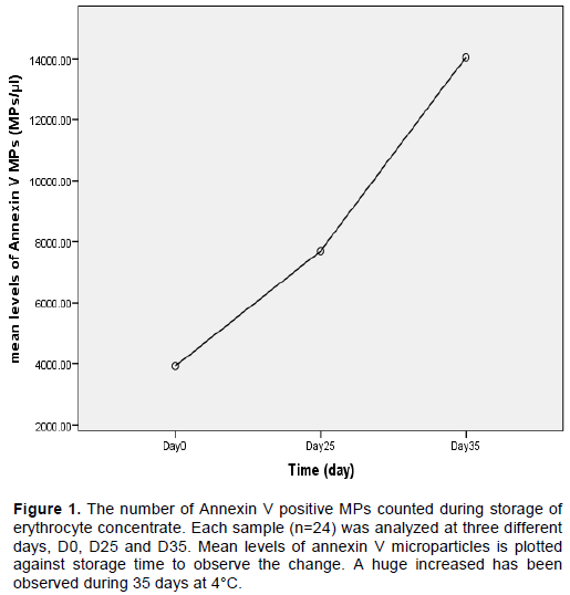
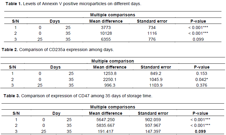
There is increasing evidence of the clinical importance of microparticles (MPs) and their role in blood transfusion-related side effects and the transmission of pathogens. The study aims to examine the red blood cell-derived MPs in blood bags during storage under standardized blood bank conditions. The samples were tested at various times to demonstrate the presence of RBC-derived MPs by flow cytometry. The quantitative assay was carried out in stored erythrocyte concentrate on days 0, 25 and 35 and their number from day 0 to 25 and 35 and the number of day 25 to day 35 were compared. The MPs were counted after being concentrated in a supernatant (labeled with the respective antibodies CD47, CD235a and Annexin V) obtained by a specific centrifugation procedure. The analysis showed that the number of Annexin V positive MPs increased between day 0 and day 35 (~ 0.001) and CD47 expression on MPs at day 25 and day 35 decreased compared to day 0 (~ 0.001). In addition, CD235a expression had shown minimal insignificant changes with an upward trend (> 0.05) during the storage period. It is concluded that monitoring the release of MPs from RBC units during storage is a sensitive approach to identifying MPs for transfusion drugs and, more broadly, for cell-based therapies.


Key words: Red blood cells, phosphatidylserine, cell-derived microparticle.


 INTRODUCTION

Red blood cells are the most commonly transfused blood component. Around 80 million red cell concentrate (EC) units are transfused worldwide each year. The introduction of blood bags, additive solutions and leukocyte filtration strategies has improved red blood  cell processing (RBCs) for transfusion purposes. They can be stored for 42 days at 4°C in a conservative standard solution of saline-adenine-glucose-mannitol (SAGM) or phosphate-adenine-glucose-guanosine-saline-mannitol (PAGGSM)  (Tzounakas   et   al.,   2016).   However,  the conditions to which erythrocytes are exposed during storage, such as temperature and anticoagulant, differ from in vivo. Current European Council guidelines do not allow erythrocyte concentrate (EC) to be stored for more than 42 days under controlled conditions. During storage, corpuscular changes occur which lead to a reduction in the number of erythrocytes in the recipient, which limits the current storage period of EC to 35 or 42 days depending on the storage medium (Pertinhez et al., 2016; Cholette et al., 2019). Although there is no conclusive data that blood stored longer is worse, some clinical practitioners (e.g. in neonatology) currently prefer to transfuse fresh blood. The main concern of the blood banks is to maintain the continuous supply of the EU and to reduce waste. During storage, EC is subject to biochemical changes, including an increase in the concentration of free hemoglobin. Potassium, lipids, lactate and the pH become acidic. Erythrocytes lose adenosine triphosphate (ATP), 2,3-diphosphoglycerate (2,3 DPG) during storage (Bardyn et al., 2017). As a result, the cell membrane undergoes changes such as asymmetry and disruption of the phospholipids, rearrangement of lipid rafts and clustering of proteins, which leads to the release of microparticles (MPs) (van Niel et al., 2018). MPs are also known as microvesicles or exosomes with a size of 1 µm or less. They are either released from apoptotic cells or stimulated after the remodeling of the plasma membrane. The size of the MPs derived from erythrocytes is around 0.15 μm (Antonelou and Seghatchian, 2016; Said et al., 2018). During the preservation period of EC at 4°C before the transfusion, storage lesions occur which lead to the breakdown or even change of the cells. As with other MPs, the exact role of erythrocyte-derived MPs is unknown and considered as storage lesions in connection with transfusions (Da Silveira et al., 2018).
 
The formation of MPs from erythrocytes is a characteristic of the aging of erythrocytes. This aging characteristic is also observed during storage in blood bags. During 120 days, almost 20% of the volume of the erythrocytes drops due to vesicle emission (Lee et al., 2018). Vesiculation is a source of erythrocytes used to remove harmful substances, that is, degraded hemoglobin, C5b-9 complement complex, band-3 neoantigen and IgG tending to aggregate inside or outside of membranes. In this way, erythrocytes prevent their early removal from the circulation (Nguyen et al., 2017; Eggold and Rankin, 2019). MPs derived from erythrocytes are removed by Kupffer cells in the human body. The release of MPs continues throughout their lifespan. Therefore, their number gradually increases with the storage time in the erythrocyte concentrate (Said et al., 2018; Melzak et al., 2018).
 
However, it is not clear how these storage lesions affect the function and survival of red blood cells after transfusion, but it is certain that storage leads to a reduction in the half-life of many transfused red blood cells. It has therefore been  speculated  that  erythrocytes experience acceleration in the normal aging process during storage (Chen et al., 2016, Belousov et al., 2018). The purpose of current research is to collect quantitative data on erythrocyte-derived MPs from the EC.


 MATERIALS AND METHODS

It is a descriptive study and included 24 blood bags. The study was designed to determine MPs on three different days (0, 25 and 35). Flow cytometry was used for the quantitative analysis of MPs. The study was carried out on 24 single donor erythrocyte concentrates. Of the 24 donors, 21 were men and 3 were women between the ages of 18 and 30. The blood bags were divided into three equal halves on 0, 25 and 35 days for the analysis of the MPs.
 
The research was carried out in the Hematology and Immunology departments of the University of Health Sciences in Lahore. It was carried out in collaboration with the Punjab Institute of Blood Transfusion, Lahore. The study was completed within six months of approval by the Ethical Review Board Committee. Donors between the ages of 20 and 40 who, after consent 10, met the donor selection criteria specified by the American Association of Blood Banking were included in the study. It was decided that leaks or discoloration are excluded in blood bags that have hemolysis. Complete blood counts (CBCs) were performed on all donors using an XT1800i automated hematology analyzer (Sysmex Middle East FZ-LLC). Blood was drawn according to standard procedures. Whole blood was taken from the antecubital vein. The sample was collected in a mother bag with quadruple bags of 450 ml (± 10%) containing 63 ml of CPD-A1 anticoagulant (citrate-phosphate-dextrose adenine). ECs were made after centrifugation of the whole blood unit. According to the instructions for the manual device, the plasma was extracted from the whole blood unit through the outlet. The volume of the erythrocyte concentrate was 150-200 ml, together with 50-60 ml of plasma, which remained in the mother bag. With the help of a manual extractor, EC was evenly distributed between the mother bag and two daughter bags. The bags were reserved for the analysis of MPson day 0, day 25 and day 35. The satellite bags were detached from the mother bag after the seal was secured. All three were stored at 4-6°C.
 
The flow cytometer was calibrated with BD CaliBRITETM beads and the MPs were counted using the FACS computer software.
 
Preparation of sample for flow cytometric analysis
 
50 ml of erythrocyte concentrate was transferred to a falcon tube (BD Biosciences) and processed for the preparation of the supernatant for each sample. In order to obtain a concentrated supernatant from MPs, a two-stage centrifugation was carried out using 4000 rpm and 4500 rpm (20 min each). The 100 µl supernatant was transferred to two labeled flow cytometric falcon tubes (tube no. 1 and 2) for each sample. Tube no. 1 was an isotype control and consists of 100 μl supernatant and 3 μlisotype controls (immunoglobulin G1 (IgG1)) IgG1FITC / immunoglobulin G2a phycoerythrin secondary (IgG2a PE). In tube no. 2, 20 μlwas PE differentiation cluster 235a (CD235a), 5 μm. APC-Annexin V and 20 μ l FITC CD47 added to identify MPs. Both tubes were incubated in the dark at 25°C (room temperature) and then at 4°C for 15 min. After the incubation, 600 μl sheath liquid was added to each tube and mixed thoroughly. Isotype control was taken as a negative control. The samples were evaluated using BD FACS caliber with Cell Quest Pro software (BD Biosciences). The above procedure was also repeated for day 25 and day 35 samples. An MP gate was placed on forward and side scatter point plots. All samples were analyzed for 20,000 events. The flow cytometer settings for the MPs counted in each sample were (detector: voltage): FSC: E00 SSC: 350 FL1: 600 FL2: 550 and FL3: 650.
 
MPs were clearly differentiated based on their size and negatively charged phospholipids. The majority of MPs population was Annexin V positive. Data analysis was performed using forward and side scatter, for which a scatter plot was selected, and then a gate R1 was placed on events where the MP population was present. The selected population was analyzed for expression of Annexin V, CD47 and CD235a and the data were documented.
 
Statistical analysis
 
Repeated measurements with ANOVA were performed to compare the levels of red blood cell MPs between different days. Mauchly's sphericity test was used to check the homogeneity of the variances between all possible pairs of days. The data violated the assumption of sphericity, so an ANOVA with repeated measurements with a Greenhouse-Geisser correction was used. The post hoc test using the Bonferroni correction was used for comparison between the days. The data (CD235a and CD47 expression, Annexin V level) are presented as mean ± SD; the P value ≤ 0.05 was considered significant.


 RESULTS

In packed erythrocytes obtained after centrifugation of whole blood units, the MPs were counted in our study using Annexin V. The mean number of MPs detected on day 0 was 3923 ± 605 / µl and reached up to 14.052 ± 1059 / µl on day 35. Statistically, the number of MPs increased linearly with the storage time (<0.001).
 
MPs were identified in the RCC sample by annexin V binding. In flow cytometry, Annexin A5 is commonly used to detect MPs due to its ability to bind negative phospholipid (phosphatidylserine). The Annexin V positive population of the microparticle count showed a fourfold increase during storage for 35 days at 4 ° C. The MP values ​​increased from 3923 ± 605 / µl on day 0 to 14.052 ± 1059 / µl on day 35, which is shown in a linear diagram (Figure 1). MPs are Annexin V-positive and recognizable in point diagrams. It has therefore been shown that phosphatidylserine is externalized on the surface of MPs (Figure 2).
 
The levels of Annexin V expression were compared between days (0, 25 and 35) and an increase was found. The difference between day 0 and day 35 was statistically significant (≤ 0.001) (Table 1). MPs were stained with CD235a (Glycophorin A) to mark its line as red blood cell-derived MPs. MPs showed an increased expression of erythrocyte-specific glycophorin A on the 0 day and the expression showed an increasing trend up to 35 days of storage. Repeated measurements ANOVA also validates this finding under the observed days mentioned (Table 2).MPs were also stained with CD47 "Marker of Self", which gives macrophages antiphagocytic signals and leads to the accumulation of MPs. CD47 expression on EDMPs decreased during storage. Statistical analysis of CD47 expression at times, day 25 and 35, compared to day 0 showed a statistically significant decrease (~ 0.001) (Table 3).


 DISCUSSION

The present study was performed to count erythrocyte-derived MPs in blood samples taken from 24 healthy volunteers. An earlier study by Dinkla and colleagues (Dinkla et al., 2014) showed that the exposure to phosphatidylserine in stored red blood cells remains quite low and begins to increase after 3 weeks of storage (Dinkla et al., 2014; Yoshinda et al., 2019). In our study, we selected the 25th day for detection. The second observation was made on day 35, the day of expiration (for CPD-A1) of blood units. MPs are counted using flow cytometry and in previous studies an increase in the number with storage time was observed (Mooberry and Key, 2016; Xie et al., 2018). The results of our study suggest an association of the storage time of erythrocyte concentrate with changes in the expression of the cell surface markers Annexin V, CD47 and CD235a on MPs derived from RBC.
 
These results are in agreement with those of Freites et al. (2020). In other studies, it was also observed that only part of the phosphatidylserine-expressing MPs was released in the early stages of storage (0 days). While later in storage (25th and 35th day) cell aging produces an increased number of damaged erythrocytes. This leads to an increase in phosphatidylserine-expressing MPs, which increases the sensitivity of Annexin V. (Aubron et al., 2013; Lelubre and Vincent, 2013; Alayash, 2018). The binding of Pannexin V to the MPs in our study shows that RBC-derived MPs expose phosphatidylserine. It was also observed that a storage time of less than 10 days had no remarkable effect in the generation of Mps. The MPs count of 7696 ± 781 / µl on the 25th day escalated on the 35th day to 14,052 ± 1059 / µl (p = 0.099), although the effect is not statistically significant. A study conducted on whole blood units with and without leukofiltration found erythrocyte-derived MPs on days 0, 1, 7, 14, 21, 28 and 35 (Saito et al., 2018). It was observed that the base number of EDMPs during storage increased significantly regardless of the filtration status. After day 21, the RDMP numbers of unfiltered products did not increase to the extent of the filtered products, and in the case of filtered products the increase after day 21 was not linear. When using a statistical test, no significant differences were observed during storage of the filtered product at any time from the 21st to the 42nd day (Chen et al., 2016).
 
The expression of CD47 on the cell surface was previously observed to decrease over time (Lelubre and Vincent, 2013; Velliquette et al., 2019). Our data support these results with the decrease in CD47 expression during a storage period of 35 days. A comparison of the expression of CD47 on MPs derived from young erythrocytes (day 0) with old erythrocytes (25th and 35th day) showed that the expression of CD47 on MPs derived from old erythrocytes was significantly lower. Almizraq et al. reported different results from our study because they found  no  significant  differences  in  CD47 expression during the storage period (Almizraq et al., 2013). They also suggested that storage in CPD-SAGM solution has a greater chance of maintaining RBC-CD47 expression during the storage period. This could further suggest that the stored CPD-SAGM erythrocytes are of better quality and their circulation and survival for a longer period after the transfusion could be anticipated. Our study was conducted on red cell concentrate stored in CPDA-1 anticoagulant. A remarkable reduction in CD47 expression on erythrocytes was observed during storage. A significant reduction in CD47 on red blood cells during storage is reported to be found both by ELISA assay (monoclonal antibody immobilization of RBC antigens [MAIRA]) and by FACSCalibur (Stewart et al., 2005).
 
CD235a is a transmembrane glycoprotein and is expressed on mature erythrocytes. After reaching the maximum CD235a level, no new surface antigens were found on the erythroid precursor (Daniele and Giovanni, 2018). Therefore, CD235a was used in our study to differentiate the lineage of MPs. The expression of CD235a on erythrocyte-derived MPs in erythrocyte concentrate showed a slight upward trend on three different  days  (0,  25  and   35),   but  this  increase  was insignificant (p = 0.376). In a study by Antonelou et al. they observed a minimal change in CD235a expression level during the 42-day storage period in leukocyte-depleted RBC concentrate (Antonelou et al., 2012; Tayer et al., 2019). Another work on MPs reported decreased expression of CD235a on day 42 of storage (Grisendi et al., 2015; Devalet et al., 2018).
 
Our study results showed that substantial changes in the form of MPs occur during the storage of blood in the form of MPs. We postulate that MPs derived from leukocytes and platelets in the whole blood unit can act synergistically with EDMPs when mediating transfusion complications. Therefore, it was concluded that blood units stored for more than 3 weeks should not be used to avoid transfusion complications in recipients. This progressive increase in MP production over the course of the storage period could be the answer to earlier observational studies in which critically ill people, including cardiac surgery, pediatric intensive care and trauma patients, were evaluated and relationships between older erythrocytes and increased mortality, multiple organ failure and sepsis (Obrador et al., 2015; Osaro et al., 2018). It has been shown that in patients undergoing cardiac surgery, transfusion of red blood cells

that have been stored for more than 2 weeks significantly increases the risk of postoperative complications and shortens survival after surgery (Loor et al., 2012; (Doctor et al., 2018).


 CONCLUSION

In summary, our results show that the storage time of erythrocyte concentrates influences the type of RBC storage lesion. In erythrocyte concentrate, the outward expression of phosphatidylserine on the cell membrane can be used as a parameter for assessing the quality. MPs derived from erythrocytes show a lower expression of CD47 on their surface, which has a negative effect on the function of the erythrocytes and the survival after the transfusion. The number of older erythrocytes in the erythrocyte concentrate can ultimately influence the in vivo efficiency of transfused erythrocytes. In order to improve the survival of the erythrocytes in the recipient and to avoid complications associated with MPs, the interval between the blood donation and the subsequent transfusion of this blood should therefore be shortened.


 CONFLICT OF INTERESTS

The authors have not declared any conflict of interests.


 REFERENCES

Almizraq R, Tchir JD, Holovati JL, Acker JP (2013). Storage of red blood cells affects membrane composition, microvesiculation, and in vitro quality. Transfusion 53(10):2258-2267.
Crossref


Antonelou MH, Tzounakas VL, Velentzas AD, Stamoulis KE, Kriebardis AG, Papassideri IS (2012). Effects of pre-storage leukoreduction on stored red blood cells signaling: a time-course evaluation from shape to proteome. Journal of Proteomics 5(76):220-38.
Crossref


Antonelou MH, Seghatchian J (2016). Update on extracellular vesicles inside red blood cell storage units: adjust the sails closer to the new wind. Transfusion and Apheresis Science 1;55(1):92-104.
Crossref


Aubron C, Nichol A, Cooper DJ,Bellomo R (2013). Age of red blood cells and transfusion in critically ill patients. Annual of Intensive Care 15;3(1):2.
Crossref


Alayash AI (2018). Oxidative pathways in the sickle cell and beyond. Blood Cells, Molecules and Diseases 1(70):78-86.
Crossref


Bardyn M, Rappaz B, Jaferzadeh K, Crettaz D, Tissot JD, Moon I, Turcatti G, Lion N, Prudent M (2017). Red blood cells ageing markers: a multi-parametric analysis. Blood Transfusion 15(3):239.


Belousov A, Malygon E, Yavorskiy V, Belousova E (2018). Simple and practical method of Additive Modernization of Preservation Solutions that does not violate the Compliance requirements and Improved the Quality, Efficiency, Safety Transfusion of Preserved RBCs. International Journal of Hematology and Blood Disorders 3(2):1-9.
Crossref


Chen D, Serrano K, Devine D (2016). Introducing the red cell storage lesion. ISBT Science Series 11(S1):26-33.
Crossref


Cholette JM, Willems A, Valentine SL, Bateman ST, Schwartz SM (2019). Recommendations on RBC Transfusion in Infants and Children with Acquired and Congenital Heart Disease From the Pediatric Critical Care Transfusion and Anemia Expertise Initiative. Pediatric Critical Care Medicine (pp. 303-312). Elsevier
Crossref


Da Silveira CL, Acker JP, Holovati JL (2018). The Effects of Liposome Treatment on Red Blood Cells during Hypothermic Storage. BiopreservBiobank 16(4):304-311.
Crossref


Daniele F, Giovanni A (2018). Induced Pluripotent Stem Cell-Derived Red Blood Cells and Platelet Concentrates: From Bench to Bedside. Cells 7(1):2.
Crossref


Devalet B, Wannez A, Bailly N, Alpan L, Gheldof D, Douxfils J, Deneys V, Bihin B, Chatelain B, Dogné JM, Chatelain C (2018). Application of a clot-based assay to measure the procoagulant activity of stored allogeneic red blood cell concentrates. Blood Transfusion 16(2):163.


Dinkla S, Peppelman M, van der Raadt J, Atsma F, Novotný VM, van Kraaij MG, Joosten I, Bosman GJ (2014). Phosphatidylserine exposure on stored red blood cells as a parameter for donor-dependent variation in product quality.Blood Transfusion 12(2):204.


Doctor A, Cholette JM, Remy KE, Argent A, Carson JL, Valentine SL, Bateman ST, Lacroix J (2018). Recommendations on red blood cell transfusion in general critically ill children based on hemoglobin and/or physiologic thresholds from the Pediatric Critical Care Transfusion and Anemia Expertise Initiative. Pediatric Critical Care Medicine 19(9):S98.
Crossref


Eggold JT, Rankin EB (2019). Erythropoiesis, EPO, macrophages, and bone. Bone 1(119):36-41.
Crossref


Freites Leal JK, Lasonder E, Sharma V, Schiller J, Fanelli G, Rinalducci S, Brock R, Bosman G (2020). Vesiculation of Red Blood Cells in the Blood Bank: A Multi-Omics Approach towards Identification of Causes and Consequences. Proteomes 8(2):6.
Crossref


Grisendi G, Finetti E, Manganaro D, Cordova N, Montagnani G, Spano C, Prapa M, Guarneri V, Otsuru S, Horwitz EM, Mari G (2015). Detection of microparticles from human red blood cells by multiparametric flow cytometry. Blood Transfusion 13(2):274.


Lee JS, Corcoran TE, Kagan V (2018).inventors; University of Pittsburgh, assignee. Red blood cell membrane-derived microparticles and their use for the treatment of lung disease. United States patent application US 10/004,764. 26.


Lelubre C, Vincent JL (2013). Relationship between red cell storage duration and outcomes in adults receiving red cell transfusions: a systematic review. Critical Care 8(17)2:R66.
Crossref


Loor G, Koch CG, Sabik JF, Li L, Blackstone EH (2012). Implications and management of anemia in cardiac surgery: current state of knowledge. The Journal of Thoracic and Cardiovascular Surgery. 1;144(3):538-46.
Crossref


Mooberry MJ, Key NS (2016).Microparticle analysis in disorders of hemostasis and thrombosis. Cytometry Part A. 89(2):111-22.
Crossref


Melzak KA, Uhlig S, Kirschhöfer F, Brenner-Weiss G, Bieback K (2018). The blood bag plasticizer di-2-ethylhexylphthalate causes red blood cells to form stomatocytes, possibly by inducing lipid flip-flop. Transfusion Medicine and Hemotherapy 45 (6):413-22.y
Crossref


Nguyen DB, Ly TB, Bernhardt I (2017). Microvesicles released from human red blood cells: Properties and potential applications. In: Novel Implications of Exosomes in Diagnosis and Treatment of Cancer and Infectious Diseases 12. IntechOpen.
Crossref


Obrador R, Musulin S, Hansen B (2015). Red blood cell storage lesion.Journal of Veterinary Emergency and Critical Care 25(2):187-99.
Crossref


Osaro E, Lukman H, Abiodun E, Charles AT, Frank U, Momodu I, Yakubu A, Zama I, Uchechukwu OF, Marafa A, Augustine O (2018). A review of the pathophysiology and consequences of red cell storage-fresh versus stored red cells-implication for optimum use of scarce allogenic blood. American Association for Science and Technology Journal of Medicine 4(2):32-50.


Pertinhez TA, Casali E, Baroni F, Berni P, Baricchi R, Spisni A (2016). A comparative study of the effect of leukoreduction and pre-storage leukodepletion on red blood cells during storage. Frontiers in Molecular Biosciences 21(3):13.
Crossref


Said AS, Rogers SC, Doctor A (2018). Physiologic impact of circulating RBC microparticles upon blood-vascular interactions. Frontiers in Physiology 12(8):1120.
Crossref


Saito S, Nollet KE, Ngoma AM, Ono T, Ohto H (2018). Platelet-, leucocyte- and red cell-derived microparticles in stored whole blood, with and without leucofiltration, with and without ionising radiation. Blood Transfusion 16(2):145-153.


Stewart A, Urbaniak S, Turner M, Bessos H (2005). The application of a new quantitative assay for the monitoring of integrin‐associated protein CD47 on red blood cells during storage and comparison with the expression of CD47 and phosphatidylserine with flow cytometry. Transfusion 45(9):1496-503.
Crossref


Tayer AH, Amirizadeh N, Ahmadinejad M, Nikougoftar M, Deyhim MR, Zolfaghari S (2019). Procoagulant Activity of Red Blood Cell-Derived Microvesicles during Red Cell Storage. Transfusion Medicine and Hemotherapy 46(4):224-30.
Crossref


Tzounakas VL, Kriebardis AG, Georgatzakou HT, Foudoulaki-Paparizos LE, Dzieciatkowska M, Wither MJ, Nemkov T, Hansen KC, Papassideri IS, D'Alessandro A, Antonelou MH (2016). Glucose 6-phosphate dehydrogenase deficient subjects may be better "storers" than donors of red blood cells. Free Radical Biology and Medicine 1(96):152-65.
Crossref


Van Niel G, D'Angelo G, Raposo G (2018). Shedding light on the cell biology of extracellular vesicles. Nature Reviews Molecular cell Biology 19(4):213.
Crossref


Velliquette RW, Aeschlimann J, Kirkegaard J, Shakarian G, Lomas‐Francis C, Westhoff CM (2019). Monoclonal anti‐CD47 interference in red cell and platelet testing.Transfusion. 59(2):730-7.
Crossref


Xie R, Yang Y, Zhu Y, Gao L, Jiang X, Sun J, Bian M, Yang J (2018). Microparticles in red cell concentrates prime polymorphonuclear neutrophils and cause acute lung injury in a two-event mouse model. International Immunopharmacology 1(55):98-104.
Crossref


Yoshinda T, Prudent M, D'Alessandro A (2019). Red blood cell storage lesion: causes and potential clinical consequences. Blood Transfusion 17(1):27.


          */?>