Full Length Research Paper
ABSTRACT
This study investigated the antioxidant and anti-inflammatory effects from the inner bark of Mimosa tenuiflora. The hydroethanol extract (HEE) and its hexane (HXF), chloroform (CLF), ethyl acetate (EAF) and hydromethanol (HMF) fractions were prepared and submitted to phytochemical screening and antioxidant activity. In vivo anti-inflammatory effect was investigated by using 12-O-tetradecanoylphorbol 13-acetate (TPA)-induced edema and myeloperoxidase (MPO) activity in mice ears. Phytochemical prospection of HEE revealed the presence of flavonoids, tannins, xantones, triterpenes, esteroids and phenols. Higher total phenol content was found in EAF and higher percentages of inhibition of 2,2-diphenyl-1-picrylhydrazyl free DPPH radical were found for HEE, EAF or HMF. Lipid peroxidation (LP) induced by 2,2'-Azobis(2-amidinopropane) dihydrochloride (AAPH) was greatly inhibited by HEE or EAF, while inhibition of FeSO4 - induced LP was higher for HMF. The coadministration of HEE (1 or 3 mg/ear) decreased edema (p<0.001) and MPO activity (p<0.05). All fractions reduced mice ear edema at the same extent, however, while EAF and HMF reduced MPO activity in mice ear at both 1 and 3 mg/ear (p<0.001 and p<0.05 respectively), CLF only at 3 mg/ear (p<0.05) and HXF did not affect this parameter. Taken together, these results demonstrate that inner bark of M. tenuifolia possesses antioxidant and anti-inflammatory effects.
Key words: Mimosa tenuiflora, oxidative stress; inflammation, edema, myeloperoxidase activity.
INTRODUCTION
Inflammation is considered a non-specific immune response that may be caused by an infectious agent, ischemia, injuries, and among others agents that can cause tissue injury associated with damage, which is generally related to free radicals (Serhan et al., 2010; Fernandes et al., 2015). The reactive oxygen (ROS) and nitrogen (RNS) species play an important role in modulating the inflammatory response, by stimulating the release of cytokines and other chemotactic agents, as wells as the expression of adhesion molecules, thus contributing to the induction and maintenance of the inflammatory response (Rotelli et al., 2003).
The use of medicinal plants for the treatment of many diseases, including inflammatory conditions, is an ancient practice, representing the therapy of choice in various communities and, therefore, has become interesting for various research areas such as botany, pharmacology and phytochemistry in order to support the popular use. In Brazil, the market for herbal medicines is growing and presents a great pharmacological potential (Lourenzani et al., 2004). Some compounds found in medicinal plants, such as phenolic compounds, are associated with a high antioxidant capacity and reduced risk for chronic degenerative diseases (Upadhyay and Dixit, 2015). Flavonoids, a subclass of polyphenols, can be found in a variety of foods and are well known for their anti-inflammatory and antioxidant properties (Johnson et al., 2015). In turn, several studies have linked the presence of some antioxidant compounds to anti-inflammatory activity, thereby increasing the interest of researchers to investigate the relationship between these compounds and their anti-inflammatory effect (García-Lafuente et al., 2009).
Among the plants used in folk medicine in Brazil, Mimosa tenuiflora (Willd.) Poir. (Leguminosae) is popularly known as “jurema-preta”, a typical tree in the Brazilian semi-arid region, also found in other regions of Latin American, from the northeast of Brazil to the southern of Mexico, which is used for therapeutic purposes, mainly skin burns and inflammation (Maia, 2004; Bitencourt et al., 2014). Lozoya et al. (1990) described antimicrobial activity for the water and ethanol extracts of the bark of M. tenuiflora. The ethanol extract of the bark of this plant was also effective against Staphylococcus aureus (Padilha et al., 2008; Bezerra et al., 2010). On the other hand, Silva et al. (2013) showed no mutagenic activity for the ethanol extract of the bark of M. tenuiflora. Rivera-Arce et al. (2007) have shown that a hydrogel containing 5% of a crude ethanol extract of the bark of this plant was useful for the treatment of venous leg ulceration disease in patients.
Anton et al. (1993) identified triterpenoid or steroid saponins and other compounds (lupeol, campesterol, stigmasterol and beta-sitosterol) in the bark of this plant. Some saponins found in the bark of M. tenuiflora were also described as immunomodulatory (Jiang et al., 1992). A study has shown that M. tenuiflora aqueous extract reduces inflammatory response caused by Titius serrulatus scorpion venom (Bitencourt et al., 2014). A recent study showed the antinociceptive and anti-inflammatory activity for the ethanol extract of the bark of M. tenuiflora (Cruz et al., 2016).
Altogether, these studies suggest that the bark of this plant has a therapeutic potential and highlight the need for more information about its pharmacological activities. In this way, the present study aimed to evaluate the antioxidant activity and anti-inflammatory effect of the hydroethanol extract and fractions of the inner bark of M. tenuiflora.
MATERIALS AND METHODS
Collection, identification and processing of plant material
The bark of M. tenuiflora was collected in April 23, 2008, in the municipality of Piranhas, Alagoas, Brazil, (09º 37 '25 "S 37º45' 24" W). A specialist (Dr. Ana Paula Prata) identified a specimen and an exsiccate was deposited in the Herbarium of Federal University of Sergipe (ASE 13166). The bark was dried at room temperature, reduced to powder and submitted to the tests.
Preparation of the hydroethanol extract and fractions
The dried bark (4.828 kg) was subjected to extraction with 90% ethanol for five days, by exhaustive maceration. After this period, it was filtered and concentrated on rotaevaporator under reduced pressure at 50°C to give the hydroethanol extract (HEE) with a yield of 1.0%. A portion of HEE was dissolved in 40% methanol and subjected to liquid-liquid extraction, to obtain the hexane (HXF), chloroform (CLF), ethyl acetate (EAF) and hydromethanol (HMF) fractions, which were subjected to phytochemical screening, quantification of total phenols and antioxidant and anti-inflammatory activities.
Phytochemical screening and determination of total phenolic compounds
In the phytochemical evaluation, the presence of phenols, tannins, flavonoids, xanthones, catechins, saponins, pentacyclic triterpenoid and free steroid using was tested by using the methods described by Matos (2009).
The total phenol content (TP) was assayed according to the methodology by Sousa et al. (2007) with adaptions. An aliquot (100 mL) to the HEE or fractions (1 mg/mL in methanol) was mixed with 6 mL of distilled water and 500 µL of Folin-Ciocalteu (1 mol/L). After, it was added 2 mL of Na2CO3 (15%) and diluted with distilled water to a final volume of 10 mL. This mixture was incubated for 120 min at 23°C and the absorbance read in a spectrophotometer (Bioespectro UV-VIS model SP22), at a wavelength of 750 nm. The content of TP was determined by interpolating the absorbance of the samples against a calibration curve using gallic acid as standard (10 to 350 mg/mL), and results were expressed as mg of gallic acid per g of extract or fraction.
DPPH• free radical scavenging activity
The antioxidant activity was evaluated using DPPH• method (Brand-Willams et al., 1995). Aliquots of a sample stock solution of 0.5 mg/mL of extract or fractions in methanol were added to a solution of DPPH• (40 μg/mL) to obtain final concentrations of 5, 10, 15, 20, 25 and 30 μg/mL of extract and fractions in a reaction volume of 3 mL. The blank was composed of a mixture of the sample analyzed and methanol; gallic acid was used as a positive control. The absorbance values at 515 nm, were measured at 1, 5 and 10 min, and then every 10 min up to 60 min. Results were expressed as percentage of remaining (REM) DPPH• calculated as follows:
DPPH•REM% = [DPPH•] T/[DPPH•] T0 × 100
In this equation, [DPPH•] T is the radical concentration after the reaction with the sample, and [DPPH•] T0 the initial concentration of DPPH•. From the DPPH•REM%, the percentage inhibition (IP) was obtained over the 60 min. The antioxidant concentration necessary to decrease the initial DPPH• concentration by 50% inhibition (EC50), as well the antioxidant activity index (AAI), were also used for the comparison of the antioxidant capacities of samples.
Lipoperoxidation
The ability to inhibit lipid peroxidation was determined by monitoring the production of thiobarbituric acid reactive substances (TBARS) in lipid-rich medium according to the description by Silva et al. (2007). A lipid solution (1% v/v) homogenizated in 20 mmol/L of phosphate buffer solution (pH 7.4) was obtained and mixed to solutions of the HEE or fractions at a concentration of 200 mg/mL. Lipid peroxidation was induced by the addition of 0.1 mL of AAPH (2,2-azobis (2-amidinopropane) dichloride, 0.17 mol/L) and 0.1 mL of FeSO4 (0.17 mol/L). For the measurement of TBARS, the homogenate (0.5 mL) was incubated with 0.5 mL of saline solution (0.9%) and two milliliters of a thiobarbituric acid/trichloroacetic acid (0.67 and 15% respectively) mixture and boiled at 95°C for 10 min. Subsequently, this mixture was cooled at room temperature and centrifuged at 4 000 x g for 10 min. The whole supernatant was taken in spectrophotometer cuvette and read at 535 nm. Trolox was used as the positive control and methanol as negative control. The results obtained were expressed as percentage of inhibition of malondialdehyde (MDA) formation.
In vivo evaluation of anti-inflammatory activity
Animals
Male Swiss mice (20-30 g) were obtained from the Animal Center of the Federal University of Sergipe. Animals were kept at 21 to 23°C with free access to food and water under a 12:12 h light/dark cycle. All experimentation was conducted in agreement with the guidelines of the Brazilian College of Animal Experimentation and the internationally accepted principles for laboratory animal use and care. It was also approved by the Ethics Committee for Animal Use in Research at the Federal University of Sergipe (52/12).
Mice ear inflammation induced by TPA
Anti-inflammatory activity of HEE and fractions was evaluated by using TPA-induced ear edema and neutrophil accumulation. Ear edema induced by TPA was performed according to the method described by De Young et al. (1989), with minor modifications by our group (Bonfim et al., 2014). Animals were divided into five groups (n=6 each): Group 1 received TPA dissolved in acetone (vehicle); group 2 received TPA concomitantly to dexamethasone (0.05 mg/ear); groups 3, 4 and 5 received TPA concomitantly to the HEE (0.3, 1 or 3 mg/ear). In the second set of experiments, treated animals received TPA concomitantly to HXF, CLF, EAF or HMF (1 or 3 mg/ear each).
Briefly, animals were anesthetized with inhalatory isoflurane and 20 mL of TPA (1 mg/ear dissolved in acetone) was topically applied to the surfaces of the mice right ear with a polypropylene tip in the absence or concomitant presence of HEE, HXF, CLF, EAF or HMF, as well as dexamethasone. Each left ear received only the vehicle (acetone) application and each animal was used as its own control. Mice were euthanized by excess of inhalatory isoflurane after six hours of the induction of inflammation and eight millimeter-diameter biopsies were obtained from ears with a metal punch. The ear mass was measured and the edema was calculated by the difference of the mass of the right ear from the left ear in each group.
After this evaluation, the biopsies were submitted to the myeloperoxidase (MPO) activity measurement. Biopsies were immediately placed in a test tube in the presence of 0.5% of hexadecyltrimethylammonium bromide in 50 mmol/L potassium phosphate buffer (pH 6.0). Each tissue sample was homogenized and centrifuged at 12 000 x g for five min. After that, samples from the supernatants were submitted to the MPO assay by using a microliter plate scanner. This was performed by mixing 10 mL of sample with 200 mL of o-dianisidine solution (0.167 mg/mL of o-dianisidine dihydrochoride and 0.0005% hydrogen peroxide) and reading the changes in absorbance at 460 nm each 15 s over a period of three minutes. The MPO activity was expressed as MPO units (UMPO) per site (biopsy). One unit of MPO activity was defined as that degrading one mmol of peroxide per minute at 25°C (Bradley et al., 1982).
Statistical analysis
Results from in vitro experiments were expressed as mean ± SD. Data from in vivo experiments were expressed as mean ± SE. Comparisons were performed by one-way analysis of variance (ANOVA) followed by the appropriated post-hoc test and the results obtained were considered significant at p < 0.05.
RESULTS
Phytochemical screening of HEE and fractions
The phytochemical screening of HEE and fractions showed that all samples contained pentacyclic triterpenes and free steroids, while saponins were presented only in EAF and HMF and phenols, taninns, flavonoids and xanthones were presented in HEE, EAF and HMF. Besides, catechins were found neither in HEE nor in fractions.
Total phenolic content and in vitro antioxidant activity of HEE and fractions
From the analysis of phenolic content in HEE it was found that this extract contains about 156 mg of phenolic compounds/g of extract (Figure 1). Also, it was found that EAF and HMF possess higher contents of phenolic compounds when compared to HEE, HXF and CLF.
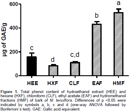
The highest antioxidant activities measured by DPPH• free radical scavenging method were observed in EAF and HMF, which did not differ statistically from HEE (Table 1). Lower antioxidant capacities were observed in HXF and CLF, when compared to other fractions or HEE. In addition, higher antioxidant activity was found for the gallic acid (control) when compared with HEE or fractions. Besides, HEE, EAF and HMF presented IP values higher than 90% and AAI above 2.0.

Lipid peroxidation induced by AAPH was greatly inhibited by HEE, EAF and trolox (95.1, 95.0 and 94.3% respectively), while CLF and HMF presented a partial inhibition (40.3 and 45.0% respectively). HXF caused only 5.9% of decrease of AAPH-induced lipid peroxidation. Differently, when the inducer of lipid peroxidation was FeSO4, HMF and trolox produced the higher inhibitory effects (63.0 and 94.3%, respectively). The percentual of inhibition of FeSO4-induced lipid peroxidation for HEE, HXF, EAF or CLF was 27.4, 38.8, 26.0 and 9.1%, respectively.
Effect of HEE and fractions of M. tenuiflora on TPA-induced ear edema and neutrophil accumulation in mice
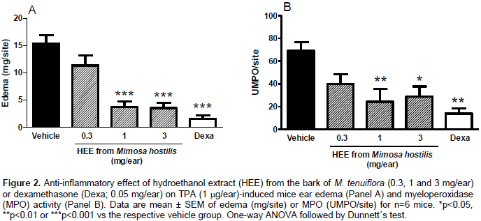
The coadministration of HEE at 1 or 3 mg/ear, but not 0.3 mg/ear, significantly reduced ear edema (p<0.001) or MPO activity (p<0.05 for 3 mg/ear and p<0.01 for 1 mg/ear) induced by TPA (Figure 2A and B, respectively).
The inhibitory activity of fractions of HEE on the edema induced by TPA is shown in Figure 3. The coadministration of HXF, CLF or EAF at doses of 1 or 3 mg/ear significantly reduced the formation of ear edema (P<0.001), when compared to vehicle group. At the same doses, HMF also decreased the ear edema (p<0.01), in comparison to vehicle group. As an anti-inflammatory control, dexamethasone (0.05 mg/ear) significantly reduced ear edema (p<0.001). The percentage of inhibition of TPA-induced ear edema formation for the dose of 3 mg/ear of AEF, HMF, CLF and HXF were respectively 75.0, 64.9, 71.4 and 79.1%.

The activity of MPO was measured in the ear biopsies as an estimative of the neutrophil content. It was demonstrated that coadministration of HXF (1 or 3 mg/ear) did not change this activity (Figure 4A). Coadministration of CLF at 3 mg/ear, but not 1 mg/ear, to mice ears significantly reduced MPO activity (p<0.05), when compared to vehicle group. Doses of EAF or HMF (1 or 3 mg/ear) also diminished (p<0.001 or p<0.05 respectively) the activity of MPO, as did dexamethasone (0.05 mg/ear; p<0.001), when compared to vehicle group. The percentage of inhibition of TPA-induced MPO activity for the dose of for 3 mg/ear of AEF, HMF and CLF were respectively 92.7, 52 and 72.0%.

DISCUSSION
In this study, the preparation of HEE from the bark of M. tenuiflora and fractions was described, as well as the phytochemical, antioxidant potential and anti-inflammatory activity evaluation. The results indicated that the bark of this plant presents topical anti-inflammatory effect that was associated to the phenolic content and antioxidant capacity of HEE and selected fractions.
Few studies have evaluated the phytochemical composition of the bark of M. tenuiflora. Anton et al. (1993) have described the presence of saponins and terpenes in the stems of M. tenuiflora. Likewise, Sukanya et al. (2009) detected the presence of phenolic substances, as the C-glycosylated flavones, O-glycosylated flavones and tannins. In the present work, by using the phytochemical screening, we demonstrated that HEE from the barks of this plant, EAF and HMF contains phenolic compounds (phenols, tannins, flavonoids and xanthones). Accordingly, the higher concentrations of phenolic compounds were found in EAF and HMF, as assessed by Folin-Ciocalteu method, although all fractions presented phenols. The highest concentrations of phenolic compounds in EAF and HMF results from the polarity of the extractor liquids that allow hydrogen bonds between the hydroxyl groups present in these compounds and the solvent, increasing its solubility. However, the HEE extracted by the high polar hydroalcohol mixture had low phenolic content when compared to EAF and HMF. The chemical diversity of the compounds, which exerts a competitive interaction with the reagents used in the assessment (Ainsworth and Gillespie, 2007) may explain this finding. No phenolic content was found in HXF and CLF in the qualitative tests and lower concentrations were found in the analytical test. We suggest that the low effective concentration of phenols when fractionated did not allow its detection by the analytical tests applied in HXF and CLF during the phytochemical screening.
Antioxidant activity against DPPH· free radical was correlated with phenolic content of HEE and fractions, because the higher the phenolic content of HEE or fraction, the best the DPPH· scavenging activity presented. According to Procházková et al. (2011), phenolic compounds are excellent radical scavengers, can donate hydrogen to the unpaired electron and remain stable due to the resonant phenoxyl ion formed. In addition, several other studies have shown that phenolic compounds significantly contribute to the antioxidant capacity of medicinal plants (Melo et al., 2010; Vaher et al., 2010; Moura et al., 2011; Silva et al., 2011). Free radical scavenging ability was higher for EAF and HMF, as evaluated by the lower values of AAI (Scherer and Godoy, 2009). These results seem to be primarily related to the abundance of flavonoids and tannins presented in these fractions.
In consonance with the phenolic content and DPPH· activity, the inhibition of lipoperoxidation caused by AAPH was more pronounced to EAF and HMF. However, FeSO4-induced lipoperoxidation did not correlate with the other antioxidant data, since lipoperoxidation was more inhibited by HMF and HXF. The FeSO4 can react with oxygen, superoxide anion radical and through the Fenton´s reaction can be dismutated to hydrogen peroxide, yielding hydroxyl radicals (Barreiros and David, 2006), which are responsible for initiating lipid peroxidation (Vasconcelos et al., 2007). Thus, these results suggest that the compounds present in HMF predominantly acts during the initiation of lipid peroxidation. Conversely, it can be suggested that EAF and HEE have compounds that acts mainly on the radicalar propagation phase. According to Silva et al. (2006), AAPH is decomposed at physiological temperature, in the presence of oxygen, and conducts to attacks to peroxyl radicals, that in turn scavenges hydrogen from lipids. These mechanist differences between AAPH and FeSO4-induced lipoperoxidation can partly explain the dissimilarities in the results between the fractions (mainly HMF and EAF) from HEE.
Besides the antioxidant activity of HEE from bark of M. tenuiflora and its fractions, in this study the anti-inflammatory activity of the extract and fractions was investigated by using TPA-induced ear inflammation. This model of cutaneous inflammation was previously described as suitable for evaluating the activity of both steroidal and non-steroidal anti-inflammatory drugs after topical administration (Schiantarelli et al., 1982). Topical administration of TPA, an active compound isolated from croton oil (Saraiva et al., 2010), concomitant with HEE led to the findings that HEE reduced the ear edema and MPO activity in mice ear, which were indicatives of decreased plasma protein and liquid extravasation, as well as neutrophil accumulation, respectively. These results clearly indicated that HEE from bark of M. tenuiflora possesses topical anti-inflammatory effect. Corroborating our findings, the ethanol extract from bark of M. tenuiflora reduced inflammation induced by carrageenan or formalin in mice, as well as produced antinociceptive effect (Cruz et al., 2016). In this study it was shown that HEE contains high phenolic content and antioxidant activity against DPPH• radical, as well as reduces lipoperoxidation induced by FeSO4 or AAPH. This antioxidant activity can be directly correlated with the anti-inflammatory activity of HEE. According to (Scalbert and Williamson, 2000) natural substances groups that scavenge free radicals, such as flavonoids, present biological effect on inflammation.
Many studies have described that phenolic compounds with antioxidant activity possess anti-inflammatory effect. A study performed with polyphenol resveratrol demonstrated that it inhibited the mRNA expression of cyclooxigenase-2, inducible nitric oxide synthase and some adhesion molecules (Rahman et al., 2006), that are important mediators of inflammation. Thus, HEE from bark of M. tenuiflora may present bioactive compounds that can interfere with the expression of these mediators, especially regarding to the inhibition of adhesion molecules, which are needed for neutrophil, as well as other leukocytes, migration to the focus of inflammation. However, we cannot discharge the possibility that other mechanisms are contributing to the anti-inflammatory effect of HEE, like its antioxidant activity.
Once the anti-inflammatory activity of HEE from bark of M. tenuiflora was established, the fractions prepared from HEE were also evaluated for this effect. Interestingly, it was observed that all fractions, when coadministrated with TPA, decreased the edema induced by this compound, but fractions with higher phenolic content and antioxidant activity caused more pronounced reduction of the MPO activity in mice ear. Comparatively, 1 mg/ear of EAF and HMF decreased the MPO activity, but for CLF it was necessary 3 mg/ear to reduce the MPO activity. Besides, for HXF, even this dose was not enough to reduce the MPO activity. Reduced MPO activity can also be related to the phenolic content of EAF and HMF and antioxidant activity. As our data indicate that these fractions contain flavonoids, tannins and phenolic compounds, the anti-inflammatory activity can be attributed to their action. In this way, it is largely known that flavonoids are able to reduce the inflammatory response through inhibition of phospholipase A2, cyclooxygenases, lipoxygenases and inducible nitric oxide synthase, besides other effects. These enzymes can generate inflammatory mediators that, in turn, may modulate the expression of adhesion molecules, such as ICAM-1 or VCAM-1 (Mutoh et al., 2000; Nijveldt et al., 2001; Raso et al., 2001; Chen et al., 2004; Peana et al., 2006; Rahman et al., 2006; Valério et al., 2009; García-Lafuente et al., 2009; Serafini et al., 2010).
Besides, the recent study by Cruz et al. (2016) has evaluated the hexane, dichloromethane, ethyl acetate and butanol fractions of the ethanol extract from bark of M. tenuiflora and found that hexane, dichloromethane and ethyl acetate caused anti-inflammatory effects in mice, although all fractions reduced nociception induced by various stimuli. However, it was not possible to establish a correlation among the polarity of the solvents used to obtain the fractions and the pharmacological effects from the study of Cruz et al. (2016), given the variability of the effects in the tests employed. This study also isolated a flavonoid called sakuranetin from M. tenuiflora, which also induced an anti-inflammatory and antinociceptive effect in mice.
The identification of phenolic compounds presented in HEE from M. tenuiflora or fractions was not yet performed, but in this study, the partition of HEE has shown that phenolic compounds may be of importance to the anti-inflammatory effects, mainly those related to the neutrophil migration.
CONCLUSION
In summary, this study demonstrated that HEE from M. tenuiflora and its fractions (mainly EAF and HMF) possess compounds with antioxidant properties and phenolic contents that seem to cause anti-inflammatory effect in mice ear. In this way, this plant may be of interest to search for new bioactive molecules for treating inflammatory conditions.
CONFLICT OF INTERESTS
The authors have not declared any conflict of interests.
ACKNOWLEDGMENTS
The authors extends their thanks to “Coordenação de Aperfeiçoamento de Pessoal de Nível Superior” (CAPES; Social Demand Program of Scholarships) for financial support. EAC is a beneficiary of CNPq productivity grant.
REFERENCES
|
|||||||||||||||||||||||||||||||||||||||||||||||||||||||||||||||||||||||||||||||||||||||||||||||||||||||||||||||||||||||||||||||||||||||||||||||||||||||||||||||||||||||||||||||||
Copyright © 2024 Author(s) retain the copyright of this article.
This article is published under the terms of the Creative Commons Attribution License 4.0


