Full Length Research Paper
ABSTRACT
Glucocorticoid-induced diabetes mellitus (GIDM) is an abnormal increase in blood glucose associated with the use of glucocorticoids in a patient with or without a prior history of diabetes mellitus. This is a common and potentially harmful problem in clinical practice, affecting almost all medical specialties, but is often diffi¬cult to detect in clinical settings. The objective of this study was to determine effect of Parkia biglobosa extract on open wound healing in dexamethasone induced hyperglycaemia. Effect of three different doses of P. biglobosa extract (25, 50 and 100 mg/kg body wt.) for 14 consecutive days on open skin wound healing before and after dexamethasone-induced hyperglycaemia was investigated; histological assessment was also conducted on the fourteenth day. The three different doses of P. biglobosa extract decreased the serum glucose concentration in pre and post-treatment dexamethasone-induced hyperglycaemic animals; the percentage reduction was greater in the 50 and 100 mg/kg of P. biglobosa-pretreated groups (14.9 and 19.21%, respectively) as compared to that of ketoconazole, where it was only 16.5%. In the post treatment groups, the percentage reduction was greater in 100 mg/kg of P. biglobosa (17.7%) as compared to that of ketoconazole, where it was only 16.6%. Histological evaluation showed that the pretreated group of animals had higher performance scores on the grading scale and improved healing when compared with the post-treated groups. There was a demonstrable reduction in the wound healing process in the pre-treatment group that was dosed dependent.
Key words: Parkia biglobosa, open skin wound healing, dexamethasone-induced hyperglycaemia, histological assessment.
INTRODUCTION
Glucocorticoids are potent anti-inflammatory and immunosuppressive drugs which are widely used to treat a wide range of diseases. A number of side effects, including new-onset hyperglycaemia in patients without a history of diabetes mellitus (Suh and Park, 2017), also severely uncontrolled hyperglycaemia in patients with known DM is also associated with them. There are two main models, that is, incisional and excision for determining three basic phases in wound healing process (inflammation, proliferation, and maturation) (Dorsett-Martin, 2004). These are simple and reproducible models which represent basic requirement assessing the effects of different external factors on skin wound healing (Regan and Barbul, 2011). The incisional (sutured) skin healing model is used for wound tensile strength measurement (Davidson, 1998) while the excisional model is more appropriate for histological evaluation due to significantly broader morphological changes which occur during the healing process. Corticosteroid induced diabetes otherwise called steroid diabetes; the most common glucocorticoids which cause steroid diabetes are prednisolone and dexamathasone. It is also a simple and inexpensive model of a complex wound healing impairment (Gal et al., 2008). The excess of either endogenous or exogenous glucocorticoids has been shown to increase gluconeogenesis and decrease tissue glucose uptake, thus resulting in hyperglycaemia, potentially inducing diabetes (Wolfsheimer, 1989).
Parkia biglobosa (Jacq.) R.Br. ex G. Don (family Fabaceae) popularly known as the “African locust bean tree”, it is a medium-sized tree growing up to 30 m in height. The plant is reported to contain carbohydrates, proteins, fats, minerals, vitamins, tannins and flavonoids. P. biglobosa have been used in Nigeria and other West Africa in rural communities to treat a variety of diseases as diabetes mellitus, malaria and pains. The hypoglycaemic effect of fermented seeds of P. biglobosa, a natural nutritional condiment that features frequently in some African diets as a spice, was investigated in alloxan induced diabetic rats (Builders, 2014) .
Therefore, the aim of this study was to establish the effect of extract of P. biglobosa on an excisional model of skin wound healing in normal healthy and corticosteroid treated as well as to evaluate the effects of various external factors on wound healing semi quantitative assessments.
MATERIALS AND METHODS
Source of plant material and identification
The stem barks of P. biglobosa were collected from Chaza village in Niger State and the stem barks were identified by a taxonomist Mallam Muazam of the Department of Medicinal Plant Research and Traditional Medicine of National Institute for Pharmaceutical Research and Development (NIPRD), Abuja, Nigeria where a voucher specimen was deposited in the herbarium for reference.
Chemicals and reagents
Chemicals and reagents were purchased from Sigma Chemical Company (St. Louis, USA).
Extraction of plant
The plant material (stem bark) was air dried under shade and then ground into coarse powder with a pestle and mortar. 200 g of the powdered bark was extracted with 2 L methanol for 48 h using a Soxhlet apparatus (Quicklet, UK). The extract was filtered through Whatmann No. 1 (Whatmann International Ltd, Maidstone, UK) paper and evaporated to dryness under reduced pressure using a rotary evaporator to yield a crude extract which was stored at 4°C until used.
Animals
Adult male Wistar rats (180-200 g) maintained at Animal Facility Centre (AFC) of the Department of Pharmacology, Faculty of Pharmacy, Bingham University were used for the study. They were fed with pelleted feed (Vital®, Jos) and water ad libitum. The rats were allowed 7 days to acclimatize before the experiments were conducted according to the permission and prescribed guidelines of the Institutional Animal Ethics Committee.
Experimental design
Eighteen rats were administered with three different doses of P. biglobosa extract (25, 50 and 100 mg/kg body wt., p.o) designated as P1, P2, P3 and six rats received ketoconazole (24 mg/kg body wt., p.o.) (Marty et al., 2000) and designated as P4 for 14 consecutive days on open skin wound healing before daily administration of a pre standardized dose of dexamethasone(1 mg/kg body wt., i.m) (Gholap and Kar, 2003), and classified as group 1. In group 2, eighteen rats were treated with three different doses of P. biglobosa extract (25, 50 and 100 mg/kg body wt., p.o) designated as P5, P6 , P7 and six rats received ketoconazole (24 mg/kg body wt., p.o.) (Marty et al., 2000) and designated as P8 for 14 consecutive days on open skin wound healing after dexamethasone-induced hyperglycaemia. Simultaneously, six rats normoglycemic animals were treated with equivalent amount of vehicle (0.2 ml of normal saline) and referred to as P9.
Anaesthesia and surgical procedures
General anaesthesia was induced by intramuscular administration of ketamine (33 mg/kg; Hameln pharmaceutical Ltd.) and xylazine (10 mg/kg; Unipex). A small incision was made above the spine through which the lower part of the belt punches pliers were slide beneath the skin. Consecutively, four round full thickness excision, 5 mm in diameter, were performed on back of each rat. The incision was then sutured (Gal et al., 2008).
Histological analysis
On days 2, 6 and 14, six rats from each group were sacrificed after surgery. Skin wounds removed and were processed routinely for light microscopy (fixating, dehydrating, embedding, and cutting). Two sections were made from each wound and stained with hematoxylin-eosin (HE- basic staining) and van Gieson (VG-collagen staining), respectively. Semi-quantitative method was used to evaluate the following histological processes and structures: reepithelization,(polymorphonuclear leucocytes, PMNL), fibroblasts new vessels, and new collagen. Sections were evaluated according to the scale: 0, 1, 2, 3, and 4 by two independent observers (Gupta and Kumar, 2015). The mean value was used for statistical comparison.
Statistical analysis
All data were expressed as mean values ±standard error of mean (SEM). Data were compared using one-or two-way analysis of variance (ANOVA). Semi-quantitative evaluation was analyzed using Mann-Whitney test. Differences were considered significant for P values <0.05.
RESULTS
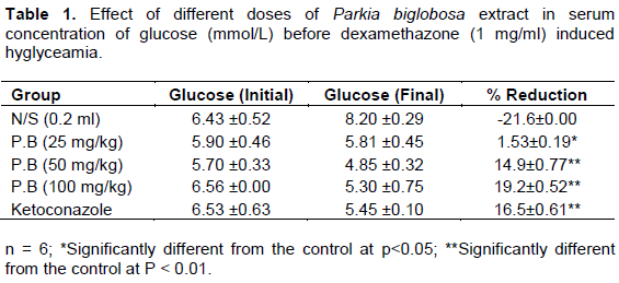
Table 1 shows that administration of dexamethasone induced increase serum glucose level however the hyperglycaemia was reversed by significant dose dependent of the extracts of P. biglobosa, this was highly significant in the groups receiving 100 mg/kg P. biglobosa extract and ketoconazole.
Dexamethasone induced higher increase in serum glucose level; high significant hypoglycaemia was observed in groups treated with 100 mg/kg P. biglobosa extract compared with the groups receiving normasaline as indicated in Table 2.
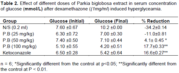
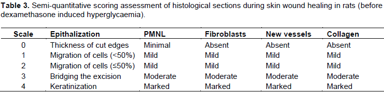
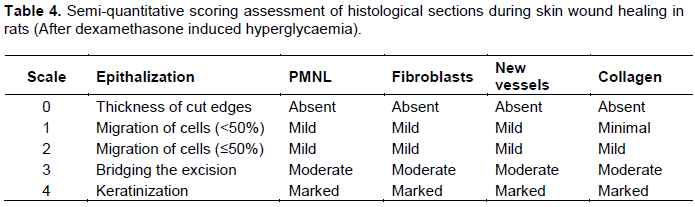
Table 3 and 4 show 4-point scale of the semi-quantitative analysis of histological section, 0 scale shows thickness of cut edges with absence of polymorphonuclear leucocytes (PMNL), fibroblasts, new vessels, and new collagen. Migration of cells <50% with mild surrounding tissues,mild subcutaneous tissues and minimal granulation tissue were described by scale 1, scale 2 was used to evaluate migration of cells >-50% mild demarcation line or granulation tissue with mild granulation tissue. Scale 3 describes bridging of the excision, moderate tissue out of the granulation tissue and moderate granulation tissue while keratinization, marked surrounding tissue and marked granulation was evaluated by scale 4.
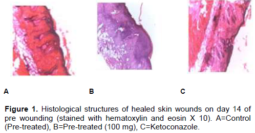
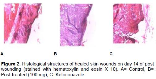
In the photomicrograph of pre-treated and post-treated groups, administration of 100 mg/kg of pre-treated and post treated P. biglobosa extract indicated increased fibroblast growth, collagen synthesis, and the healing process as illustrated in Figures 1 and 2.
DISCUSSION
In this study, rat was used as an experimental animal model, since rat skin represents one of the most common models used in experimental studies concerning the skin wound healing. It is a useful model because we can study the healing of three different tissue types (epidermis, dermis and striated muscle) and it is only epidermis that has the capability to regenerate (Vidinský et al., 2006).
In the pre-treated groups, the antihyperglycemic activity of the P. biglobosa extract in dexamethasone-treated animals may be due to decrease peripheral insulin resistance or by suppression of enzymes involved in hepatic gluconeogenesis as well as by stimulation of glucose uptake and use in peripheral tissues similar to study conducted by Shanmugasundaram et al. (1983) and Persaud et al. (1999). Decrease in serum glucose level may also be mediated through an increase in insulin release from pancreas according to the research carried out by Caro and Amatruda (1982).
Corticosteroid-induced diabetes mellitus mechanism involves insulin resistance, caused by the alteration in binding of insulin to its receptor (receptor defect) or by the impairment of the intracellular response to insulin (Rizza et al., 1982). Hyperglycaemia inhibits wound healing process associated with prolonged inflammatory phase (Naguib et al., 2004) defected angiogenesis (Goren et al., 2006) and diminished fibroblast proliferation (Hehenberger and Hansson, 1997).
In order to effectively manage chronic wounds, periodic assessment of the healing process is necessary (Mullins et al., 2005). A semi‑quantitative score was adopted in this study for scoring the degree of changes observed on an ordinal scale, namely, low, medium or high grade. Even though quantitative scoring system is highly specific and standardized due to difficulty to objectify the exact interval between two values, semi‑quantitative scoring systems remain in wide use in the world of the biomedical research (Lemo et al., 2010).
Corticosteroid induced diabetic wound-healing indicated a reduction in the contraction of open wounds, decreased capillary volume, decreased number of olymorphonucleocytes, increased edema, decreased number of fibroblasts, decreased neovascularization, and increased rate of infection. This reduction is related to a delay in the appearance of inflammatory cells and to a reduction in fibroplasia, a new connective tissue matrix, collagen synthesis and deposition, and wound breaking strength (Bitar, 1998).
Wound healing is composed of three stages namely inflammation, proliferation and remodeling (Whaley and Burt, 1996). The proliferative stage typically demonstrates angiogenesis, collagen deposition, granulation tissue formation, epithelialization and wound contraction. In angiogenesis, new blood vessels are formed from endothelial cells (Cotran, 1999).
Increased collagen deposition, regeneration, and well-aligned tissue observed in both pre and post-treated groups are in accordance with a study, which reported prohealing parameters (Muhammadu et al., 2016).
The influence of various factors on the wound healing was evaluated histologically on the fourteenth day similar to study conducted by Whelan et al. (2003). In the present study the most significant changes occur during the first week of wound healing, this correlates with the study conducted by Medrado et al. (2003).
Differentiation of fibroblasts into myofibroblasts was observed during the healing process. Myofibroblasts synthesize extracellular matrix components such as collagen types I and III. Indicator for the assessment of wound healing is fibroblast proliferation (Park et al., 2005). The major cell type found in the granulation of wound tissues is fibroblasts. They play important role in wound healing including secretion of a series of growth factors that facilitates angiogenesis, proliferation and matrix deposition (Mansbridge et al., 1999).
Studies conducted on the phytochemical screening of P. biglobosa showed the presence of phytoconstituents such as anthraquinones, tannins, flavonoids, terpenes, saponins, phenols and steroids (Builders et al., 2016); these may be attributed to the wound healing activity of P. biglobosa. The enhanced wound healing activity of this extract could be related to a function of either the individual or the additive effects of the phytoconstituents which is similar to research conducted by Liu et al. (2013). These bioactives have been reported to possess pharmacological properties such as antimicrobial, antioxidant, analgesic, and anti-inflammatory activities which promote the wound-healing process mainly due to wound contraction and increased rate of epithelization (Liu et al., 2013).
CONCLUSION
The wound healing activity of P. biglobosa extract in a simple experimental model has been established. Further studies are in progress in our laboratory to isolate and characterize the relevant bioactive components and elucidate the mechanisms of actions of these active ingredients.
CONFLICT OF INTERESTS
The authors have not declared any conflict of interests.
ACKNOWLEDGEMENT
The authors are grateful to all the staff of animal care unit for their contribution to this work.
REFERENCES
|
Bitar MS (1998). Glucocorticoid Dynamics and Impaired Wound Healing in Diabet Mellitus. American Journal of Pathology 152:454-457. |
|
|
Builders MI (2014). Parkia biglobosa (African Locust Bean Tree), World Journal of Pharmaceutical Research 3:1672-1682. |
|
|
Builders MI, Builders PF, Ogundeko TG (2016). Anti-ulcer activity of African locust bean tree in rats. International Journal of Phytotherapy 6:11-19. |
|
|
Caro JF, Amatruda JM (1982).Glucocorticoid-induced insulin resistance. Journal of Clinical Investigation 69:866-875. |
|
|
Cotran RS (1999). Tissue repair: cellular growth, fibrosis and wound healing. In: Robbins Pathologic Basis of Disease 6th Edition, eds. Cotran RS, Kumar V, Collins TWB Saunders Co Philadelphia, London, New York, St. Louis, Sydney, Toronto p.89-112. |
|
|
Davidson JM (1998). Animal models for wound repair. Archives of Dermatological Research 290:1-11. |
|
|
Dorsett-Martin WA (2004). Rat models of skin wound healing: A review. Wound Repair and Regeneration 12:591-599. |
|
|
Gal P, Kilik R, Mokry B, Vidinsky T, Vasilenko S, Mozes N, Bobrov Z, Tomori Z, Bober J, Lenhardt L (2008). Simple method of open skin wound healing model in corticosteroid-treated and diabetic rats: tandardization of semi-quantitative and quantitative histological assessments. Veterinarni Medicina 53(12):652-659. |
|
|
Gholap SM, Kar A (2003). Efficacy of some plant extracts in regulating corticosteroid induced hyperglycaemia in mice. Pharmaceutical Biology 41:315-318. |
|
|
Goren I, Muller E, Pfeilschifter J, Frank S (2006). Severely impaired insulin signaling in chronic wounds of diabetic ob/ob mice: a potential role of tumor necrosis factor-alpha. The American Journal of Pathology 168:765-777. |
|
|
Gupta A, Kumar P (2015). Assessment of the histological state of the healing wound. Plastic and Aesthetic Research (2):239-242. |
|
|
Hehenberger K, Hansson A (1997). High glucose-induced growth factor resistance inhuman fibroblasts can be reversed by antioxidants and protein kinase Cinhibitors. Cell Biochemistry and Function 15:197-201. |
|
|
Lemo N, Marignac G, Reyes Gomez E, Lilin T, Crosaz O, Ehrenfest DM (2010). Cutaneous reepithelialization and wound contraction after skin biopsies in rabbits: a mathematical model for healing and remodelling index. Veterinarski Arhiv 80:637 652. |
|
|
Liu H, Lin S, Xiao D, Zheng X, Gu Y, Guo S (2013). Evaluation of the Wound Healing Potential of Resina Draconis (Dracaena cochinchinensis) in Animal Models. Evidence-Based Complementary and Alternative Medicine, pp. 1-10. |
|
|
Mansbridge JN, Liu K, Pinney RE, Patch R, Ratcliffe A, Naughton GK (1999). Growth factors secreted by fibroblasts: role in healing diabetic foot ulcers. Diabetes, Obesity and Metabolism 1:265-279 |
|
|
Marty MS, Crissman JW, Carney EW (2000). Evalution of the male pubertal onset assay to detect testosterone and steroid biosynthisis inhibitor in CD rats. Toxicological Science 60:285-295. |
|
|
Medrado AR, Pugliese LS, Reis SR, Andrade ZA (2003). Influence of low level laser therapy on wound healing and its biological action upon myofibroblasts. Lasers Surgical and Medical 32:239-244. |
|
|
Muhammadu AA, Arulselvan P, Cheah PS, Abas F, Fakurazi S (2016). Evaluation of wound healing properties of bioactive aqueous fraction from Moringa oleifera Lam on experimentally induced diabetic animal model. Drug Design, Development and Therapy 10:1715-1730. |
|
|
Mullins M, Thomason SS, Legro M (2005). Monitoring pressure ulcer healing in persons with disabilities. Rehabilitation Nursing 30:92 99. |
|
|
Naguib G, Al-Mashat H, Desta T, Graves DT (2004). Diabetes prolongs the inflammatory response to a bacterial stimulus through cytokine dysregulation. Journal of Investigative Dermatology 123:87-89. |
|
|
Park SG, Shin H, ShinYK, Lee Y, Choi EC, Park BJ, Kim S ( 2005). The novel cytokine p43 stimulates dermal fibroblast proliferation and wound repair. The American Journal of Pathology 166:387-398. |
|
|
Persaud SJ, Al-Majed H, Raman A, Jones PM (1999). Gymnema sylvestre stimulates insulin release in vitro by increased membrane permeability. Journal of Endocrinology 163:207-212. |
|
|
Regan MC, Barbul A (2011). Cellular biology of wound healing. In: Fischer J.A. (ed.): Surgical Basic Science. Mosby Yearbook, St. Louis; p. 68-88. |
|
|
Rizza RA, Mandarino LJ, Gerich JE (1982). Cortisol induced insulin resistance in man:Impaired glucose production and stimulation of glucose utilization due to post-receptor defect of insulin action. Journal of Clinical Endocrinology and Metabolism 54:131-138. |
|
|
Shanmugasundaram KR, Panneerselvam C, Samudram P, Shanmugasundaram ERB (1983). Enzyme changes and glucose utilization in diabetic rabbits: the effect of Gymnema sylvestre, R.Br. leaf extract. Journal of Ethnopharmacology 7:265-273. |
|
|
Suh S, Park MK (2017). Glucocorticoid-Induced Diabetes Mellitus: An Important but Overlooked Problem. Endocrinology Metabolism 32:180-189. |
|
|
Vidinský B, Gál P, Toporcer T, Longauer F, Lenhardt Ľ, Bobrov N, Sabo J (2006) Histological study of the first seven days of skin wound healing in rats. Acta Veterinaria. Brunensis 75:197-202Whaley K, Burt AD Inflammation (1996). Healing and Repair. In: Muir's Textbook of Pathology, MacSween, R.M.N. and K. Whaley (Eds.). 13th Ed., Arnold, London; p. 112-165. |
|
|
Whelan HT, Buchmann EV, Dhokalia AA , Kane MP, Whelat NT, Wong- riley MT, Eells JT, GouldLI, HammamiehR, Das R, Jett M (2003). Effect of NASA light-emitting diode irradiation on molecular changes for wound healing in diabetic mice. Journal Clinical Laser Medical and Surgical 21:67-74. |
|
|
Wolfsheimer KJ (1989). Insulin resistant diabetes mellitus. In: Kirk RW, ed., Current Veterinary Therapy. Philadelphia, WB Saunders Co; p. 1012-1019. |
|
Copyright © 2024 Author(s) retain the copyright of this article.
This article is published under the terms of the Creative Commons Attribution License 4.0


