Full Length Research Paper

ABSTRACT
In equine clinics, skin injuries are some of the most frequently treated injuries, this research evaluated the healing potential and antioxidant activity of Abarema cochliacarpos in order to develop a pharmaceutical formulation for treating skin lesions in horses. Firstly, the antioxidant activity of several fractions was evaluated using the 2,2-diphenyl-1-picrylhydrazyl radical method and the thiobarbituric acid test. The determination of phenolic compounds was done by means of the Folin-Ciocalteau test. To assess the wound healing activity the dry powdered stem bark and the aqueous, ethanolic and propylene glycol extracts from the same plant, material were incorporated in a gel formulation and evaluated in equines with induced wounds. Results show that all obtained extracts and their fractions have antioxidant action. Regarding the healing evaluation in equines, the extracts tested and the dry powdered stem bark showed statistically significant activity in the contraction of wounds. Histological analysis revealed the powdered stem bark as the best treatment to induce healing of cutaneous lesions in horses. Experimental studies in animals with different dosages and formulations, besides the isolation of its chemical components, as a tool in the discovery of new healing agents or optimization of the existing ones is recommended.
Key words: Abarema cochliacarpos, antioxidant activity, wound healing activity.
INTRODUCTION
Abarema cochliacarpos (Fabaceae) is a native tree from Brazil (popularly called in this country by "barbatimão") found mainly in the Atlantic forest and Caatinga. Many traditional communities in northeast Brazil use its bark in folk medicine (Agra et al., 2008; Pereia et al., 2013). An ethnopharmacological survey in a Brazilian traditional community has documented popular uses of this species to treat inflammatory disorders, including ulcerations, and for wound healing purposes (Tenório et al., 2016).
The stem bark used in the preparation of herbal medicines, comprise chemical compounds that include alkaloids, flavonoids, terpenes, stilbenes, steroids, protease inhibitors, and tannins. The latter class of secondary metabolites comprises the major components of A. cochliacarpos and have been regarded as the main responsible compounds for its antioxidant activity, as they can scavenge free radicals (Shimizu et al., 2009).
Plant extracts thus represent an important alternative to traditional medicine for the treatment of wounds, because they can act on the surface of the lesion, and through anti-inflammatory activity, create a micro-environment that facilitates fibroplasia. The lesions may heal faster because the wound is decontaminated by the extract’s antimicrobial effects, or through changes of pH in the wound, which create an unfavorable environment for the growth of micro-organisms (Dias et al., 2013).
In equine clinics, skin injuries are some of the most frequently treated injuries, affecting mainly the locomotor limbs. They occur due to the animals’ active behavior and rapid reactions, especially in horses involved in sporting activities (Paganela et al., 2009). Horses have sudden reactions that can lead to limb trauma. In addition, inadequate facilities and grazing are also crucial factors for the occurrence of these lesions. Incorrect treatments and the tendency to chronicity of the lesions also hamper the cicatricial process (Viana et al., 2014). Given the interest in development of herbal medicines for the treatment of wounds, and the high incidence of equine skin lesions. The research aims to assess the phytochemical profile, antioxidant activity, and effects of powdered stem bark, natrosol gel formulation obtained from aqueous, ethanolic and propylene glycol extracts of the stem barks of A. cochliacarpos, on healing of skin wounds in horses.
MATERIALS AND METHODS
Study type and setting
The experiment involved both in vitro and in vivo laboratory evaluation. The research was conducted in the multidisciplinary research laboratory of the Centro Universitário Cesmac, located in Maceió, Alagoas; Brazil, and in the Clinical Veterinary School of the same institution, which is located in the city of Marechal Deodoro, Alagoas, Brazil.
Ethical aspects
This study was approved by the Ethics Committee for the use of animals (CEUA) of the Federal University of Alagoas with opinion substantiated n° 43/2012.
Collection and identification of botanical material
The specimen was collected in the municipality of Marechal Deodoro, Alagoas, Brazil. Three samples were collected for botanical identification the specimen was identified at the local of collection by a botanist and samples from stem and leaves were stored in the Herbarium of the Environment Institute, in Alagoas, under MAC registration number: 25.370. The stem bark was dried in the shade at room temperature and powdered afterwards by means of a mill that was used in the preparation of the extract. The stem bark powder was then stored in a tightly sealed dark container. Aliquots of this material were used for incorporation into the gel of natrosol, production of extracts, and phytochemical screening.
Preparation of extracts
Aqueous extract
Extracts from the bark of the stems of A. cochliacarpos were obtained by decoction over 30 min, in a proportion of 100 g of dry powdered bark for each 1000 ml of distilled water. The aqueous extract was then stored at room temperature until it was incorporated into the natrosol gel via the method of Verza (2006).
Propylene glycol extract
To obtain this extract, the powdered material was macerated with an 80% mixture of propylene glycol in distilled water for 24 h. After this period, the extract was filtered in a filter paper (Whatman No 1). The propylene glycol extract was further stored at room temperature until its incorporation into the natrosol gel via the method of Verza (2006).
Ethanolic extract
The ethanolic extract from the barks of the stem of A. cochliacarpos was prepared by a steeping method, in which the plant material was first crushed, then placed in a percolator tube containing absolute ethanol for 72 hours. After this period, the extract was filtered in (Whatman No 1 filter paper). This procedure was repeated until the plant material was exhausted. A portion of the liquid sample was then concentrated in a rotary evaporator under reduced pressure until a crude ethanolic extract (BSE) was obtained (Weber, 2013). The other part of the liquid sample was stored at room temperature until it was incorporated in the natrosol gel.
Preparation of natrosol gel and obtaining gel from A. cochliacarpos
The natrosol gel was prepared according to previously described methodology (Wettasinghe and Shahidi, 1999). After the preparation of the natrosol gel, each extract was incorporated into the natrosol gel in a 10% v/w concentration for a seven-day stability test.
The Crude Ethanolic Extract (CEE) of the A. cochliacarpos stem bark was fractioned through a liquid-liquid partition method. The extract was dissolved in mixture of methanol and water at a ratio of 6:4, respectively. From the methanol/water phase, the process of partition was initiated using the solvents hexane, chloroform and ethyl acetate, resulting in Hexane Fraction (HF), Chloroform Fraction (CF) and Ethyl Acetate Fraction (EAF), respectively. The fractions were then subjected to HPLC-DAD (High Performance Liquid Chromatography-Diode Array Detector), TLC (thin layer chromatography), phytochemical prospecting, evaluation of antioxidant activity, and determination of phenolic compounds.
Phytochemical screening
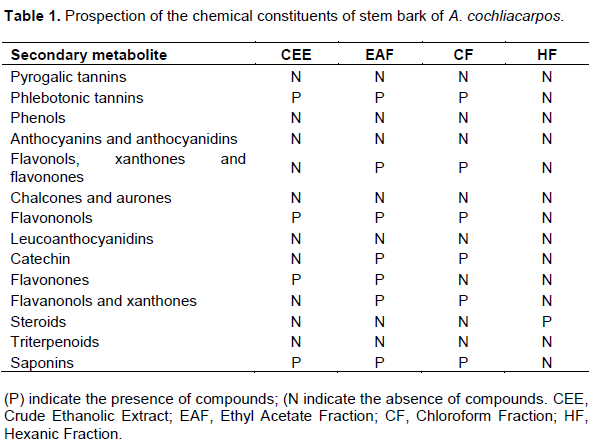
Phytochemical screening was performed for searching the following metabolites: pyrogalic tannins, phlebotonic tannins, phenols, anthocyanins, anthocyanidins, flavonols, xanthones, flavonones chalcones, aurones, flavononols, leucoanthocyanidins, catechin, flavonones, flavanonols, xanthones, steroids, triterpenoids, and saponins (Matos, 1997).
Analysis of antioxidant activity by radical 2.2-diphenyl-1-picrilidrazila
In the analysis of antioxidant activity by radical 2.2-diphenyl-1-picrilidrazila (DPPH), vegetable samples, HF, CF and AEF were dissolved in 95% ethanol, applied in chromatoplates (Gel silicic F254, disabled) and eluted in solvent system containing ethyl acetate/formic acid (8:2). After elution, chromatoplates were immersed in ethanolic solution of DPPH 0.3 µgmL-1 for 10 seconds. The appearance of yellow spots underneath the purple coloration is indicative of antioxidant activity.
Absorbance values were converted to antioxidant activity percentages (AAO%) using the formula: AAO% = 100-{[ABSA-ABSB) X 100]/ABSC}, where ABSA represents the absorbance of the sample, ABSB is the absorbance of the blank, and ABSC is the absorbance of the control sample. Quantitative assessment of antioxidant activity was done by monitoring the free radical DPPH consumption and by measuring the decrease in absorbance of solutions at different concentrations (Eurides et al., 2007). Samples from BSE, HF, CF and AEF were diluted in triplicate in ethanol, with final concentrations of 250, 125, 50, 10 and 5 μg/ml. Reactions were carried out at room temperature for 30 minutes. Readings of the absorbances were subsequently made at 518 nm.
Evaluation of antioxidant activity by the Thiobarbituric acid method
For evaluation of the antioxidant activity by the Thiobarbituric acid method (TBA), homogenized egg yolk solution was used as a rich medium in lipids. The procedure was performed triplicate, in accordance with the methodology described by Ferreira (2010). Five test tubes were set up containing 0.5 ml of egg yolk (10% w/v) and 0.1 ml of the plant samples was dissolved in the least amount of methanol and the volume diluted to 1 ml with water. Three concentrations were tested for each BSE solution: 100, 500 and 1000 μg/ml.
Next, 2.2 chloride '-azo-bis (2-amidinopropano)-ABAP (0.07 mol/L) was added to each tube to induce lipid peroxidation, followed by a 20% solution of acetic acid (pH 3.5) and 1.5 ml of TBA (0.8% w/v) solution in sodium dodecyl sulfate-SDS (1.1% w/v). To allow observation of the complete lipid peroxidation, the antioxidants 3.5-di-tert-butyl-4-α-tocopherol and butylated hydroxytoluene BHT were used as positive standards in the same conditions to which plant extracts were subjected. The vials were placed in a water bath at 95°C under stirring over 60 min. After cooling, 5 ml of 1-butanol were added to each tube, and the tubes were centrifuged at 3000 rpm for 10 min.
To allow observation of the complete lipid peroxidation, the antioxidants 3.5-di-tert-butyl-4-α-tocopherol and butylated hydroxytoluene BHT were used as positive standards in the same conditions to which plant extracts were subjected.” Should be changed to: The antioxidants, 3.5-di-tert-butyl-4-α-tocopherol and butylated hydroxytoluene (BHT) were used as positive standards in the same conditions to which plant extracts were subjected, allowing hence, the observation of the complete lipid peroxidation. Absorbance of organic supernatant layer was measured in a spectrophotometer at a wavelength of 532 nm. The values obtained were applied in the following formula to determine the antioxidant content of plant samples in percentage (IA%): IA% = (1-A/C) x 100, where C is the absorbance of the control and the fully oxidized, the arithmetic mean of the sample tested absorbances .
Determination of phenolic compounds
The vegetable samples CEE, HF, CF, and EAF were evaluated at a concentration of 2.0 mg/ml. 0.5 ml of a 2N Folin-Ciocalteau reagent solution was added to 0.5 mL of each sample, followed by 1.0 ml of water (in triplicate). After stirring for 2 min, 0.5 ml of 10% (w/v) aqueous sodium carbonate (Na2CO3) was added to the tube. Then the samples were incubated for 2 h at room temperature (while protected from light). Absorbance values were obtained by readings at 750 nm, using the Folin-Ciocalteau reagent in methanol as blank sample. In order to build a calibration curve, gallic acid solutions were used at concentrations of 0.15, 0.1, 0.05, 0.025, 0.01, and 0.005 μg/ml. Total phenol values were expressed as gallic acid equivalents (Junqueira, 1999).
EC calculation
Results of in vitro tests were expressed in EC50 values (Nascimento et al., 2014), which is the concentration required to produce half (50%) of maximum effect estimated at 100% for the plant extract.
In vivo assay
Animal selection
Two male and three female horses were selected from the farm of the CESMAC Veterinary Medicine School. All animals were of mixed race, aged between 2.5 and 3 years, weighing from 270 to 320 kg, and were clinically healthy and well-nourished. The animals were fed with a balanced diet (corn + wheat + soy) at a rate of 2.5 kg/day, bulky (Tifton Grass) 28 kg/animal/day and water ad libitum. The animals were accommodated in individual stalls covered with sand.
Surgical procedure
After anesthesia, administered intravenously using 10% xylazine (0.5 mg/kg/EV), the animals were trichotomized, and a skin antisepsis was made with 1% povidone-iodine. Then, five circular and symmetrical wounds were made aseptically in the thoracic region, at the lateral to dorsal midline of each animal with the help of scalpel blade No. 23, after local anesthesia with lidocaine hydrochloride 2%, according to the method of Andriguetto and Perly (2002). The five wounds were made with the help of a punch of 3 cm, measuring approximately 3.0 × 3.0 cm, with a distance of 5 cm between them. The damage included the epidermis, dermis, and subcutis. After the surgical procedure, the wounds were measured with Vernier Calipers 200 × 0.05 mm/8 × 1/128, France.
Clinical evaluation of wounds
Clinical evaluations were performed visually by the same individual every 72 h, observing the presence of hyperemia, edema, pain, discharge, itching, crust, contraction, granulation tissue, and scar tissue in accordance with Ardisson et al. (2002).
Morphometric analysis of wounds
For the acquisition of the area of wounds, measurements of diameters at the time of biopsy to 0, 3, 7 and 14 days post-op were carried out. All wounds were measured with the aid of calipers (Andriguetto and Perly, 2002) and from these elements, the following equation was formulated: where A represents the area (cm²), and r the radius.The degree of contraction, expressed in percentage, was measured by the equation proposed by Ramsey et al. (1995) where Wo = initial wound area and Wi = wound area on the day of the biopsy: 100 x (Wo-Wi)/Wo =% contraction.
Treatment for the wounds
Wounds were treated daily at 24 h intervals, using powdered stem barks as well as aqueous, ethanolic and propyleneglycol extracts of A. cochliacarpos in a gel formulation. Four wounds were treated with A. cochliacarpos and the remaining wound was treated with Dakin (0.5% NaOCl). Treatments were applied on wounds of different positions for each animal. Throughout the experiment, bandages were not applied to the wounds.
Histological procedures
The animals underwent incisional biopsy of the skin for bilateral microscopic analysis of the healing process. Microscopic assessments were carried out at the end of the trial period. Material collection was performed for histopathological analysis, with tissue removal foursquare measuring about 2 x 2 cm, in order to include the entire diameter of the wound, as well as tissue adjacent to injury. The collected material was fixed in 10% buffered formalin for 24 h, being subjected to routine histological processing, included in paraffin and then cut into 6 cm. set microtome Then, the cuts were stained by hematoxylin and eosin and then examined using a light microscope (Barroso et al., 2010).
Microscopic analysis of the healing of each of the wounds was descriptive, qualitative evaluating the morphology of the inflammatory process, granulation tissue, cellularity, neovascularization and fibroplasia, as well as the evolution of the healing process throughout the ages (Barroso et al., 2010).
Statistical analysis
The results of the area and contraction of the wounds had been expressed on average ± shunting line standard, submitted to analyses of variance, and also to Tukey’s test, considering significant comparative values to the level of 5% of significance. Statistical analysis was performed using SPSS to determine the linear regression and the coefficient of determination (R2), which best explain the antioxidant potential of plant samples.
RESULTS
Preparation and fractionation of the ethanolic extract
From the crude extract (598 g) of stem bark, approximately 35 grams (5.85% yield) of the CF, 15 grams of EAF (2.50% yield) and 2 g of HF (0.33% yield) were obtained through primary fractionation. Fractionation of the crude extract with solvents of increasing polarity allows inferring the classes of substances extracted in the different fractions, in accordance with the known solubilities and polarities presented by various substances (Müller, 2006).
Phytochemical prospection
The phytochemical screening revealed different classes of secondary metabolites including phlebotonic tannins, flavonols, xanthones, flavones, catechins, steroids, and saponins, as shown in Table 1.
Quantitative assessment by DPPH assay
A significant radical scavenging activity was found for the CEE extract at low concentrations as shown in Figure 1. Furthermore, fractionation of the crude extract showed that the activity was retained by the EAF (Figure 2) since higher scavenging of radicals could be attained at lower concentrations. In contrast, similar scavenging activity for CF (Figure 3) and HF fractions (Figure 4) were achieved with higher concentrations and indicates that the compounds responsible for the activity in the crude extract had greater affinity by ethyl acetate solvent.


Determination of phenolic compounds
Total phenolic content found in the screening was of 181.8 mg Gallic acid equivalents/g of the sample with regard to CEE extract, and 50% of this content was found in AEF.
Antioxidant content percentage
The most common technique used to measure lipid peroxidation is the test of Thiobarbituric Acid (TBA), a spectrophotometric method that measures the concentration peroxidation products. The final product measured is the malondialdehyde or reactive substances formed from barbituric acid. In TBA test, the CEE extract presented the highest antioxidant capacity when compared to BHT at concentrations of 1000, 500 and 100 μg/ml, as shown in Table 2.

Evolution of the wound areas
The wound areas gradually decreased with the evolution of time over the course of 14 days of treatment (Figure 5). After 24 h from the beginning of the treatment, hyperemic areas were observed in the wounds, with zones of intense vascularization. The edges were well defined geometrically; however, the presence of edema and crusting areas were sometimes displayed at the outer regions of the wounds. Throughout the course of the experiment, those wounds treated with A. cochliacarpos presented irregular thick crusts and dryness.

The reduction of the wound area occurred due to the mechanism of contraction and the centripetal movement of the wound border towards the center, in order to reduce the area to be covered by the proliferating epithelium. The diameter and contraction data as a function of the different treatments are shown in Table 3. Based on results shown in Table 3, it was attested that the dry powdered stem bark as well as the ethanolic and propylene glycol extracts had the best performances in relation to reducing diameters and areas of induced wounds, with a wound healing activity comparable to Dakin's solution (p<0.05).

Histological analysis
Histological evaluation of wounds on the 14th day revealed significant differences regarding the inflammatory response and deposition of granulation tissue between the aqueous, ethanolic, propylene glycol extracts and powdered bark of A. cochliacarpos. The best healing occurred using the bark powder, followed by propylene glycol and ethanolic extracts. Because the treated animals exhibited less ulceration associated with neutrophilic inflammation and a greater organization of connective tissue, as illustrated by Figure 6A, B and C.

The water extract and the Dakin fluid were less effective treatments. Wounds treated with these substances presented greater inflammatory response, sharp ulceration associated with moderate neutrophilic inflammation, and disordered of connective tissue. These changes are not expected with that intensity on the 14th day. Histological features of the healing provided by these extracts are shown in Figure 6D and E. The presence of mononuclear infiltrates (macrophages and lymphocytes) and granulomas was observed as expected. The inflammatory phase was characterized by the presence of inflammatory cells in scar tissue, especially polymorphonuclear leukocytes (PMN) and macrophages. The wound treated with powdered stem bark accelerated re-epithelialization by the end of 14th day, which indicates the end of the healing process and suggesting greater efficacy of the product. All lesions presented a moderate to severe eosinophilic inflammatory reaction Figure 6F. Probably due to the response to induced tissue injury in the dermis of horses.
DISCUSSION
The DPPH assay is a widely used antioxidant procedure that asserts the ability of a compound to scavenge free radicals (Bendini et al., 2007). Several advantages have contributed to its dissemination as a first choice method of evaluation for antioxidant potential of foods (Floegel et al., 2011), plant extracts(Mensor et al., 2001) or isolated compounds (Villeño et al., 2007) which include its low cost, ease of handling and valid accuracy (Kedare and Singh, 2011).
The extracts obtained herein were able to scavenge the DPPH radical and act as potential antioxidants. This property correlates to the presence of some classes of secondary metabolites in the stem barks of A. cochliacarpos. As we have shown by the preliminary phytochemical screening, tannins and flavonoids are present in the stem bark of this species, and may be regarded as responsible for the antioxidant properties of the extracts since they are widely acknowledged as potent radical scavengers (Hatano, 1995; Nanjo et al., 1996; Cai et al., 2006).
Polyphenolic compounds comprise a large class of secondary metabolites featuring more than one phenolic ring in their core structure, which is derived from shikimate or polyketide metabolic pathway (Quideau et al., 2011). Their antioxidant mechanisms have been known for some time and relate to the presence of the phenolic ring, which provides the ability to scavenge free radicals and form itself stable free radicals, by hydrogen-atom transfer, or cation radicals, via single-electron transfer (Leopodini et al., 2011; Aquino et al., 2017). The stability of the radical formed is explained by the delocalization of the unpaired electron through the framework of phenolic rings existent in the molecular structure of these secondary metabolites (Quideau et al., 2011; Leopodini et al., 2010).
Wound healing is a complex and stepwise process that comprises different but complementary biochemical and cellular events in order to provide anatomical reconstruction and physiological regeneration of damaged tissue in most cases (Broughton et al., 2006). We believe in the view point that describes the wound healing as a four-stage process: coagulation, inflammation, proliferation and remodeling. The first stage, coagulation, takes place immediately after injury and involves the formation of blood clot to avoid excessively bleeding. Secondly, recruitment of inflammatory cells ensues due to the release of chemoattractant substances so that bacterial dissemination is prevented and removal of damaged tissue is carried out. At the same time, proliferation occurs and includes re-epithelialization and angiogenesis which altogether leads to wound contraction. Finally, this is followed by remodelling of the extracellular matrix and deposition of collagen to restore tensile strength of the newly formed tissue (Velnar et al., 2009).
Wound contraction involves a reduction in the size of the wound from the day of operation until complete epithelialization occurs. The qualitative histological criteria analysed in order to assay the degree of wound healing comprise the presence of inflammatory cells and necrotic tissue, the extent of epithelialization and angiogenesis, and the organization of connective tissue(Thakur et al., 2011). The results in Table 3 confirm increased rate of wound contraction. These observations are further supported by histopathological evidences that confirmed enhanced epithelialization by ethanolic and propylene glycol extracts, as well as the powdered stem bark, when compared to the control.
The inflammatory stage features the infiltration of mononuclear and polynuclear leukocytes at the skin continuity solution in order to provide defense against micro-organisms and to promote debridement of necrotic tissue(Koh et al., 2011). Even though inflammation is indispensable to wound healing, the migration of neutrophils to the wound site has marked influence in increasing the levels of Reactive Oxygen Species (ROS) locally. This can induce tissue damage to healthy cells by destroying their membranes through lipid peroxidation or by causing damage to essential macromolecules such as proteins and nucleic acids. This ultimately might delay or impair wound healing (Schäfer and Werner, 2008).
There are reports of the use of A. cochliacarpos to alleviate inflammatory diseases, through studies using experimental models in vitro and in vivo (Sánchez-Fidalgo et al., 2013). Furthermore, the antiulcer effect of the aqueous extract of stem bark of A. cochliacarpos was tested in experimental alcohol gastric ulcers and had efficacy in promoting the healing of colonic lesions and reducing neutrophil infiltration, which may be related to the decrease of proinflammatory cytokines and down-regulation of inflammatory COX-2 (cyclooxygenase-2) and iNOS proteins (inducible Nitric Oxide Synthase), as well as JNK activation (c-Jun N-terminal Kinase)(Da Silva et al., 2010).
In addition, A. cochliacarpos has pharmacological potential against Gram-positive bacteria, mainly of the genus Staphylococcus, and can be exploited in the future to obtain bioactive compounds with antibacterial action(Tenório et al., 2016). The hydroethanolic extract from stem barks of A. cochliacarpos for the treatment of rats with induced burns and infected with strains of S. aureus reported that the animals showed complete healing in 21 days (Soares et al., 2013).
The relevance of the antioxidant activity in wound healing is well described (Süntar et al., 2012). Since phenolic compounds such as flavonoids and tannins are present in A. cochliacarpos extracts, and as these agents influence one or more phases of healing process, the wound healing activity might be related to these compounds. Several plant species from the Brazilian Caatinga ecoregion are known for their high total phenolic content, and hence are used for medicinal purposes by traditional communities to treat inflammatory conditions and for healing wounds (Oliveira et al., 2013). The phytochemistry of A. cochliacarpos was investigated in more detail by nuclear magnetic resonance studies that pointed to the presence of proanthocyanidins, mainly catechins (Da Silva et al., 2010).
Polyphenols are able to prevent lipid peroxidation directly by scavenging free radicals or by delaying the onset of cell necrosis and improving vascularity, which contributes to increase collagen viability, reduce cell damage and promote DNA synthesis(Getie et al., 2002). Moreover, tannins are known for promoting capillary vasoconstriction, which decrease vascular permeability and cause a local anti-inflammatory effect in addition to their action as scavengers of reactive species, which greatly contributes to their antioxidant potential and hence favours the healing (Lopes et al., 2005). As radicals can damage cell structures including membrane lipids, proteins, enzymes, and nucleic acids, the antioxidant role of tannins might be one of the most important components of wound healing (Edwin et al., 2008).
The solubility of polyphenos is determined by the chemical nature of the plant sample, as well as the polarity of the solvents used for extraction procedure. In addition, other factors such as the ratio between solid and solvent, and the particle size of the sample may influence the content of polyphenols (Dai and Mumper, 2010). Ethanol extracts of barks usually present a higher content of polyphenols in their composition when compared to aqueous extracts of the same plant parts, as shown by several investigations (Igbinosa et al., 2011; Olajuyigbe and Afolayan, 2011; Iloki-Assanga et al., 2015; Pawar and Dasgupta, 2016). Since the wound healing activity of A. cochliacarpos might be connected to the antioxidant activity of its chemical constituents, this may partially explain the lower efficacy of the aqueous extract.
CONCLUSION
The crude ethanolic extract and the ethyl acetate fractions of the A. cochliacarpos showed antioxidant action in quantitative and qualitative DPPH and TBA assays. After partition, the ethyl acetate fraction was particularly distinctive, with 95.45% of antioxidant activity occurring in a concentration of 50 µg/ml for DPPH. These results are likely due to the presence of flavonoids, as well as hydrolysable and condensed tannins, indicated by the chromatographic profile and phytochemical screening. The ethyl acetate fraction also presented the highest content of phenolic compounds: 0.010 mg of Gallic acid/g of the sample.
This study confirmed microscopically and macroscopically that the healing process in horses was mediated by the use of A. cochliacarpos ethanolic and propylene glycol extracts, as well as the powdered stem bark, being the latter the best in terms of decrease in diameter of wounds and wound contraction. Thus the enhanced ability of wound healing in equines with the extracts could be explained on the basis of antioxidant effects of the chemical constituents of A. cochliacarpos and the in vivo model in equines showed enhanced rate of wound contraction and drastic reduction in healing time than control, which might be due to enhanced re-epithelialization.
CONFLICT OF INTERESTS
The authors have not declared any conflict of interests.
REFERENCES
|
Agra MF, Silva KN, Basilio IJLD, Freitas PF, Filho-Barbosa JM (2008). Levantamento das plantas medicinais usadas na região Nordeste do Brasil. Revista Brasileira de Farmacognosia 18(3):472-508. |
|
|
Andriguetto JM, Perly L (2002). Nutrição animal: as bases e os fundamentos da nutrição animal: os alimentos. 4. ed. São Paulo: Nobel 395 p. |
|
|
Ardisson L, Godoy JS, Ferreira LAM, Stehmann JR, Brandão MGL (2002). Preparação e caracterização de extratos glicólicos enriquecidos em taninos a partir das cascas de Stryphnodendron adstringens (Mart.) Coville (Barbatimão). Revista Brasileira de Farmacognosia 12(1):27-34. |
|
|
Aquino VVF, Costa JGM, Angélico EC, Medeiros RS, Lucena MFA (2017). Metabólitos secundários e ação antioxidante de Croton heliotripifolius e Croton blanchetianus. Acta Brasiliensis 1:28-31. |
|
|
Barroso JEM, Ximenes FHB, Leite CR, Mustafa VS, Borges JRJ, Castro MB, Godoy RF (2010). Comparação entre os efeitos de diferentes tratamentos na cicatrização de pele por segunda intenção em ovinos. Acta Veterinaria Brasilica 4(4):298-302. |
|
|
Bendini A, Cerretani L, Carrasco-Pancorbo A, Gómez-Caravaca AM, Segura-Carretero A, Gernández-Gutiérrez, A, Lercker G (2007). Phenolic molecules in virgin olive oils: a survey of their sensory properties, health effects, antoxidant activity and analytical methods. An overview of the last decade. Molecules 12(8):1679-1719. |
|
|
Broughton G, Janis J, Attinger C (2006). The basic science of wound healing. Plastic and Reconstructive Surgery 117(7):12S-34S. |
|
|
Cai Y-Z, Sun M, Xing J, Luo Q, Corke H (2006). Structure-radical scavenging activity relationships of phenolic compounds from traditional chinese medicinal plants. Life Sciences 78(25):2872-2888. |
|
|
Da Silva M, Sánchez-Fidalgo S, Talero E, Cárdeno A, Da Silva MA, Villegas W, Brito ARMS, De La Lastra CA (2010). Anti-inflammatory intestinal activity of Abarema cochliacarpos (Gomes) Barnaby & Grimes in TNBS colitis model. Journal of Ethnopharmacology 128(2):467-475. |
|
|
Dai J, Mumper RJ (2010). Plant Phenolics: Extraction, analysis and their antioxidant and anticancer properties. Molecules 15(10):7313-7352. |
|
|
Dias AS, Lima ACB, Santos ALML, Rabelo TK, Serafini MR, Andrade CR, Araujo BS (2013). Redox properties of Abarema cochliacarpos (Gomes) Barneby & Grime (Fabaceae) stem bark ethanol extract and fractions. Natural Product Research 27(16):1479-1483. |
|
|
Eurides D, Mazzanti A, Belleti ME, Silva LAF, Fioravante MCS, Neto NST, Junior PLS (2007). Morfologia e morfometria da reparação tecidual de feridas cutâneas de camundongos tratadas com solução aquosa de barbatimão (Stryphynodendron barbatiman martius). Revista da FZVA 3:30-40. |
|
|
Edwin S, Edwin Jarald E, Deb L, Jain A, Kinger H, Dutt KR (2008). Wound Healing and Antioxidant Activity of Achyranthes aspera. Pharmaceutical Biology 46(12):824-828. |
|
|
Ferreira AO (2010). Guia prático da farmácia magistral. 4ª ed. v.1. São Paulo: Pharmabooks. |
|
|
Floegel A, Kim DO, Chung SJ, Koo SI, Chin OK (2011). Comparison of ABTS/DPPH assays to measure antioxidant capacity in popular antioxidant-rich US foods. Journal of Food Composition and Analysis 24(7):1043-1048. |
|
|
Getie M, Gebre-Mariam T, Rietz R, Neubert RH (2002). Evaluation of the release profiles of flavonoids from topical formulations of the crude extract of the leaves of Dodonea viscosa (Sapindaceae). Die Pharmazie 57(5):320-322. |
|
|
Hatano T (1995). Constituents of natural medicines with scavenging effects on active oxygen species-tannins and related polyphenols. Natural Medicines 49:3577-363. |
|
|
Igbinosa OO, Igbinosa IH, Chigor VN, Uzunuigbe OE, Oyedemi SO, Odjadjare E. E, Okoh AI, Igbnosa EO (2011). Polyphenolic contents and antioxidant potential of stem bark extracts from Jatropha curcas (Linn).International Journal of Molecular Sciences 12(5):2958-2971. |
|
|
Iloki-Assanga SB, Lewis-Luján LM, Lara-Espinoza CL, Gil-Salido AA, Fernandez-Ângulo D, Rubio-Pino JL, Haines DD (2015). Solvent effects on phytochemical constituent profiles and antioxidant activities, using four different extraction formulations for analysis of Bucida buceras L. and Phoradendron californicum. BMC Research Notes 8:1-14. |
|
|
Junqueira LC, Carneiro J (1999). Histologia Básica. 9. ed. Rio de Janeiro: Guanabara Koogan 427p. |
|
|
Kedare SB, Singh RP (2011). Genesis and development of DPPH method of antioxidant assay. Journal of Food Science and Technology 48(4):412-422. |
|
|
Koh TJ, Dipietro LA (2011). Inflammation and wound healing: the role of the macropage. Expert Reviews in Molecular Medicine 13:1-12. |
|
|
Leopodini M, Russo N, Toscano M (2011). The molecular basis of working mechanism of natural polyphenolic antioxidants. Food Chemistry 125:288-306. |
|
|
Leopodini M, Rondinelli F, Russo N, Toscano M (2010). Pyranoanthocyanins: a theretical investigaton on their antioxidant activity. Journal of Agricultural and Food Chemistry 58(15):8862-8871. |
|
|
Lopes GC, Sanches AC, Nakamura CV, Dias Filho BP, Hernandes L, De Mello JC (2005). Influence of extracts of Stryphnodendron polyphyllum Mart. and Stryphnodendron obovatum Benth. on the cicatrisation of cutaneous wounds in rats. Journal of Ethnopharmacology 99:265-272. |
|
|
Matos FJA (1997). Introdução à Fitoquímica Experimental. 2ª ed. Fortaleza, Edições: UFC. |
|
|
Mensor LL, Menezes FS, Leitão GG, Reis AS, Santo TC, Coube CS, Leitão SG (2001). Screening of Brazilian plant extracts for antioxidante activity by the use of DPPH free radical method. Phytotherapy Research 14:127-130. |
|
|
Muller JB (2006). Avaliação das atividades antimicrobiana, antioxidante e antinociceptiva das folhas da Luehea divaricata Martius. Santa Maria: Universidade de Santa Maria. 100 p. Dissertação de Mestrado em Ciências farmacêuticas. Santa Maria. |
|
|
Nascimento FG, Faqueti A, Wihelm JF, Wittkowski C, Tomczak FD, Borges SL, Yunes RA, Franchi-JR GC, Nowill AE, Filho VC, Machado MS, Freitas RA, Malheiros A (2014). Seasonal influence and cytotoxicity of extracts, fractions and major compounds from Allamanda schottii. Revista Brasileira de Farmacognosia 24:545-552. |
|
|
Nanjo F, Goto K, Seto R, Suzuki M, Sakai M, Hara Y (1996). Scavenging effects of tea catechins and their derivatives on 1,1-diphenyl-2-picrylhydrazil radical. Free Radical Biology and Medicine 21(5):895-902. |
|
|
Olajuyigbe OO, Afolayan AJ (2011). Phenolic content and antioxidant property of the bark extracts of Ziziphus mucronata Willd. subsp. mucronata Willd. BMC Complementary and Alternative Medicine 11:1-8. |
|
|
Oliveira RF, Antunes CGC, Santos GKM, Oliveira CS, Silva PRC, Rocha EM, Fernandez LG, Trindade RC (2013). Use of Abarema cochliacarpos (Gomes) Barneby & J. W. Grimeson the skin burn treatment of wistar Rattus norvegicus. Revista Brasileira de Farmácia 94:302-306. |
|
|
Paganela JC, Ribas LM, Santos CA, Feijó LS, Nogueira CE, Fernandes CG (2009). Abordagem clínica de feridas cutâneas em equinos Clinical approach in equine skin wounds. RPCV 104:13-18. |
|
|
Pawar SS, Dasgupta D (2016). Quantification of phenolic content from stem-bark and root of Hugonia mystax Linn. using RP-HPLC. Journal of King Saud University - Science 30:293-300. |
|
|
Pereia C, Moreno CDS, Carvalho C (2013). Usos farmacológicos do Stryphnodendron adstringens (mar.) barbatimão. Revista Panorâmica On-Line 15:127-137. |
|
|
Quideau S, Deffieux D, Douat-Casassus C, Pouységu L (2011). Plant polyphenols: chemical properties, biological activities, and synthesis. Angewandte 50(3):586-621. |
|
|
Ramsey DT, Pope ER, Wagner-Mann C, Berg JN, Swaim SF (1995). Effects of three occlusive dressing materials on healing of fullthickness skin wounds in dogs. American Journal of Veterinary Research 56(7):941-949. |
|
|
Soares JA, Barros M, Gonçalez WP, Crisci AR, Jorge MHS (2013). Avaliação da atividade cicatrizante da Caesalpinia Férrea ex. TUL. var ferrea e da Aloe Vera (L.) Burm. f. em lesões cutâneas totais em ratos. Perspectivas online 11: 33-42. |
|
|
Süntar I, Akkol EK, Nahar L, Sarker SD (2012). Wound healing and antioxidant properties: do they coexist in plants? Free Radicals and Antioxidants 2(2):1-7. |
|
|
Schäfer M, Werner S (2008). Oxidative stress in normal and impaired wound repair. Phamacological Research 58(2):165-171. |
|
|
Shimizu BJ, Eurides D, Beletti ME, Freitas PMC, Mota FCD, Dias RMG, Chang R (2009). Extrato de barbatimão a 5% em gel de hidroxietilcelulose aplicado em feridas cutâneas, produzidas experimentalmente em camundongos.Brazilian Journal of Veterinary Research and Animal Science 15:21-27. |
|
|
Tenório RFL, Nascimento MS, Lima Filho JVM, Maia MBS, Coelho M COC (2016). Atividade antibacteriana in vitro do extrato de Abarema cochliacarpos (GOMES) Barneby & J.W.Grimes contra bactérias isoladas de feridas cutâneas de cães. Ciência Animal Brasileir 17:252-259. |
|
|
Thakur R, Jain N, Pathak R, Sandhu SS (2011). Practices in wound healing studies of plants. Evidence Based Complementary and Alternative Medicine 2011:1-17. |
|
|
Velnar T, Bailey T, Smrkolj V (2009). The Wound Healing Process: An Overview of the Cellular and Molecular Mechanisms. Journal of International Medical Research 37:1528-1542. |
|
|
Verza SG (2006). Avaliação das variáveis analíticas dos métodos de determinação do teor de taninos totais baseados na formação de complexos com substâncias protéicas e derivados da polivinilpirrolidona. Porto Alegre: Universidade Federal do Rio Grande do Sul. 164 p. Dissertação de Mestrado em Ciências Farmacêuticas. |
|
|
Viana LFS, Wenceslau AA, Costa SCL, Figueiredo MAF, Andrade FS, da Silva Dias de, Ferreira ML (2014). Tratamentos complementares para ferida com tecido de granulação exuberante em um equino - Relato de caso. Brazilian Journal of Veterinary Medicine 36(4):417-420. |
|
|
Villeño D, Fernández-Pachón MS, Moyá ML, Troncoso AM, García-Parrilla MC (2007). Radical scavenging ability of polyphenolic compounds towards DPPH free radical. Talanta 71:230-235. |
|
|
Weber LD (2013). Composição química, atividade bacteriana e antioxidante de óleo essencial e diferentes extratos vegetais de Prunus myrtifolia (L.). Cascavel: Universidade Estadual do Oeste do Paraná. 77 p. Dissertação de Mestrado em Manejo de Recursos Naturais. |
|
|
Wettasinghe M, Shahidi F (1999). Evening primrose meal: a source of natural antioxidants and scavenger of hydrogen peroxide and oxygen-derived free radicals. Journal of Agricultural and Food Chemistry 47(5):1801-1812. |
|
Copyright © 2024 Author(s) retain the copyright of this article.
This article is published under the terms of the Creative Commons Attribution License 4.0


