Full Length Research Paper

ABSTRACT
Crude methanolic extracts of Crinum latifolium plant (Leaves) was assayed to identify various pharmacological properties. Antimicrobial potential of crude methanolic extracts of C. latifolium was accomplished by most commonly used disc diffusion method against a wide range of Gram positive (+ve) and Gram negative(-ve) bacteria. Extracts showed slight antimicrobial activity against Gram positive (+ve) bacteria while surprisingly showed significant antimicrobial activity against Gram negative (-ve) bacteria Escherichia coli. In contrast to vincristine sulphate, the crude methanolic, n-hexane soluble, petroleum ether soluble and chloroform soluble extracts showed slight to moderate cytotoxic properties with LC50 value of 7.06, 48.978, 242.83 and 153.93 µg/ml respectively. Plant extract showed significant (P<0.05) anti-inflammatory properties, that is, 16.21 and 20.55%10 mg/ml for hypotonic solution and heat induced condition respectively. So, this plant extract demands further research for revealing all its potency to have new safe drug for the entire respective field of medical science.
Key words: Crinum latifolium, zone of inhibition, brine shrimp lethality bioassay, anti-inflammatory.
INTRODUCTION
Cancer or tumor is the most common cause of death in both developed and developing countries. There are many methods are available to describe how cancer spread throughout the body. One method showed cancer is preliminary effect on specific part of our body and then invade to the other parts of our body very quickly and ultimately causes death of the patient (Evan, 2002; Ueda et al., 2002). So it is very necessary to identify or diagnosis of cancer at early stage otherwise if it is spread other part of the body then difficult to treat. However, there are several approaches of cancer treatments are available including surgery, radiation therapy and chemotherapy. All of these approaches are aimed to destroy cancerous cell from the body. Each approach possesses several side effect (Kintzios et al., 2004). That is why it is now demand of the present era to discover drug with fewer side effect. There are many chemotherapeutic agents is being invented to treat various cancer. They utilize sometimes in individual form or in conjunction with other drugs in the form of chemotherapy. But most of these drugs are synthetic and shown numerous side effects. We know that plant is always the safer source for treating any kind of disease. By considering this universal truth our present study was undertaken to discover drug from natural source with fewer side effect for treating different types of cancer.
Again, Antibiotic resistance has become a great concern of treating infectious disease globally which offers great challenges for clinicians and pharmaceutical industry (Bauer et al., 2003). Many of our currently used antibiotics have become less active against a wide range of pathogen due to emergences of drug resistance. On the other hand, newly discovered drug possess many unwanted side effect. So the analysis of medicinal plants to explore antimicrobial agents will be a fruitful task in generating new way of treatment (Shahidi, 2004; Runyoro et al., 2006). That is why our present study was undertaken.
Another outcome of our present research is to determine anti-inflammatory potentials of our plant part. In the perspective of inflammatory disease it is established that stabilization of lysosomal membrane limiting the inflammatory response through inhibiting the release of lysosomal constituents such as bactericidal enzymes and proteases which cause further tissue inflammation and damage upon extracellular release (Rajendran et al., 2008). It is evidence that RBC membrane represents the lysosomal membrane. So, if the drugs effect on the stabilization of erythrocyte membrane could be resembled to the stabilization of lysosomal membranes (Omale et al., 2008). Anti-inflammatory agent causes the red blood cells membrane stabilization, subjected to hypotonic stress, through the release of hemoglobin (Hb) from RBCs (Naibi et al., 1985). Therefore, the stabilization of red blood cells hypotonic solution induced condition represent useful technique for the assessing the anti-inflammatory activity of various plant extractives (Oyedapo et al.,1999).
Our present research was conducting on Crinum latifolium, which is an herb belonging to the family Amaryllidaceae that arises from an underground bulb. It is locally known as sukhdarsan. Phytochemical screening of leaves reveals the presence of a wide variety of compounds such as alkaloids, phenolic compounds, tannins, flavonoids, terpenoids, amino acids, steroid saponins, and antioxidants. Traditionally Bulbs are used as a rubefacient for rheumatism. Juices of the leaves are used for earaches. Crushed and toasted bulbs are used for piles and abscesses to hasten suppuration (Dewan et al., 2013). The purpose of our current study is to analyze antimicrobial, cytotoxic and membrane stabilizing potentials of the plant methanolic extract.
MATERIALS AND METHODS
Collection and identification of plant material
The fresh leaves of C. latifolium were collected from Noakhali, a coastal region of Bangladesh on 26th July, 2012 and were taxonomically identified by taxonomist and botanist of Bangladesh National Herbarium, Mirpur, and Dhaka. Their given Accession number was -37751.
Plant extracts preparation and isolation
The leaves were collected by hand plucking from plant and cleaned of debris. The leaves were then air-dried by using mechanical graded e aluminum foil and finally kept at room temperature for 14 days (Atata et al., 2003). From which 400 gm of pounded material was taken into a suitable clean, flat-bottomed glass container and extracted with 1600 ml of 80% methanol. Then the container with plant part in powder form was made air tight by using mechanical graded e aluminum foil and finally kept at room temperature for 14 days. During this time the sample mixture were shacked and stirred at regular interval of time. The mixture was then passed through Markin cloth in order to obtain maximum quantity of extract. It was then filtered through Whatman filter paper and allowed to evaporate at a convenient rotary evaporator. The filtrate (Methanol extract) was then placed in a water bath. After a certain period of time the extract converted into a brownish black color residue, properly preserved at 4º C temperature, which was then used as a sample for further study.
Antimicrobial activity
To determine the antimicrobial potential of this plant, antimicrobial screening was performed by using disk diffusion method with slight modification for convenience. Many of the recent work was done by this method we used here (Bauer et al., 1966; Prabhu et al., 2011; Pratibha et al., 2012).
Test organism
Gram positive (Staphylococcus aureus) and Gram negative (Escherichia coli, Salmonella typhi, Pseudomonas aeruginosa) bacteria were used as a test organisms for antimicrobial activity. The strains of these organisms were collected from the Department of Microbiology, Noakhali Science and Technology University, Sonapur-3814, Noakhali, Bangladesh and are sub-cultered in nutrient broth and nutrient agar culture media.
Media preparation
To prepare fresh cultures and to test the sensitivity of the materials against micro-organism we used Nutrient agar medium (DIFCO). For preparing the media specified amount of nutrient agar was taken in a conical flask and distilled water was added to it to make the required volume of 1000 ml. For perfect dissolution the contents were heated in a water bath with continuous steering and the pH (at 25°C) was maintained at 7.2-7.6 using NaOH or HCl. The tip of the flask was mounted with a flag of cotton and aluminum foil and subjected to sterilization by autoclaving machine at a pressure of 15 lbs/sq inch, for 25 min at 125ºC temperature. About 10 ml and 5 ml of the medium was then transferred into screw cap test tubes to prepare plates and slants respectively and lower the temperature to 45-50°C. The slants were used for making fresh culture of microorganisms that were in turn used for sensitivity study.
Application of discs, diffusion and incubation
Freshly prepared sample discs and commercially available standard antibiotic disc were transferred to each petri dish. The plates were then inverted and kept in a refrigerator for about 24 h at 4°C to allow sufficient diffusion of the materials from the discs to the surrounding area of the medium. The dishes were then incubated at 37°C for 24 h to allow optimal growth of microorganism.
Measurement of zone of inhibition
Antibacterial activity of test sample was measured by calculating zone of inhibition (Scalbert, 1991), which can be expressed in millimeter or centimeter unit by using suitable antibiotic zone scale. Different antibiotics discs (Ampicillin, Imipenem, Penicillin and Cefixitime) and sterile filter paper disc with respective solvent (methanol) of 25 µl were used as positive and negative control respectively. If the test sample possesses any antimicrobial activity, it will reduce the growth of the microorganisms and a clear, distinct zone of inhibition will be appeared surrounding the medium.
Brine shrimp lethality bioassay
The measurement of toxicity plays a vital role in drug discovery and is a useful tool in biological, especially ecological investigations (Opler et al., 2002). It also serves as a tool for screening plant extracts of possible medicinal value. In this study, we used simple brine shrimp bioassay test of Meyer with slight modification by using Artimia salina as test organism, which was collected from a pet shop (Meyer et al., 1982).
Brine shrimp hatching
Sea water was prepared by dissolving 38 g sea salt (pure NaCl) in one liter of distilled water, which is then filtered to get clear solution of 3.8% concentration (Krishnaraju et al., 2006). In a suitable plastic or glass vessel sea water was taken and shrimp eggs were added to one side of the vessel and allowed to hatch for 24 h till the mature nauplii were found. Continuous oxygen and light supply were provided to support the hatching process.
Sample preparation
All the test samples were taken in vials and dissolved in 100 µl of pure dimethyl sulfoxide (DMSO) to get stock solutions. Then 50 µl of solution was taken in the first test tube containing 5 ml of simulated seawater and 10 shrimp nauplii. Thus, final concentration of the prepared solution in the first test tube was 400 µg/ml. Then a series of solutions of varying concentrations were prepared from the stock solution by serial dilution method. In every case, 50 µl samples were added to test tube and fresh 50 µl DMSO was added to vial.
Negative control group test
100 µl of DMSO was added to each of three pre-marked glass vials containing 5 ml of simulated sea water and 10 shrimp nauplii to use as negative control groups.
Positive control group test
Here we used vincristine sulphate (VINCRIRST ®, Techno Drugs Ltd., Bangladesh) as a positive control. Measured amount of vincristine sulphate was dissolved in DMSO to get an initial concentration of 40 µg/ml from which serial dilutions were made using DMSO to get 20, 10, 5, 2.5, 1.25, 0.625, 0.3125, 0.15625 and 0.078125 µg/ml respectively. Then the positive control solutions were added to the pre-marked vials containing ten living brine shrimp nauplii in 5 ml simulated sea water to get the positive control groups (Islam et al., 2009).
Counting of nouplii
After 24 h, the number of survived nauplii in each vial was counted by using magniflying glass. From this data the percent (%) of mortality of brine shrimp nauplii was calculated for each concentration.
Membrane stabilizing activity
The membrane stabilizing activity of the extractives was assessed by evaluating their ability to inhibit hypotonic solution hemolysis of human erythrocytes following the method developed by Omale et al. (2008).
Statistical analysis
All the above assays were conducted in triplicate and repeated threes for consistency of results and statistical purpose. The data were expressed as Mean±SD and analyzed by one way analysis of variance (ANOVA) followed by Dunnett ‘t’ test using SPSS software of 10 version. P<0.05 was considered statistically significant.
RESULTS
Antimicrobial activity
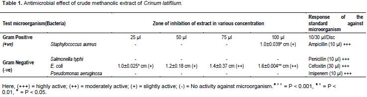
From the experiment, we observed that, crude methanolic extracts of C. latifolium showed slight activity against Gram positive (+ve) S. aureus bacteria. On the other hand, it showed good antibacterial properties against Gram negative (-ve) E. coli bacteria. The overview of the results is shown in Table 1.
Findings of brine shrimp lethality bioassay
By using brine shrimp bioassay, developed by Meyer we could understand the cytotoxic potential and anti-tumor properties. In our current study we used various solvent soluble extracts of C. latifolium. Different solvent soluble extracts showed various rate of mortality at different concentration. By plotting the log of concentration against percent of mortality for all test sample, we found a linear correlation. On the basis of this correlation the LC50 (the concentration at which 50% of mortality of brine shrimp nauplii occurred) was determined for each solvent soluble extracts. We also found that, there was no rate of mortality obtained, in case of control study. The overview of the results is shown in Table 2.

Anti-inflammatory activity
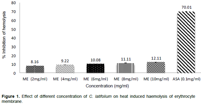
The anti-inflammatory activities of the Crude methanolic extracts of C. latifolium are showed in Tables 3 and 4. The crude methanolic extracts dose dependently increased in anti-inflammatory study, whereas 10 mg/ml concentration most significantly showed 16.21 and 20.23% inhibition of hemolysis respectively by hypotonic solution and heat induced hemolysis. Acetyl salicylic acid was used as standard in membrane stabilization. ASA (0.10 mg/mL) revealed 70.01 and 56.32% inhibition of hemolysis, respectively induced by hypotonic solution and heat induced hemolysis correspondingly.


DISCUSSION
Antimicrobial activity
The medicinal properties of the plants lie in a several chemical group such as tannins, flavonoids, alkaloids and phenolic compound. Many parts of the plant especially leaves possess antimicrobial properties due to presence of tannins and flavonoids (Scalbert, 1991; Chung et al., 1998). Plants also synthesize huge amount of aromatic compound among which phenols or their oxygen-substituted derivatives are predominant (Geissman, 1963). These compounds provide protection against microbes for the plant (Cowan, 1999). This is great to see our plant extract showed to have phytochemicals responsible for anti-microbial effect (Dewan et al., 2013).
May be that is why Extracts showed slight antimicrobial activity against Gram positive (+ve) bacteria while Surprisingly showed significant antimicrobial activity against Gram negative (-ve) bacteria E. coli.
Cytotoxic activity
Cancer-related research is conducted all over the world for discovering new hopes for patient suffering with cancer. These studies frequently able to originate biologically active agents from plants used and will be used for treating different carcinoma (Mukherjee et al., 2001). In addition, it is important to understand the mechanisms of anticancer agents for future application in cancer therapy (Half et al., 2009). Our present study investigated the cytotoxic activity of the methanolic extract of C. latifolium. It was found that many of the phytochemicals provide protection against cancer due to poly-phenyl antioxidant and anti-inflammatory effect. Several studies also suggest that these phytochemical provide protection against colorectal plus other types of cancer (Michaud et al., 2000; Greenberg et al., 1994; Birt et al., 2001). Our plant part also contain polyphenol so this plant was one will be one of the most trusted source for discovering anticancer drug, that was so far established through our present study as our plant methanolic extract showed remarkable cytotoxic activity.
Membrane stabilizing activity
C. latifolium methanolic extract inhibited hypotonic solution and heat induced hemolysis of erythrocyte at varying percentage that was comparable with membrane stabilizing activity shown by standard acetyl salicylic acid. As through the standard anti-inflammatory drug showed higher stabilization activity than the experimental plant methanolic extract, but our plant extract will be the existing source of anti-inflammatory activity with fewer or no side effects. The moderate membrane stabilizing activity shown by our plant methanolic extract may be due to the presence of flavonoid contents. It has been established by many experimental study that plants with flavonoids shown profound stabilizing effects on lysosomes both in vitro and in vivo laboratory condition (Middleton, 1996).
CONCLUSION
From the above experiments we could terminated that the crude methanolic and various solvent soluble extracts of C. latifolium (leaves) showed slight to moderate cytotoxic activities. We also confreres that, it also revealed excellent antibacterial and membrane stabilizing activities (Figures 1 and 2).

CONFLICT OF INTEREST
The authors have not declared any conflict of interest.
ACKNOWLEDGEMENT
The authors would like to express their heartfelt gratitude, indebtedness, profound appreciation to all honorable teacher, staff and supervisor Md. Mizanur Rahman Moghal, Assistant Professor, Department of Pharmacy, Mawlana Bhashani Science and Technology University, for their continuous support, untiring inspiration, scholastic supervision, constructive criticism, affectionate feeling and optimistic counseling throughout the project work.
REFERENCES
|
Atata RF, Sani A, Ajewole SM (2003). Effect of Stem Bark Extracts of Enantia chloranta on some clinical isolates. Niger. Soc. Exp. Biol. 15(2):84-92. |
|
|
Bauer AW, Kirby WMM, Sheriss JC, Turck M (1966). Antibiotic susceptibility testing by standarised single method. Am. J. Clin. Pathol. 45:493-496. |
|
|
Bauer J, Rojas R, Bustamante B (2003). Antimicrobial activity of selected Peruvian medicinal plants. J. Ethnopharmacol. 88:199-204. |
|
|
Birt DF, Hendrich S, Wang WQ (2001). Dietary agents in cancer prevention: flavanoids and isoflavonoids. Pharmacol. Ther. 90:157-177. |
|
|
Chung KT, Wong TY, Wei Y, Huang YW, Lin Y (1998). Tannins and human health. A review. Crit. Rev. Food Sci. Nutr. 8:421-464. |
|
|
Cowan MM (1999). Plant products as antimicrobial agents. Clin. Microbiol. Rev. 12:564-582. |
|
|
Dewan SMR, D Abhijit (2013). Investigation of in vitro thrombolytic potential and phytochemical nature of Crinum latifolium leaves growing in coastal region of Bangladesh. Int. J. Biol. Pharm. Res. 4(1):1-7. |
|
|
Evan WC (2002). Trease and Evans pharmacognosy. London: WB Saunders. P 585. |
|
|
Geissman TA (1963). Flavonoid compounds, tannins, lignins and related compounds. In: Florkin M and Stotz EH (eds). Pyrrole Pigments, Isoprenoid Compounds and Phenolic Plant Constituents, New York, USA: Elsevier Press. P 265. |
|
|
Greenberg ER, Baron JA, Tosteson TD (1994). A clinical trial of antioxidant vitamins to prevent colorectal cancer. N. Engl. J. Med. 331:141-147. |
|
|
Half E, Arber N (2009). Colon cancer: Preventive agents and the present status of chemoprevention. Exp. Opin. Pharmacother. 10:211-219. |
|
|
Islam MK, EtiI Z, Chowdury JA (2009). Cytotoxic Studies on Two Meliaceae plants: Chukrasia Tabularis and Aglaia Roxburghiana. J. Sci. Res. 1(2):399-403. |
|
|
Kintzios SE, Barberaki MG (2004). Plants that fight cancer. Boca Raton: CRC Press. |
|
|
Krishnaraju AV, Rao TVN, Sundararaju D, Vanisree M, Tsay HS, Subbaraju GV (2006). Biological Screening of Medicinal Plants Collected from Eastern Ghats of India Using Artemia salina (Brine Shrimp Test). Int. J. Appl. Sci. Eng. 4(2):115-125. |
|
|
Meyer BN, Ferrigni NR, Putnam JE, Jacobsen LB, Nichols DE, McLaughlin JL (1982). Brine shrimp: a convenient general bioassay for active plant constituents. Planta Med. 45:31-34. |
|
|
Michaud DS, Feskanich D, Rimm EB (2000). Intake of specific carotenoids and risk of lung cancer in two prospective U.S. cohorts. Am. J. Clin. Nutr. 72:990-997. |
|
|
Middleton JE (1996). Biological properties of plant flavonoids: An overview. Int. J. Pharmacogn. 34:344-348. |
|
|
Mukherjee A, Basu S, Sarkar N, Ghosh A (2001). Advances in cancer therapy with plant based natural products. Curr. Med. Chem. 12:1467-1468. |
|
|
Naibi RA, Sukumar Sethuraman V, Suluchana N, Sadique J (1985). Satellite symposium on traditional medicine as Asian congress of Pharmacology. Tamil University of Thanjavur. 140. |
|
|
Opler A, Mizell R, Robert A, Cervantes-Cervantes M, Kincaid D, Kennelly EJ (2002). Use of a brine shrimp assay to study herbal teas in the classroom. Am. Biol. Teach. 64: 8596-8604. |
|
|
Oyedapo OO, Sab FC, Olagunju JA (1999). Bioactivity of fresh leaves of Lantana camara. Biomed. Lett. 59:175- 183. |
|
|
Omale J, Okafor PN (2008). Comparative antioxidant capacity, membrane stabilization, polyphenol composition and cytotoxicity of the leaf and stem of Cissus multistriata. Afr. J. Biotechnol. 7:3129-3133. |
|
|
Prabhu S, Britto SJ, Thangavel P, Raj L (2011). Antibacterial and Antioxidant Activity of Leaves of Canavalia Mollis Wight and Arn. (Horse Bean). Int. J. Pharm. Sci. Drug Res. 2(1):95. |
|
|
Pratibha N, Sushma D, Gupta, Rajinder K (2012). Screening for Antioxidant and Antibacterial potential of common medicinal plants in the treatment of Acne. Int. J. Drug Dev. Res. 4:65-71. |
|
|
Rajendran V, Lakshmi KS (2008). In vitro and In vivo anti-inflammatory activity of leaves of Symplocos cochinchnensis (Lour) Moore ssp laurina. Bangladesh J. Pharmacol. 3(2):121-124. |
|
|
Runyoro D, Matee M, Olipa N, Joseph C, Mbwambo H (2006). Screening of Tanzanian medicinal plants for anti-Candida activity. BMC Complement Altern. Med. 6:11. |
|
|
Scalbert A (1991). Antimicrobial properties of tannin. Phytochemistry 30:3875-3883. |
|
|
Shahidi BH (2004). Evaluation of antimicrobial properties of Iranian medicinal plants against Micrococcus luteus, Serratia marcescens, Klebsiella pneumonia and Bordetella bronchoseptica. Asian J. Plant Sci. 3:82-6. |
|
|
Ueda J, Tezuka Y, Banskota AH, Tran Q, Harimayay, SaikiI (2002). Antiproliferative activity of vietnamese medicinal plants. Biol. Pharm. Bull. 25:753-60. |
|
Copyright © 2024 Author(s) retain the copyright of this article.
This article is published under the terms of the Creative Commons Attribution License 4.0


