Full Length Research Paper
ABSTRACT
The resistance of microorganisms to current antimicrobials, and the deleterious effects caused by the excessive free radical manufacturing in the human body and their relationship with increasing global incidence of cancer, has led to a continuous search for new chemical agents that can contribute to the fight against these ills. The objective of the present work was to evaluate the antimicrobial, antioxidant and cytotoxic activities and determine the chemical profile of ethyl acetate extract of ten species of the family Melastomataceae. Antimicrobial activity was assessed by the methods of disk diffusion in agar and microdilution in broth (MIC-µg/mL). Antioxidant activity was measured by DPPH free radical capture assay while toxicity was evaluated with Artemia salina Leach. Cytoxicity was evaluated by in vitro tests with THP-1-cells. Identification of the classes of metabolites was performed using chemical reagents, while quantification of total phenols (EGA/g) and total flavonoids (EQ/g) was done by spectrophotometry. The extract of Clidemia capitellata exhibited activity against Micrococcus luteus with MIC = 62.5-μg/mL. The extract of C. hirta had the highest sequestering activity of DPPH free radicals (63.54-%). The toxicological assay revealed high toxicity for Miconia alborufescens extract (CL50 61.6-μg/mL). Cytotoxic activity of extracts for THP-1-cells was observed through visualization of apoptotic bodies and cell death. Phytochemical analysis detected the presence of condensed tannins, terpenes, steroids and polyphenols, and the absence of alkaloids. The assays performed provided promising results, suggesting the continuation of new chemical-pharmacological evaluations and the isolation of the active principle of the extracts.
Key words: Bacteria, Artemia, total phenols, THP-1 cells, toxicity.
INTRODUCTION
Excessive and indiscriminate use of antibiotics has generated increased bacterial resistance to conventional antibiotics. Increased estimates for cancer incidence and excessive free radical manufacturing by the human body have become concerns of health authorities throughout the world. Due to their diverse metabolic capacities, plants represent a potential alternative for the isolation of new drugs (Hosseinzadeh et al., 2015). Of the drugs on the market for the treatment for infectious diseases, 75% are of natural origin or analogous derivatives (Newman and Cragg, 2016).
There are approximately 374,000 species of plants on the planet (Christenhusz and Byng, 2016), of which 46,403 are distributed among the several biomes and ecosystems of Brazil (Flora do Brasil 2020, 2017). The Atlantic Forest is the most diversified phytogeographical domain in Brazil, with more than 15,001 plant species. Melastomataceae is the fifth largest family of angiosperms in Brazil with 66 genera and 1,367 species, and is one of the most represented in the Atlantic Forest, which is home to 582 species (BFG, 2015).
The Melastomataceae family presents a variety of secondary metabolite classes, including flavonoid derivatives, tannins, terpenes, fatty acids and anthocyanidins (Bonfim-Patricio et al., 2001; Yoshida et al., 2005; Grayer et al., 2008). Some studies with representatives of this family have already demonstrated that they possess analgesic, antipyretic, neuroprotective, anti-parasitic, immunological, antioxidant, anti-tuberculotic, anti-inflammatory, antitumor, antimicrobial and anticancer potentials (Pavan et al., 2009; Tan et al., 2012; Balamurugan et al., 2013; Nono et al., 2014; Nguta et al., 2016).
The objective of the present work was to investigate the antimicrobial, antioxidant and cytotoxic activity and to determine the chemical profile of crude extracts obtained with ethyl acetate from the leaves of Miconia albicans, M. alborufescens, M. amoena, M. ciliata, M. fallax, Clidemia capitellata, C. hirta, C. sericea, Tibouchina francavillana and T. lhotzkyana, belonging to the family Melastomataceae.
MATERIALS AND METHODS
Aerial parts of ten plant species of the family Melastomataceae [Miconia albicans (Sw.) Triana (14025); Miconia ciliata (Rich.) DC. (14036); Miconia fallax DC. (14022); Miconia amoena Triana (14024); Miconia alborufescens Naudin (14023); Clidemia hirta (L.) D.Don (13982); Clidemia sericea D.Don (13517); Clidemia capitellata (Bonpl.) D.Don (7685); Tibouchina lhotzkyana (C.Presl) Cogn. (13981) and Tibouchina francavillana Cogn.] (13984) were collected in an Atlantic Forest remnant in the municipality of Alagoinhas, state of Bahia, Brazil (12°10’42.62”S, 38°24’39.52’W). The plants were identified and exsiccates deposited in the Alagoinhas, Bahia, collection of herbarium da Bahia State University. The leaves were dried in an drying oven at 50°C and pulverized manually. Extraction was performed by percolation in ethyl acetate (PA), with three successive extractions at intervals of 72 h. The extract was obtained after filtration and evaporation of the solvent, and then preserved at 4°C until use.
In vitro evaluation of antimicrobial activity
Antimicrobial activity was evaluated by the method of disk diffusion in agar according to the recommendations of Clinical and Laboratory Standards Institute (CLSI; 2003), with fungal bacterial lineages belonging to American Type Culture Collection (ATCC) — Staphylococcus aureus (ATCC 6538), Pseudomonas aeruginosa (ATCC 15442), Escherichia coli (ATCC 94863), Micrococus luteus (ATCC 10240) and Bacillus subtilis (ATCC 6633) — and the fungus Aspergillus niger (16404). The bacteria cultures were cultivated in Müeller-Hinton Agar at 37ºC for 24 hours while the fungus was cultivated in Sabouraud Dextrose Agar at 37°C for 48 h.
Aliquots of 10 μl of plant extract (100.0 mg/ml) were applied to sterile filter-paper disks (6.0 mm in diameter). The microorganisms were then cultured in Muller-Hinton Agar and, as a control, DMSO with chloramphenicol (0.1%), for bacteria and with Cyclopiroxolamine (0.1%) for the fungus. The bacterial and fungal plates were incubated at 37°C for 24 and 48 h, respectively. Antimicrobial activity was evaluated by inhibition halo measurement.
Minimal inhibitory concentration (MIC)
The Minimal Inhibitory Concentration (MIC) was determined based on CLSI document M7-A6 (CLSI, 2003), with some modifications. The microorganisms were cultured on plates with 96 wells and evaluated in the presence of different concentrations of extract (500 to 3.90 µg/ml) for 24 h at 37°C, with chloramphenicol (0.1%) and DMSO (4%) as controls. Wells without turbidity were considered to contain active extracts.
In vitro analysis of antioxidant activity
The antioxidant capacity of the extracts was determined by photocolorimetric assay of the free radicle 2,2-diphenyl-1-picrylhydrazyl (DPPH), using plates with 96 wells, with adaptations (Brand-Willians et al., 1995). The extracts were diluted in ethanol (6.0 mg/ml), and then 50 µl of this solution was added to microplate wells containing 150 µl of ethanol. Serial dilutions were performed to produce extract concentrations ranging from 3.0 to 0.045 mg/ml. Next, 100 µL DPPH solution (0.5 mM) was added to each well. After 1 h of reaction at room temperature, and in the absence of light, absorbance readings were made using a UV-Vis spectrophotometer at 492 nm. All tests were performed in triplicate. The percentage of free radical sequestration (% FRS) was determined using the equation:
%FRS = {[(Abscontrol – (Abssample – Abswhite)] x 100} /Abscontrol}
Where Abscontrol is the absorbance of DPPH in the presence of ethanol; Abssample is the absorbance of extract after the reaction with DPPH; and Abswhite is the absorbance of ethanol (Moreira et al., 2005).
Phytochemical screening
The phytochemical profile of the extracts was determined using chemical reactions to identify alkaloids (Petruczynik, 2012) and terpenes and steroids (Harbone, 1998) by means of thin layer chromatography (TLC) using a 0.2 mm silica gel (F254) in aluminum (MERCK). Total phenols were determined by spectrophotometry using the Folin-Ciocalteu reaction, and read with a UV-Vis spectrophotometer at 620 nm, with adaptations (Singleton et al., 1999). The calibration curve for gallic acid was obtained using the same conditions for the preparation of the extracts (y = 3.8883x + 0.1141. R² = 0.9954). The results were determined by the interpolation of the absorbance of the samples with the standard curve of gallic acid expressed in milligrams of gallic acid per gram of crude extract (mg EGA/g of extract). Total falvonoids was determined by reaction with aluminum chloride (AlCl3) (Arvouet-Grand et al., 1994), which was measured by absorbance using a UV-Vis spectrophotometer at 492 nm. The results were expressed in milligrams of quercetin per gram of crude extract (mg EQ/g of extract). The calibration curve for quercetin was obtained under the same preparation conditions (y = 1.2078x – 0.1175. R² = 0.9889). The tests were performed in triplicate.
Toxicity assays with Artemia salina leach
In vitro evaluation of the toxicity of extracts was performed according to the methodology proposed by Meyer et al. (1982). The extracts were categorized according to Clarkson et al. (2004) as: CL50 > 1000 μg/ml are nontoxic; CL50 > 500 μg/ml are of low toxicity; CL50 between 500 and 100 μg/ml are moderately toxic; and CL50 < 100-μg/ml are highly toxic.
Ten Artemia nauplii extracts were used in each well in three different conditions - 10, 100 and 1000 μg/ml - with 10 other nauplii exposed to marine water and another 10 exposed to DMSO (1.0%) as positive controls. All wells were submitted to artificial lighting and the tests were performed in triplicate. The nauplii were counted after 24 and 48 h of exposure. The mean lethal concentration (CL50) was determined using the program GraphPadPrism 5.0.
Lineages and cell culture
The acute monocytic leukemic cell line (THP-1) was donated by the Immunology Laboratory of the Institute of Health Sciences (ICS) of the Federal University of Bahia. The cells were cultured in 5.0 mL of RPMI medium supplemented with 10% v/v fetal bovine serum (PBS) and 20 µg/ml of penicillin/streptomycin (1%) obtained from Cultilab. Cells were seeded and cultured to confluency in 25 cm² culture bottle at 37°C in an atmosphere of 5% CO2 and controlled humidity.
Preliminary determination of in vitro cytotoxicity to THP-1 cells
Cytotoxicity of plant extracts to the THP-1 cell lineage was evaluated using an inverted microscope (20x). Cells (1×10⁶ /-well) were pated in 1.0 ml RPMI complete medium per well in 24 well plates. The cells were then incubated in the presence of crude plant extract (100 μg/ml), diluted in DMSO for 24 h at 37°C, and then evaluated morphologically. Wells used for control contained RPMI cells and wells containing cells with RPMI and DMSO (0.5%).
Statistical analysis
Analyses were based on the mean in triplicate ± standard deviation of the mean, (one-way ANOVA; p<0.05), using GraphPadPrism 5.0, Minitab 18 and Excel 2013.
RESULTS
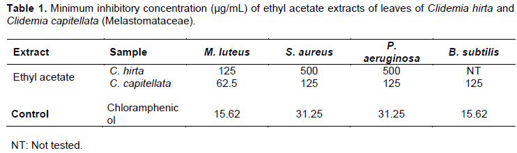
For the diffusion disk tests, Clidemia hirta exhibited activity against M. luteus, S. aureus and P. aeruginosa, with halos of 13.0 ± 1.02, 9.0 ± 0.57 and 10.0 ± 1.52 mm, respectively, while Clidemia capitellata exhibited activity against M. luteus, S. aureus, P. aeruginosa and B. subtili, with halos of 12.0 ± 0.57, 10.0 ± 0.57, 9.0 ± 0.57 and 11.0 ± 1.73 mm, respectively. For MIC (Table 1), the greatest activity was for extract of C. capitellata against the bacteria strain M. luteus (MIC 62.5 µg/ml).
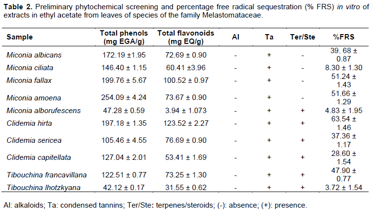
For antioxidant activity (DPPH sequestration capacity), the extract of C. hirta was most effective (63.54 ± 1.46%), followed by M. amoena (51.66 ± 1.29%) and M. fallax (51.24 ± 1.43%). All extracts gave positive reactions in tests for total phenols, total flavonoids and tannins, but were negative for alkaloids. Half of the extracts were positive for the presence of terpenes or steroids (Table 2). The extracts of Miconia alborufescens (CL50 61.6 µg/ml) and C. hirta (CL50 75.8 µg/ml) exhibited high toxicity for Artemia salina. The extracts of M. albicans (CL50 512.7 µg/ml), C. sericea (CL50 831.7 µg/ml) and Tibouchina lhotzkyana (CL50 537.0 µg/ml) exhibited low toxicity. All other extracts were inactive. Tests against the monocytic cell lineage THP-1 revealed that 90% of the extracts were capable of stimulating the formation of apoptotic bodies and induce cell death, except for C. sericea.
DISCUSSION
The phytochemical data for C. hirta, C. capitellata and the other extracts were consistent with other studies (Gordon et al., 2011; Abdellaoui et al., 2014; Tracanna et al., 2015; Scalco and Munhoz, 2016). The absence of alkaloids in the extracts confirms this as a chemotaxonomic characteristic of the family Melastomataceae (Silva et al., 2010). Differences between the present study and previous studies in the content of phenols and flavonoids may be a reflection of differing factors, including the extraction solvent, seasonality and the collection site.
The absence of growth inhibition halos in the agar diffusion tests was observed for most of the extracts in ethyl acetate, with the exception of C. hirta and C. capitellata. In concordance with the results of the present study, Meléndez and Capriles (2006) found antimicrobial activity for methanolic extract of leaves of C. hirta against fifteen bacterial strains, among them S. aureus and M. luteus, but no such activity for C. capitellata.
Dianita et al. (2011) demonstrated bactericidal activity of the ethyl acetate extract of the leaves of C. hirta against P. aeruginosa and bacteriostatic activity for E. faecalis.
For antioxidant activity, the data show that extracts with significant free radical scavenging activity also had a high content of polyphenols and total flavonoids, suggesting a direct connection between antioxidant action and the presence of these compounds. The phenolic constituents, among them flavonoids and phenolic acids, are known for their antioxidant effect in biological systems due the arrangement of their function groups, in particular hydroxyl groups, capable of neutralizing or sequestering reactive species (Fabri et al., 2009). Santos et al. (2017) reported the presence of compounds of this nature in different extracts of Miconia, and Lopez et al. (2016) for C. hirta, and Gordon et al. (2011) for C. rubra. Plant extracts that demonstrated toxicity for Artemia salina, according to the literature, possess cytotoxic compounds in concentrations sufficient to potentiate antitumor (Mclaughlin et al., 1991), antiplasmodic (Amarante et al., 2011) and actions against bacterial (Zuque et al., 2004) and fungal lineages (Niño et al., 2006). The evaluation of the THP-1 lineage revealed that the extracts stimulated the formation of apoptotic bodies and cell death.
Antiproliferative effects- induction for the formation of apoptotic bodies and cell death- are an interesting approach in the treatment of leukemia (Kapoor and Kakkar, 2012). Research shows that most antitumor drugs cause cancer cells to die (Park et al., 2013) through induction of the apoptotic process (Onrubia et al., 2012). This, thus, suggests that the preliminary data of this evaluation of leaf extracts of species of the family Melastomataceae are promising in the search for new antitumor agents.
The presence of non-toxic extracts for A. salina and, concomitantly, cytotoxicity against THP-1 cells, may be indicative of the chemical constituents of the plant acting against antitumor cells without affecting the health cells in the body. However, toxicity in A. salina cannot be directly translated into human toxicity since the physiological media are different, and thus further analysis is needed to determine the effects on different human cell lines. Similar results for M. fallax ethyl acetate extract in THP-1 cells were observed by Cunha et al. (2008), for the ethanol extract and for two triterpenes isolated, ursolic acid and oleanolic acid; Aristizabal et al. (2013) for the butanolic extract of C. hirta in Vero cells and by Narasimham et al. (2017), using Dalton's lymphoma ascites cells (DLA), extracts of C. hirta leaves in petroleum ether and in chloroform.
CONCLUSION
The extracts from the species of Melastomataceae evaluated in the present work can be considered promising as potential sources of chemical agents with antimicrobial, antitumor and antioxidant properties.
Furthermore, C. hirta should be emphasized for exhibiting activity in all the tests performed. New chemical-pharmacological evaluations and the isolation of the active principle of the extracts should be performed to demonstrate the relationships and mechanisms involved in their antibacterial, antioxidant and cytotoxic activity
CONFLICT OF INTERESTS
The authors have not declared any conflict of interests.
ACKNOWLEDGMENTS
The author extends their thanks to FAPESB for a Master’s scholarship to the first author; the Bahia State University; the Natural Substances Study Group of the Bahia of the Institute of Chemistry of the Federal University of Bahia; the team of the Organic Chemistry Laboratory of Bahia State University, Campus I, Salvador, Bahia; the team of the Laboratory of Immunology and Molecular Biology, Institute of Health Sciences, Federal University of Bahia.
REFERENCES
|
Abdellaoui SE, Destandau E, Krolikiewicz-Renimel I, Cancellieri P, Toribio A, Jeronimo-Monteiro V, Landemarre L, André P, Elfakir C (2014). Centrifugal partition chromatography for antibacterial bio-guided fractionation of Clidemia hirta roots. Separation and Purification Technology 123:221-228. |
|
|
Amarante CB, Muller AH, Póvoa MM, Dolabela MF (2011). Phytochemical study bioassay-guided by tests of toxicity on Artemia salina and antiplasmodial activity from stem of aninga (Montrichardia linifera). Acta Amazonica 41(3):431-434. |
|
|
Aristizabal LSR, Marín D, González FJJ (2013). Actividad ictiotóxica y citotóxica de extractos de plantas Chrysobalanaceae, Melastomataceae, Rubiaceae y Rutaceae, de la flora Colombiana. Scientia et Technica 18(3):548-552. |
|
|
Arvouet-Grand A, Vennat B, Pourrat A, Legret P (1994). Standardisation d'un extrait de propolis et identification des principaux constituants. Journal de Pharmacie de Belgique 49(6):462-468. |
|
|
Balamurugan K, Nishanthini A, Mohan VR (2013). Anticancer activity of ethanol extract of Melastoma malabathricum L. leaf against Dalton Ascites Lymphoma. Journal of Pharmaceutical Sciences and Research 5(5):111-114. |
|
|
BFG - The Brazil Flora Group (2015). Growing knowledge: an overview of seed plant diversity in Brazil. Rodriguésia 66:1085-1113. |
|
|
Bonfim-Patricio MC, Salatino A, Martins AB, Salatino MLF, Wurdack JJ, (2001). Flavonoids of Lavoisiera, Microlicia and Trembleya (Melastomataceae) and their taxonomic meaning. Biochemical Systematics and Ecology 29(7):711-726. |
|
|
Brand-Willians W, Curvelier ME, Berset C (1995). Use of afree radical method to evaluate antioxidant activity. Food Science and Technology 28(1):25-30. |
|
|
Christenhusz MJM, Byng JW (2016). The number of known plants species in the world and its annual increase. Phytotaxa 261(3):201-217. |
|
|
Clarkson C, Maharaj VJ, Crouch NR, Grace OM, Pillay P, Matsabisa MG, Bhagwandin N, Smith PJ, Folb PI (2004). In vitro antiplasmodial activity of medicinal plants native to or naturalized in South Africa. Journal Ethnopharmacology 92:177-191. |
|
|
CLSI - Clinical and Laboratory Standards Institute (2003). Methods for dilution antimicrobial susceptibility tests for bacteria that grow aerobically. NCCLS document M7-A6:23(2). P. 81. |
|
|
Cunha WR, Silva MLA, Santos FM, Montenegro IM, Oliveira ARA, Tavares HR; Leme Dos Santos HS, Bizário JCS (2008). In vitro inhibition of tumor cell growth by Miconia fallax. Pharmaceutical Biology 46(4):292-294. |
|
|
Dianita R, Ramasamy K, Rahman NAB (2011). Antibacterial activity of different extracts of Clidemia hirta (L.) D. Don leaves. Planta Medica 77:12. |
|
|
Fabri RL, Nogueira MS, Braga FG, Coimbra ES, Scio E (2009). Mitracarpus frigidus aerial parts exhibited potent antimicrobial, antileishmanial, and antioxidant effects. Bioresource Technology 100(1):428-433. |
|
|
Flora do Brasil 2020 em construção (2017). Lista de espécies da flora do Brasil. Jardim Botânico do Rio de Janeiro. |
|
|
Gordon A, Schadow B, Quijano CE, Marx F (2011). Chemical characterization and antioxidant capacity of berries from Clidemia rubra (Aubl.) Mart. (Melastomataceae). Food Research International 44(7):2120-2127. |
|
|
Grayer JR, Thabrew MI, Hughes RD, Bretherton S, Lever A, Veitch NC, Kite GC, Lelli R, Simmonds MSJ (2008). Phenolic and terpenoid constituents from the Sri Lankan Medicinal Plant Osbeckia aspera. Pharmaceutical Biology 46(3):154-161. |
|
|
Harbone JB (1998). Phytochemical methods. London: Chapman & Hall 288p. |
|
|
Hosseinzadeh S, Jafarikukhdan A, Hosseini A, Armand R (2015). The application of medicinal plants in the traditional and modern medicine: a review of Thymus vulgaris. International Journal of Clinical Medicine 6:635-642. |
|
|
Kapoor R, Kakkar P (2012). Protective role of morin, a flavonoid, against high glucose induced oxidative stress mediated apoptosis in primary rat hepatocytes. PLoS One 7(8):e41663. |
|
|
Lopez T, Corbin C, Falgueires A, Doussot J, Montguillon J, Hagège D, Hano C, Laine E (2016). Secondary metabolite accumulation, antibacterial and antioxidant properties of in vitro propagated Clidemia hirta L. extracts are influenced by the basal culture medium. Comptes Rendus Chimie 19(9):1071-1076. |
|
|
Mclaughlin JL, Chang CJ, Smith DL (1991). "Bench-top" bioassays for the discovery of bioactive natural products: an update. In: Rahman A. (Org.), Studies in Natural Product Chemistry. Amsterdam, NED: Elsevier. pp. 383-409. |
|
|
Meléndez PA, Capriles VA (2006). Antibacterial properties of tropical plants from Puerto Rico. Phytomedicine 13 (4):272-276. |
|
|
Meyer BN, Ferrigni NR, Putnam JE, Jacobsen LB, Nichols DE, Mclaughlin JL (1982). Brine Shrimp: A convenient general bioassay for active plant constituents. Planta Medica 45(5):31-34. |
|
|
Moreira DL, Leitão, SG, Gonçalves JLS, Wigg MD, Leitão GG (2005). Antioxidant and antiviral properties of Pseudopiptadenia contorta (Leguminosae) and of quebracho (Schinopsis sp.) extracts. Química Nova 28(3):421-425. |
|
|
Narasimham D, Bindu YH, Cheriyamundath S, Raghavan R, Kumari MK, Chandrasekhar T, Madassery J (2017). Evaluation of in vitro anticancer and antioxidant activities from leaf extracts of medicinal plant Clidemia hirta. International Journal of Pharmacy and Pharmaceutical Sciences 9(4):149-153. |
|
|
Newman DJ, Cragg GM (2016). Natural products as sources of new drugs from 1981 to 2014. Journal of Natural Products 79(3):629-661. |
|
|
Nguta JM, Appiah-Opong R, Nyarko AK, Yeboah-Manu D, Addo PGA, Otchere I, Twum AK (2016). Antimycobacterial and cytotoxic activity of selected medicinal plant extracts. Journal of Ethnopharmacology, 182:10-15. |
|
|
Niño J, Narváez DM, Mosquera OM, Correa YM (2006). Antibacterial, antifungal and cytotoxic activities of eight Asteraceae and two Rubiaceae plants from colombian biodiversity. Brazilian Journal of Microbiology 37(4):566-570. |
|
|
Nono RN, Teponno RB, Quassinti L, Bramucci M, Vitali LA, Petrelli D, Lupidi G, Tapondjou AL (2014). Antimicrobial, antioxidant, anti-inflammatory activities and phytoconstituents of extracts from the roots of Dissotis thollonii Cogn. (Melastomataceae). South African Journal of Botany 93:19-26. |
|
|
Onrubia M, Cusido RM, Ramirez K, Hernandez-Vasquez L, Moyano E, Bonfill M, Palazon J (2012). Bioprocessing of plant in vitro systems for the mass production of pharmaceutically important metabolites: paclitaxel and its derivatives. Current Medicinal Chemistry 20(7):880-910. |
|
|
Park HY, Kim G, Know TK, Hwang HJ, Kim ND, Yoo YH, Choi YH (2013). Induction of apoptosis by fucoidan in human leukemia U937 cells through activation of p38 MAPK and modulation of Bcl-2 family. Marine Drugs 11: 2347-2364. |
|
|
Pavan FR, Sato DN, Higuchi CT, Santos ACB, Vilegas W, Leite CQE (2009). In vitro anti-Mycobacterium tuberculosis activity of some Brazilian "Cerrado" plants. Brazilian Journal of Pharmacognosy 19(1):204-206. |
|
|
Petruczynik A (2012). Analysis of alkaloids from different chemical groups by diferrent liquid chromatography methods. Central European Journal of Chemistry 10(3):802-835. |
|
|
Santos MAF, Silva MAP, Santos ACB, Bezerra JWA, Alencar SR, Barbosa EA (2017). Atividades biológicas de Miconia spp. Ruiz & Pavon (Melastomataceae Juss.). Gaia Scientia 11(1):157-170. |
|
|
Scalco CDN, Munhoz CL (2016). Estudo fitoquímico e avaliação da toxicidade aguda dos extratos brutos das plantas Alternanthera brasiliana (L.) Kuntze, Chenopodium ambrosioides L. E Miconia albicans sw. Triana. Journal of Agronomic Sciences 5(2):181-194. |
|
|
Silva JDS, Acosta POA, Nascimento FCN, Silva AJR (2010). Análise fitoquímica das partes aéreas de Rhynchanthera grandiflora (Aubl.) DC. (Melastomataceae). In: Reunião Regional da SBPC, Boa Vista, Roraima, Brasil. |
|
|
Singleton VL, Orthofer R, Lamuela-Raventós RM (1999). Analysis of total phenols and other oxidation substrates and antioxidants by means of folin-ciocalteu reagent. Methods in Enzymology 299:152-178. |
|
|
Tan HP, Wong DZH, Ling SK, Chuah CH, Kadir HA (2012). Neuroprotective activity of galloylated cyanogenic glucosides and hydrolysable tannins isolated from leaves of Phyllagathis rotundifolia. Fitoterapia 83(1):223-229. |
|
|
Tracanna MI, Fortuna AM, Cárdenas AVC, Marr AK, Mcmaster WR, Gómez-Velasco A, Sánchez-Arreola E, Hernández LR, Bach H (2015). Anti-leishmanial, anti-inflammatory and antimicrobial activities of phenolic derivatives from Tibouchina paratropica. Phytotherapy Research 29(3):393-397. |
|
|
Yoshida T, Ito H, Hipolito IJ (2005). Pentameric ellagitannin oligomers in melastomataceous plants-chemotaxonomic significance. Phytochemistry 66(17):1972-1983. |
|
|
Zuque ALF, Watanabe ES, Ferreira AMT, Arruda ALA, Resende UM, Bueno NR, Castilho RO (2004). Avaliação das atividades antioxidante, antimicrobiana e citotóxica de Couepia grandiflora Benth. (Chrysobalanaceae). Revista Brasileira de Farmacognosia 14(2):129-136. |
|
Copyright © 2024 Author(s) retain the copyright of this article.
This article is published under the terms of the Creative Commons Attribution License 4.0


