Full Length Research Paper
ABSTRACT
This study was designed to investigate the anti-inflammatory and anti-oxidant potentials of leaf extracts of Zea mays (maize) and Sorghum bicolor (sorghum) with a view of utilizing them as herbal tea on the management and treatment of oxidant and inflammatory disorders. The powdered plant materials: (Z. mays, (30 g), S. bicolor (30 g), mixture (15 g of Z. mays + 15 g of S. bicolor) were soaked separately in 80% (v/v) ethanol, in sealed containers at room temperature (25 ± 1°C) for seven days with frequent stirring. The suspensions were filtered using double-layered cotton material. The filtrate was evaporated at 40°C under reduced pressure in Rotary Evaporator (BuchiRotavapor RII coupled to a Butchi Vacuum Pump V-700). The extracts were screened for the presence of phytochemicals and the antioxidant constituents of the extracts were also quantified. The antioxidant properties of the extracts were investigated using two different mechanisms: the scavenging of DPPH radical and iron reduction Ferric reducing antioxidant power (FRAP). The anti-inflammatory properties of the extracts were assessed using three models: inhibition of heat-induced haemolysis, inhibition of albumin denaturation and the membrane stabilizing potentials of the extracts. The results revealed that the ethanolic extracts of Z. mays, S. bicolor and the mixture of both contained appreciable quantities of antioxidant constituents. The ethanolic extract of S. bicolor contain the highest quantities of phytoconstituents: phenolics, flavonoids, Vitamin C and E. The ethanolic extract of Z. mays, S. bicolor and the mixture scavenged DPPH radical maximally at 42, 38 and 36% respectively and the reducing power concentrations of Z. mays, S. bicolor and the mixture were 289.94 ± 18.1 µg/ml, 253.071 ±18.91 µg/ml, and 286.81 ± 20.49 µg/ml respectively. The results of the membrane stabilizing activities revealed the order of stability as Z. mays >S. bicolor > mixture. The percentage inhibition of heat-induced heamolytic activities of Z. mays, S. bicolor and the mixture was 21.0, 19.7, and 82.2%, respectively. The order of inhibition of albumin denaturation of the extract was Mixture >S. bicolor > Z. mays. The ethanolic extract of Z. mays showed the highest antioxidant potential while that of the mixture demonstrated the highest anti-inflammatory potentials. Z. mays, S. bicolor and the mixture elicited potent and appreciable anti-inflammatory and antioxidant potentials.
Key words: Antioxidant, anti-inflammatory, Sorghum bicolor, Zea mays, phytochemicals, ethanolic extract.
INTRODUCTION
Reactive oxygen species (ROS) are generated or produced during normal metabolic activities and are involved in the pathogenesis of various diseases such as cancer, ageing, coronary heart diseases, arthritis, atherosclerosis, cataract, inflammation, diabetes and neurodegenerative disorders (Oyedapo et al., 2015). Inflammation is defined as a part of complex biological response of vascular tissues to harmful stimuli, such as pathogens, damaged cells or irritant, it is characterized by redness, edema, fever, pain, and loss of function. The migration of leukocytes from the venous systems to the site of damage and the release of cytokines are known to play a crucial role in the inflammatory response. These chemicals increase blood flow to the area of infection, resulting in redness and warmth. Some of the chemicals cause leakage of fluid into the tissues resulting in swelling. Hence, the inflammatory process may stimulate nerves, cause pain, swelling and redness (Akinwumi and Oyedapo, 2015).
Relationship between oxidative stress and inflammation has been documented by many authors. The mechanism of inflammatory injury involves oxidative stress induced by ROS from activated neutrophils and macrophages. Also ROS extend inflammation by promoting release of cytokines which also stimulate additional production of free-radicals. In many inflammatory disorders, there is an excessive activation of phagocytes and production of free radicals which increase vascular permeability, protein denaturation and membrane alteration (Umapathy et al., 2010; Sakat et al., 2010). Evidences indicated that oxidative stress plays a pathogenic role in chronic inflammatory diseases. Damage of oxidative stress such as oxidized proteins, glycated products, and lipid peroxidation results in neuron degenerations mostly reported in brain disorders (Popa-Wagner et al., 2013).
The immune system can be modified by diet, pharmacologic agents, environmental pollutants, and naturally occurring food chemicals. A number of flavonoids such as hesperidin, apigenin, luteolin, and quercetin are reported to possess anti-inflammatory and analgesic effects. Flavonoids may affect specifically the function of enzyme systems critically involved in the generation of inflammatory processes, especially tyrosine and serine-threonine protein kinases (Pan et al., 2017). The inhibition of kinases is due to the competitive binding of flavonoids with ATP at catalytic sites on the enzymes. These enzymes are involved in signal transduction and cell activation processes involving cells of the immune synthase, cyclooxygenase, and lipooxygenase, which are system. It has been reported that flavonoids are able to inhibit expression of isoforms of inducible nitric oxide responsible for the production of a great amount of nitric oxide, prostanoids, leukotrienes, and other mediators of the inflammatory process such as cytokines, chemokines, or adhesion molecules. Flavonoids also inhibit phosphodiesterases involved in cell activation (Tunon et al., 2009).
Recently, there has been an upsurge of interest in the therapeutic potential of plants as antioxidants in reducing oxidative tissue injuries and inhibiting inflammatory process (Osuntokun et al., 2020). Medicinal plants have been known to be an important potential source of therapeutics. This involves the use of medicinal plants not only for the treatment of diseases but also as potential material for maintaining good health and conditions (Oladeji, 2016). Low harmful effects and multiple actions can also justify this interest. Various parts of plants such as leaf, stem, bark, root, etc. are being used to prevent, revert abnormalities back to normal (Patrick et al., 2018).
S. bicolor is an important staple food in developing countries of the semi-arid tropics. It is the world’s fifth most important cereal, with higher protein content than Zea mays. It is particularly important as human food resources and folk medicine in Asia and Africa. Z. mays commonly known as maize is an important traditional basic commodity contributing a significant production of food requirement for the Nigerian population, cultivated in almost all the agro-ecological zones of Nigeria (Kernels et al., 2013). Numerous studies have been carried out on the grains of these plants but there is little scientific reports on their leaves, hence the study compare the anti-inflammatory and anti-oxidant potentials of ethanolic extracts of Z. mays (maize) and S. bicolor and their mixture.
MATERIALS AND METHODS
Collection and identification of plant materials
Fresh leaves of yellow maize (Z. mays) were collected within May to June, 2019 from a farm along Ife-Ibadan Road, Ajebandele Ile-Ife, Osun State Nigeria. Dried sheath of S. bicolor (red variety) were purchased from local herb sellers at Gbongan Market, Aiyedaade Local Government, Osun State. The plant materials used were identified and authenticated at IFE Herbarium (Voucher number: IFE 17946 and IFE 17947 for Z. mays and S. bicolor respectively), Department of Botany Obafemi Awolowo University, Ile-Ife, Nigeria.
Reagents and chemicals
All reagents used in the study were of analytical grade and were purchased from British Drug House (BDH) limited Poole England. The analytical grade solvents (sulphuric acid, heptane, sodium hydrogen carbonate, hydrochloric acid, glacial acetic acid, ethanol, sodium nitrite, and ethanol) were purchased from Sigma Aldrich, London. Aluminium chloride (AlCl3), Sodium sulphate (Na2SO4), Copper sulphate (CuSO4.5H2O), Sodium hydroxide (NaOH), Folin- Ciocalteau’s Phenol reagent, Acetic acid, ascorbic acid, 2,21-bipyridyl, Ferric chloride, Glutamic acid, Tannic acid, Sodium carbonate (Na2CO3), Aluminium chloride (AlCl3), Sodium nitrite, Rutin, 2,2- diphenyl -1-picrylhydrazyl (DPPH) were also obtained from Sigma Aldrich, London.
Preparation of ethanolic leaf extract of S. bicolor, Z. mays and the mixture
The ethanolic extracts of S. bicolor, Z. mays and the mixture of both plant materials (1:1 w/w) were prepared according to a modified method of Oyedapo and Amos (1997). Powdered plant materials of Z. mays (30 g), S. bicolor (30 g) and the mixture (15 g of Z. mays + 15 g of S. bicolor) were soaked separately in 80% (v/v) ethanol, in sealed containers at room temperature (25 ± 1°C) for seven days with occasional stirring. The suspensions were filtered using double-layered cotton material. The residues were re-extracted with same solvent, 80% (v/v) ethanol until the filtrate became colorless. The combined filtrate was evaporated to dryness at 40°C under reduced pressure in Rotary Evaporator (BuchiRotavapor RII coupled to a Butchi Vacuum Pump V-700).
Phytochemical screening of ethanolic extracts of S. bicolor, Z. mays and the mixture
The ethanolic extracts of S. bicolor, Z. mays and the mixture were screened for the presence of secondary metabolites (flavonoids. alkaloids, saponins, tannins, anthraquinone and cardiac glycosides) using the standard procedures (Gini and Jothi, 2013; Morakinyo et al., 2018).
Quantitative evaluation of phytochemicals
Estimation of total flavonoid content of the ethanolic extract of S. bicolor, Z. mays and the mixture
The estimation of the flavonoid content was carried out according to the spectrophotometric method of Sun et al. (1999) based on the formation of the aluminum-flavonoid yellow complex. To the varying volumes (1 mg/ml, 0- 1.0 ml) of the extracts in separate test tubes, distilled water was added to make it up to 2 ml. This was followed by addition of 5% (v/v) sodium nitrite (0.3 ml). After 5 min, 0.3 ml of 10% (w/v) aluminum chloride and 2 ml of 4% (w/v) sodium hydroxide were added. The absorbance was measured at 500 nm against the blank using visible Spectrum lab 23A spectrophotometer. The standard (rutin) calibration curve was prepared by pipetting 0.0, 0.2, 0.4, 0.6, 0.8 and 1.0 ml rutin solution (1 mg/ml) in triplicates into clean test tubes. The volumes were made up to 2 ml with distilled water and treated as above. The absorbance of the standard was plotted against the concentration. The concentrations of the flavonoids were obtained from the standard calibration curve and expressed as milligram rutin equivalent per gram dry matter (mg RE/g DM).
Estimation of phenolic contents of the ethanolic extract of S. bicolor, Z. mays and the mixture
The total phenolic content of the ethanolic extracts was carried out according to the spectrophotometric method of Singleton et al. (1999) with tannic acid (0.5 mg/ml) as standard. The reaction mixture contained varying volumes of the extracts (stock concentration 0.5 mg/ml) made up to 1 ml with distilled water, Na2CO3 [7.5% (w/v), 1.5 ml] and Folin-Ciocateau’s phenol reagent [1.5 ml, 10% (v/v)]. The mixture was incubated for 1 h, 30 min in the dark at room temperature. The absorbance was read at 725 nm against the reagent blank (distilled water) using visible Spectrum lab 23A spectrophotometer. The standard (tannic acid) calibration curve was prepared by pipetting (0.0, 0.2, 0.4, 0.6, 0.0, and 1.0 ml) tannic acid solution (0.5 mg/ml) in triplicate into clean test tubes. The volumes were made up to 1 ml with distilled water and treated as above. The absorbance of the standard was plotted against the concentration The concentrations of the phenolics were obtained from the standard calibration curve and expressed as milligram tannic acid equivalent per gram of dry matter (mg TAE/g DM).
Determination of vitamin C concentration from powdered S. bicolor, Z. mays and the mixture
The concentrations of Vitamin C (Ascorbic acid) of the dried leaves of the extracts were estimated according to the earlier method of Omaye et al. (1979) as modified and reported by Fajobi et al. (2017) with slightly modification.
Extraction of vitamin C from powdered S. bicolor, Z. mays and the Mixture
Dried powered samples (1 g) were soaked in 20 ml of 2% (w/v) meta phosphoric acid and stirred on a magnetic stirrer for over 2 h, 30 min. The suspensions were filtered following by centrifugation at 3000 rpm for 10 min using Bosch model R-8C-DX centrifuge. The supernatants were collected and used for estimation of the concentration of vitamin C.
Estimation of vitamin C concentration from powdered S. bicolor, Z. mays and the mixture
Typically, extract (1 ml) was pipetted into separate clean dry test tubes in triplicates; 1.5 ml of 10% (v/v) acetic acid was added and mixed gently. This was followed by the addition of 0.5 ml of Folin - Ciocalteu’s Phenol reagent (1:10 dilution). The mixture was incubated at room temperature (25 ± 1°C) for 15 min. The absorbance was read at 760 nm using visible Spectrum lab 23A spectrophotometer against the reagent blank. The standard ascorbic acid (100 µg/ml) was treated as above with varied concentrations. The absorbance of standard was plotted against the concentration. The vitamin C concentration of the extracts was obtained from the extrapolated standard curve and expressed as mg/g of sample.
Determination of vitamin E concentration from powdered S. bicolor, Z. mays and the mixture
The extraction of vitamin E was carried out carried our according to the method described by Santosh et al. (2013) and the content was estimated as reported by Fajobi et al. (2017) with slight modification.
Extraction of vitamin E from powdered S. bicolor, Z. mays and the mixture
The powdered plant material (1 g) was separately mixed with 20 ml of 80% (v/v) ethanol for 30 min, heated gently at 85°C in Gen Lab model water bath and stirred. The suspension was allowed to cool down, filtered and the residue was re-extracted and shaken vigorously for 10 min and 20 ml of 1.25% (w/v) sodium sulphate was added. The mixture was shaken vigorously, allowed to separate into layers (organic and aqueous). The organic layer was collected, stored and used for the estimation of the vitamin E concentration.
Estimation of Vitamin E concentration from powdered S. bicolor, Z. mays and the Mixture
To the organic layer (Vitamin E) extract (1 ml) was added, 0.12% (w/v) 2, 2-bipyridyl (1 ml) and 0.2 ml of 0.12% (w/v) ferric chloride and followed by thorough mixing. The absorbance was read at 493 nm using visible Spectrum lab 23A spectrophotometer against reagent blank. Vitamin E was used as standard and treated as above. The absorbance of the standard was plotted against the concentration. The vitamin E concentration was obtained from standard calibration curve and expressed as mg/g of sample.
Evaluation of antioxidant activities of ethanolic extract of S. bicolor, Z. mays and the Mixture
Assay of DPPH free-radical scavenging
DPPH radical scavenging ability of the ethanolic extracts of S. bicolor, Z. mays and the mixture was determined using the stable radical 2,2-diphenyl-1-picrylhydrazyl hydrate (DPPH) according to the method of Blois (1985) as modified by Aina and Oyedapo (2013). Briefly, to 2 ml of different concentrations (0, 50, 100, 150, 200, 250, 300, 350 µg/ml) of the plant was added 1 ml of 0.3 mM DPPH in methanol. The mixture was mixed and incubated in dark for 30 min. The absorbance was read at 517 nm against reagent blank containing 2 ml methanol in place of the extract. Ascorbic acid (0, 50, 100, 150, 200, 250, 300, 350 µg/ml) was used as standard and treated as above. The percentage inhibition of free radical by the extracts/standard was calculated using this formular.

Assay of reducing anti-oxidant power of ethanolic extract of S. bicolor, Z. mays and the mixture
The reducing antioxidant power of the extracts was carried out according to the method of Nebavi et al. (2013). The principle of the assay is the ability of the extract to reduced Fe3+ to Fe2+ The Fe2+ solution possess and exhibit specific color that is equivalent to potential of the samples. Briefly, different concentrations of each of the extracts (0- 250 µg/ml) were mixed with 2.5 ml of phosphate buffer (0.2 M, pH 6.6) and 2.5 ml 1% (w/v) potassium ferricyanide. The mixture was incubated using Bosch incubator at 50°C for 20 min. A portion (2.5 ml) of trichloroacetic acid 10% (w/v) was added to the mixture to stop the reaction, and followed by centrifugation at 3000 rpm for 10 min using Bosch model R-8C-DX centrifuge. The upper layer of solution (2.5 ml) was mixed with distilled water (2.5 ml) and FeCl3 (0.5 ml, 0.1%), and the absorbance was measured at 700 nm using visible Spectrum lab 23A spectrophotometer. Increased absorbance of the reaction mixture indicated increased reducing power. Vitamin C (Ascorbic acid) was used as standard control.
Evaluation of anti-inflammatory activities of ethanolic extract of S. bicolor, Z. mays and the mixture
The anti-inflammatory activities of ethanolic extracts of S. bicolor, Z. mays and mixture were investigated by using three models namely: (a) Inhibition of heat-induced haemolysis, (b) Inhibition of albumin denaturation and (c) Red blood membrane stability.
Preparation of erythrocyte suspension
Erythrocyte suspension was prepared according to a procedure that was based on the method of Shinde et al. (1999) and Oyedapo et al. (2010) with slight modifications. Blood was collected from healthy rats by cardiac puncture into anticoagulant bottle containing 3.8% (w/v) trisodium citrate. The blood was centrifuged at 2000 rpm using Bosch model R-8C-DX centrifuge for 5 min in heparinized centrifuge tube, and washed three times with equal volume of normal saline 0.9% (w/v) NaCl until the supernatant became clear. The blood volume was measured and reconstituted to 2% (v/v) suspension with isotonic buffer solution (10 mM sodium phosphate buffer pH 7.4).
Inhibition of albumin denaturation
Inhibition of albumin denaturation was carried out according to the modified methods of Okoli et al. (2009) and Gunathilake et al. (2018) with some modifications as described by Aina and Oyedapo (2013). The reaction mixture (3 ml) consisted of 1.0 ml of 1% (w/v) of bovine albumin, normal saline (1.0 ml) and 1.0 ml of the extract. The mixture was mixed, and incubated in water bath at 37°C for 15 min, and then heated at 60°C for 10 min using Gen. Lab model water bath. After cooling, the turbidity was measured at 660 nm using visible Spectrum lab 23A spectrophotometer. The phosphate buffer was used as blank. The percentage inhibition of albumin denaturation was calculated by using the following formula:
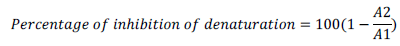
here A1 = absorption of the control sample and A2 = absorption of the test sample
Inhibition of haemolysis of red blood cell (RBC)
The assay was carried out according to the procedure of Okoli et al. (2009) as reported by Gunathilake et al. (2018) with slight modification. Briefly, the assay volume contained 0.5 mlextract, 1.0 ml 2% (v/v) RBC suspension and 1.5 ml hyposaline [0.42% (w/v) NaCl]. The mixture was incubated at 65°C for 10 min in a shaking Gen Lab model water bath following by centrifugation at 2500 rpm for 3 min using Bosch model R-8C-DX centrifuge. The supernatant was collected and the absorbance of the supernatant was measured at 540 nm using visible Spectrum lab 23A spectrophotometer. The percentage inhibition of haemolysis was calculated using the following equation.

Where A1 = absorbance of the control, and A2 = absorbance of test sample
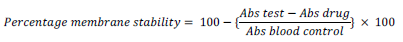
Assay of membrane stabilizing activity
The assay was based on the earlier procedures of (Oyedapo et al. 2010) and with slight modifications. Briefly, the assay mixture contained 1.0 ml hyposaline [0.42 v% (w/v) NaCl], 0.5 ml phosphate buffer (0.15 M, pH 7.4), varying concentrations of each extracts (0-300 µg/ml) which was made up to 3.0 ml with normal saline following by the addion of 0.5 ml of 2% (v/v) erythrocytes. The reaction mixture was incubated at 56°C for 30 min using Bosch incubator and centrifuged at 2500 rpm for 10 min using Bosch model R-8C-DX centrifuge. The absorbance of the supernatant was read at 560 nm using visible Spectrum lab 23A spectrophotometer against reagent blank. The drug control was prepared as above but without RBC while the blood control contained all the reagents and extract. Ibruprofen (1 μg/ml) was used as the standard drug and treated as the extract. The blood control represents 100% lysis or zero percentage stability.
Statistical analysis
Data was expressed as mean ± SEM of n = 3 readings. The mean values of S. bicolor and Z. mays extracts and the mixture were compared. Value of (p < 0.05) was taken as statistically significant.
RESULTS
Yields of extracts
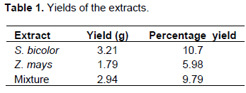
The extraction of 30 g of each powdered leaf (S. bicolor, Z. mays and mixture) yielded 3.21, 1.79, and 2.94 g extract representing 10.7, 5.98, and 9.8% of the starting materials, respectively. It was observed that S. bicolor contained more ethanol soluble content than Z. mays and the mixture (Table 1).
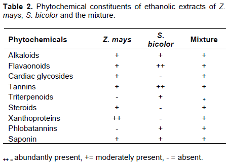
Phytochemical constituents of Z. mays, S. bicolor and the mixture
In Table 2, is the summary of the phytochemical constituents of ethanolic extracts of Z. mays, S. bicolor, and the mixture of Z. mays and S. bicolor. The results revealed the presence of alkaloids, flavonoids, cardiac glycosides, tannins, steroids, xanthoproteins, phlobatannin, saponins, and triterpenoid, which were confirmed by specific detecting reagents.
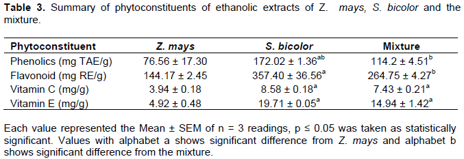
Concentrations of phytoconstituents
The results revealed that the total phenolic contents of Z. mays, S. bicolor, and the mixture were 76.56 ± 17.30 mg TAE/g, 172.02 ± 1.36 mg TAE/g, and 114.2 ± 4.51 mg TAE/g extract, respectively. The flavonoid contents of the extracts of Z. mays, S. bicolor, and the mixture were 144.17 ± 2.45 mg RE/g, 357.40 ± 36.56 mg RE/g, and 264.75 ± 4.27 mg RE/g extract, respectively. The vitamin C contents of Z. mays, S. bicolor, and the mixture were 3.94 ± 0.18 mg/g, 8.58 ± 0.18 mg/g, and 7.43 ± 0.21 mg/g extracts, respectively. The vitamin E content of Z. mays, S. bicolor, and the mixture were 4.92 ± 0.48 mg/g, 19.71 ± 0.05 mg/g and 14.94 ± 1.42 mg/g extract, respectively (Table 3).
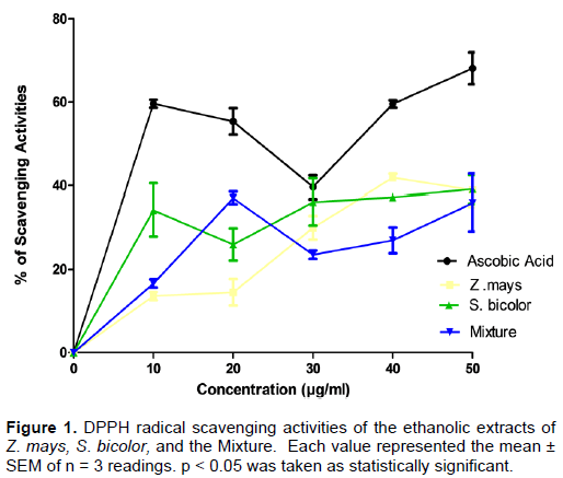
Diphenyl-2-Picrylhydrazyl (DPPH) radical scavenging activity of the extracts
The result of Figure 1 revealed that the ethanolic extract of Z. mays, S. bicolor, and the mixture scavenged DPPH radical maximally at 42, 38 and 36% respectively. The DPPH activity of the standard (ascorbic acid) and the extracts was concentration dependent. The free radical scavenging activities increased with increase in concentration.
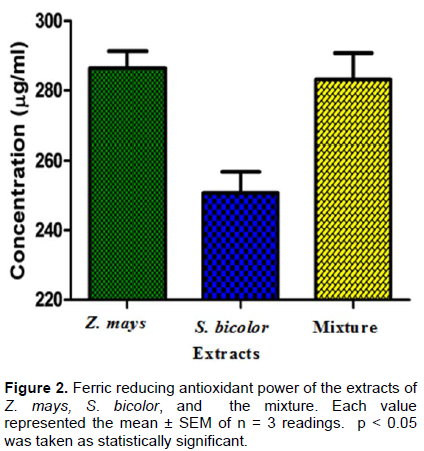
Ferric reducing antioxidant power of Z. mays, S. bicolor and the mixture
The results revealed that the ferric reducing power of the extracts increased as the concentration increased (Figure 2). The reducing power of Z. mays, S. bicolor and the mixture were 289.94 ± 18.1 µg/ml, 253.071 ±18.91 µg/ml, and 286.81 ± 20.49 µg/ml respectively. The ability of the extracts to reduce Fe3+ to Fe2+ was observed to be concentration dependent since there was increase in absorbance as the concentration increased.
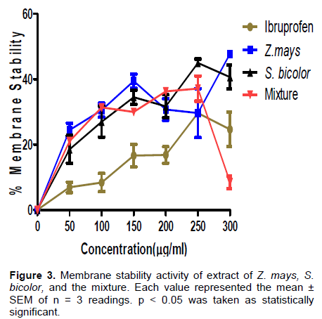
Membrane stabilizing activity of Z. mays, S. bicolor and the mixture
The results revealed that the extracts as well as the standard drug (Ibuprofen) protected the red blood cell (RBC) exposed to both heat and hypotonic induced lyses at concentrations ranged between 50 and 300 μg/ml (Figure 3). The results revealed that S. bicolor extract exhibited monophasic mode of protection, Z. mays extract exhibited both monophasic and biphasic mode of protection. It implied that at low concentrations of the extracts, the RBC membranes are protected while at higher concentrations the extracts become toxic to the cells. Moreover, the standard drug (Ibuprofen) and the mixture exhibited biphasic mode of protection. It was observed further that, the extract of Z. mays elicited highest activity. The order of the stability by the extracts was Z. mays >S. bicolor > mixture.
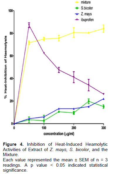
Inhibition of heat-induced blood hemolysis
The inhibition of heat-induced hemolysis abilities of the different ethanolic extracts are illustrated in Figure 4. The percentage inhibition of the extracts of Z. mays, S. bicolor and the mixture was 21.0, 19.7, and 82.2%, respectively. The ethanolic extract of the mixture elicited a significantly (p < 0.05) highest hemolysis inhibition activity when compared with the extracts of S. bicolor and Z. mays.
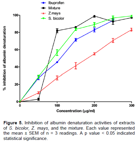
Inhibition of albumin denaturation
The inhibition of albumin denaturation by the ethanolic extracts is illustrated in Figure 5. It was observed that the ethanolic extract of the mixture exhibited a significantly higher level of inhibition compared to other extracts, while extract of Z. mays showed the lowest inhibition levels. The order of the inhibition of the extracts was Mixture >S. bicolor > Z. mays.
DISCUSSION
Inflammation is a complex series of events and functionally protective response that develop when an organism is injured either by mechanical or chemical agents by a self-destructive process (Sadique et al., 1989; Morakinyo et al., 2018). Several reports have shown that plants play vital roles in the maintenance of health due to the presence of their phytochemicals. These are bioactive components of plants in fruits and vegetables and are linked to reducing the risk of chronic diseases. The phytochemicals vary in distribution within the plant. They contribute to the aroma, flavor, colour and protect the plant from internal and external factors within the environment (Kyung-Baeg et al., 2016).
Phytochemical screening of ethanolic extract of Z. mays revealed the presence of alkaloids, flavonoids, cardiac glycoside, tannins, steroids, xanthroproteins, saponins with the absence of phlobatannins. The results agreed with earlier observations of Thoudam et al. (2011) who observed the presence of the above phytoconstituents except anthocyanins. The ethanolic extract of S. bicolor tested positive for alkaloids, flavonoids, tannins, saponins. The mixture of both Z. mays and S. bicolor was further observed to give positive tests for the presence of alkaloids, flavonoids, cardiac glycosides, tannins, steroids, xanthroproteins, saponins and phenols. The presence or absence of a phytoconstituent as well as its quality depends on conditions such as time of collection, place of collection, periods of collection and of course the methods of extraction.
Flavonoids are widely distributed groups of polyphenolic compounds in plant kingdom especially in fruits, vegetables, nuts and teas possess and exhibit potent and appreciable broad pharmacological properties. Flavonoids are gradually gaining increasingly applications not only in prophylaxis and therapeutic but also as nutraceutics and functional foods for improved health and well-being. The ability of flavonoids to act as strong antioxidant with potential to scavenge free radicals directly or indirectly by augmenting cellular antioxidant defenses make the molecule a promising candidate to contribute immensely to the health of the brain. Also, the ability of flavonoid to cross the blood brain barrier as well as their receptive safety in the central nervous system qualifies them to be a suitable candidate for promotion of brain health (Fajobi et al., 2017, Akinmoladun et al., 2019; Fajobi et al., 2020, 2021).
In this study, quantification of antioxidants constituents (phenolics, total flavonoids, vitamin C and E) revealed that ethanolic extracts of Z. mays and S. bicolor were rich in vital phyto-constituents that possess and exhibit potent and appreciable antioxidant activities. Analyses of the constituents showed that the phenolic and flavonoids contents of the ethanolic extracts of S. bicolor were higher when compared to ethanolic extract of Z. mays and mixture.
DPPH radical scavenging assay is a method employed to screen the free radical scavenging potentials of plants extracts, fractions and isolated compounds. It is based on the ability of the compound to decolorize DPPH in the presence of antioxidants due to their hydrogen donating ability (Egemole et al., 2007). The study revealed that the activities of the standard (ascorbic) and the ethanolic extract of Z. mays were observed to exhibit a higher percentage DPPH radical scavenging activity when compared with the ethanolic extracts of S. bicolor and the mixture. The ability of the extracts to scavenge free radicals might be due to the presence of flavonoids, alkaloids, tannins, which act as antioxidants. The mechanisms might involve absorbing and neutralizing free radicals, quenching singlet and triplet oxygen, and or decomposing of peroxides. The result agreed with that of Madaan et al. (2011). The reducing power of ethanolic extracts of Z. mays, S. bicolor and mixture was investigated using ascorbic acid as standard due to the presence of free hydroxyl group in its structure thereby preventing the oxidation of free radicals. The reducing power is a result of the ability of substances to convert Fe3+ to Fe2+. Ferric reducing antioxidant power (FRAP) measures the ability of antioxidants to reduce ferric 2,4,6-tripyridyl-s-triazine complex to intensively blue colored ferrous complex in acidic medium. The study revealed that ethanolic extract of Z. mays had the highest potential followed by the mixture and then, ethanolic extract of S. bicolor.
Inflammation generally refers as a way the body response to an injury, it is a protective measure involving immune cells, blood vessels, and molecular mediators of the body tissues to the harmful stimuli, such as pathogens and damage cells (Grivennikov et al., 2010). Different methods such as inhibition of phosphatases, aminotransferases, cotton pellet granulation techniques, inhibition of heat-induced hemolysis, inhibition of albumin denaturation, membrane stabilizing, platelet aggregation, have been used to study the anti-inflammatory potentials of drugs or agents (Oyedapo et al., 2010). In this study, heat-induced hemolysis, protein denaturation, and membrane stabilizing activities were used to investigate the anti-inflammatory activities of ethanolic extracts of Z. mays, S. bicolor and the mixture.
The results revealed that the percentage of red blood cell (RBC) membrane stabilizing activities of the ethanolic extracts were concentration dependent and was active as that of the standard anti-inflammatory drug (Ibruprofen). This could be as a result of the extracts being rich in flavonoids. Flavonoids-rich compound have been shown to possess high anti-inflammatory ability. The mode of response of erythrocyte to the ethanolic extract of S. bicolor was monophasic which implied that the extract protected the RBC at all the concentrations of the extract investigated. The extract of Z. mays, the mixture and the standard drug (Ibruprofen) elicited biphasic mode of protection. The results agreed with the earlier observations of Oyedapo et al. 2010) in their investigations of RBC membrane stabilizing potentials of extracts of root extracts of Theobroma cacao and Lantana camara and its fraction.
The stabilization of erythrocyte membrane exposed to both hypotonic and heat induced lyses by the extracts revealed that the extracts elicited a dose dependent membrane stabilizing activity over the concentration ranges in this study. The results compared favourably with the activity of ibuprofen, the standard non-steroidal anti-inflammatory drug. The potency of the extracts could be attributed to the synergistic (Oyedapo et al., 2010). This is in agreement with earlier reports of Akinwunmi and Oyedapo (2015) that reported the activity of Monodora myristica seeds in stabilizing the erythrocyte membrane exposed to both heat and hypotonic induced lyses was dose-dependent and might be due to the presence of naturally occurring flavonoids. Sakat et al. (2010) also reported the dose-dependent stabilizing activity of Oxalis corniculata on erythrocyte membrane exposed to heat induced lysis.
The heat-induced hemolysis inhibition activities of the extracts are shown in Figure 4. The extracts significantly reduced (p < 0.05) the hemolysis of red blood. The inhibition of the red blood cell by the extracts was in the order: mixture > Z. mays > S. bicolor. The mixture exhibited a significantly highest (p < 0.05) hemolytic inhibitory activity when compared with that of Z. mays and S. bicolor. The observations agreed with the earlier observations of Gunathilake et al. (2018) in their studies of in vivo anti-inflammatory properties of selected Green leafy vegetables.
Moreover, the inhibition of albumin denaturation by the extracts revealed that the mixture exhibited a significantly highest (p < 0.05) level of inhibition compared to other extracts and the standard, whereas Z. mays extract elicited the lowest inhibitory activity. The order of the inhibition of the extracts was mixture > S. bicolor > Z, mays which was also correlated with the observations of Gunathilake et al. (2018). The anti- inflammatory potentials extracts of S. bicolor, Z. mays and mixture might be attributed to the phytochemicals present in it.
In conclusion, the ethanolic extracts of Z. mays, S. bicolor and the mixture were found to be rich in biologically active phytocontituents which include flavonoid, phenolics, Vitamin C and E. The extracts of Z. mays, S. bicolor and the mixture possess and exhibit potent, appreciable and significant antioxidant and anti- inflammatory properties. The extracts of leaves of Z. mays possess highest membrane stabilizing activity as compared to those of S. Bicolor and the mixture. The extract of the mixture possessed and exhibited the highest albumin denaturation inhibition activity and hemolytic inhibition. It is suggested that both plants could be employed for the management of inflammatory and oxidant disorder either singly or in combination.
CONFLICT OF INTERESTS
The authors have not declared any conflict of interests.
REFERENCES
|
Aina IO, Oyedapo OO (2013). In vitro investigations into the antioxidant and anti-inflammatory potentials of the fractions and ethanolic extract of Cyclosorus afer (christ.) ching, stalks. Ife Journal of Science (15) 2:235-249. |
|
|
Akinmoladun CA, Farombi TH, Farombi EO (2019).Food for the brain health: flavonoids. Encyclopedia of Food Chemisty 3:370-386. |
|
|
Akinwunmi KF, Oyedapo OO (2015). In vitro anti-inflammatory evaluation of african nutmeg (Monodora myristica) seeds. European Journal of Medicinal Plants 8(3):167-174. |
|
|
Blois MS (1985). Antioxidant determination by use of stable free radicals. Nature 181(4617):1199-1200. |
|
|
Egemole AN, Odekanyin OO, Akinpelu BA, Akinwumi KF, Oyedapo OO (2007). An examination of the extracts of Icacina tricantha Oliv. Nigerian Journal of Biochemistry and Molecular Biology 22:39-42. |
|
|
Fajobi OA, Fasakin OW, Oyedapo OO (2017). Phytochemicals, antioxidant potentials and 2,2-diphenyl-1-picrylhydrazyl (DPPH) radical scavenging activity of Piper guineense (Schumach Thonn) seed. African Journal of Plant Science 11(4):99-104. |
|
|
Fajobi OA, Emma-Okon BO, Oyedapo OO (2020). In vitro antioxidant, anti-inflammatory and thrombolytic properties of leaf extract and fractions of Pterocarpus mildbraedii Harms. Ife Journal of Science 22(3):065-074. |
|
|
Fajobi OA, Emma-Okon BO, Oyedapo OO (2021). Haematopoietic potential of methanolic leaf extract of Pterocarpus mildbraedii in Male Wistar Rats. International Journal Medicinal Plants 115: 983-991. |
|
|
Gini TG, Jothi GJ (2013). Preliminary phytochemical screening of whole plant extracts of Peperomia pellucida (Linn.) HBK (Piperanceae) and Marsilea quadrifolia Linn. (Marsileaceae). International Journal of Pharmacognosy and Phytochemical Research 5(3):200-214. |
|
|
Grivennikov SI, Greten FR, Karin M (2010). Immunity, inflammation, and cancer. Cell 140(6):883-899. |
|
|
Gunathilake KK, Ranaweera HP, Vasantha R (2018). In vitro Anti-inflammatory properties of selected green leafy vegetables. Biomedicines 6(107):10-3390. |
|
|
Kernels BT, Masciangelo S, Michelett A, Ferretti G (2013). Carotenoids, phenoliccompounds and antioxidant capacity of five local Italian corn (Zea mays L.). Journal Nutrition Food Science 3(7):6-12. |
|
|
Kyung-Baeg R, Hyoyo K, Seungwoo S, Young-Soo K, Jung AL, Kim MO, Eunsun J, Jongsung L, Deokhonn P (2016). Anti-inflammatory effects of Zea mays L. husk extracts. BMC Complementary and Alternative Medicine 16:298. |
|
|
Madaan R, Kumar S, Bansal G, Sharma A (2011). Estimation of total phenols and flavonoids in extracts of Actaea spicata roots and antioxidant activity studies. Indian Journal of Pharmaceutical Sciences 73(6):666. |
|
|
Morakinyo AE, Oyedapo OO, Akinpelu BA, Fajobi AO, Fasakin WO, Komolafe IJ, Akinlalu AO (2018). Evaluation of Antioxidant, Anti-inflammatory and Phytochemical Constituents of Aframomum melegueta Aqueous Leaf Extract. International Journal of Medicinal Plants 112: 888-903. |
|
|
Nebavi SM, Ebrahimzadeh MA, Nabavi SF, Jafari M (2013). Free Radical Scavenging Activity and Antioxidant Capacity of Eryngium Caucasicum Trautv and Froripia subpinnata. Pharmacology Online 3:19-25. |
|
|
Okoli CO, Ezike A, Akah P, Udegbunam SO (2009). Studies on Wound healing and antiulcer activities of extract of aerial parts of Phyllanthus niruri L. (Euphorbiaceae). American Journal of Pharmacology and Toxicology 4(4):118-126. |
|
|
Oladeji O (2016). The characteristics and roles of medicinal plants: some important medicinal plants in Nigeria. An Indian journal 12(3):102. |
|
|
Omaye ST, Turnbull JD, Sauberlich HE (1979). Selected methods for the determination of ascorbic acid in animal cells, tissues and fluids. The Journal of Biological Chemistry 62:3-11. |
|
|
Osuntokun OT, Olumekun VO, Ajayi1 AO, Omotuyi IO, Olonisakin A (2020). Assessment of in-vitro antioxidant/enzymes inhibitory potentials of Aframomum melegueta (Roscoe) K.Schum (Grains of Paradise) leaf, stem bark, seed bark and seed extracts. Archives of Current Research International (2):40-57. |
|
|
Oyedapo OO, Akinpelu BA, Akinwumi KF, Adeyinka MO, Sipeolu FO (2010). Red blood cell membrane stabilizing potentials of extracts of Lantana camara and its fractions. International Journal of Plant Physiology and Biochemistry 2(4):46-51. |
|
|
Oyedapo OO, Amos S (1997). Further investigation into the activities of the root extract of Plumbago zaylanica. Phytotherapy Research (London) 11(1)62-63. |
|
|
Oyedapo OO, Makinde AM, Gbemisola IM, Abimbola EO, Kemi AF, Akinpelu BA (2015). Biological activities (anti-inflammatory and anti-oxidant) of fractions and methanolic extract of Philonotis hastata (duby wijk & margadant). African Journal Traditional Complement Alternative Medicine 12(4):50-55. |
|
|
Pan P, Wang Y, Leng X, Deng J, Wang C (2017). 90 protective effects of cistanches herba aqueous extract on cisplatin-induced premature ovarian failure in mice. African Journal Tradition Complement Alternative Medicine 14(6):90-101. |
|
|
Patrick AE, Quansah TK, Karikari AA, Osei-Safo D, Kennedy KE, Isaac KA, Alexander K (2018). Medicinal plants used in the treatment of mental and neurological Disorders in Ghana. |
|
|
Popa-wagner A, Mitran S, Sivanesan S, Chang E, Buga AM (2013) ROS and brain diseases: The good, the bad and the ugly. Oxidave Medicine and Cellular Longetivity. |
|
|
Sadique J, Al-Rqobahi WA, Bughaith MF, El-Ginaly RR (1989). The bioactivity of certain medicinal plants on the stabilization of the RBC system. Fitoterapia 60(6):525-532. |
|
|
Sakat S, Juvekar AR, Gambhire MN (2010). In vitro antioxidant and anti-inflammatory activity of methanol extract of Oxalis corniculata Linn. International Journal of Pharmacy and Pharmaceutical Sciences 2(1):146-155. |
|
|
Santosh R, Nitin K, Baijnath P, Vandana H, Arun GB (2013). Neuroprotective effect of Vitamin E supplementation in wistar rat treated with acrylamide. Toxicology International 19(1):1-8. |
|
|
Shinde UA, Phadke AS, Nair AM, Mungantiwar AA, Dikshit VJ, Saraf MN (1999). Membrane stabilizing activity-a possible mechanism of action for the anti-inflammatory activity of Cedrus deodara wood oil. Fitoterapia 70(3):251-257. |
|
|
Singleton VL, Orthofer R, Lamuella-Raventos M (1999). Analysis of total phenols and otheroxidation substrates and antioxidants by means of |
|
|
Folin-Ciocalteau's phenol reagent. Methods of Enzymology 299:152-178. |
|
|
Sun PX, Yie LK, Zhang ZL, Hu M, Lu L (1999). Colometric determination of the total content of the flavonoids in epicedium capsules. Journal of Shenyang Pharmaceutical University 16:68-70. |
|
|
Thoudam B, Kirithika T, Ramya J, Usha K (2011). Phytochemical constituents and antioxidant activity of various extracts of corn silk (Zea mays). Research Journal of Phamaceautical, Biological and Chemical Sciences 2(4):986-993. |
|
|
Tunon MJ, Garcia-Mediavilla MV, Sanchez-Campos S, Gonzalez-Gallego J (2009). Potential of flavonoids asanti-inflammatory agents: modulation of pro-inflammatory gene expression and signal transduction pathways. Current Drug Metabolism 10(3):256-71. |
|
|
Umapathy E, Ndebia EJ, Meeme A, Adam B, Menziwa P, Nkeh-Chungag BN, Iputo JE( 2010). An experimental evaluation of Albucasetosa aqueous extract on membrane stabilization, protein denaturation and white Photon 903 blood cell migration during acute inflammation. Journal of Medicinal Plants Research 4(9):789-795. |
|
Copyright © 2024 Author(s) retain the copyright of this article.
This article is published under the terms of the Creative Commons Attribution License 4.0


