Full Length Research Paper

ABSTRACT
Cardiovascular diseases represent the major cause of death and morbidity in the world. An uncontrolled activation of the coagulation cascade and platelet aggregation may lead to the formation of thrombi. Antithrombotic drugs have limitations and may produce side effects, and consequently, alternative therapies have been extensively investigated. Thus, the aim of this work was to evaluate anticoagulant and antiplatelet effects of extracts (prepared in methanol, dichloromethane, ethyl acetate or acetone) of the Brazilian alga Acanthophora spicifera and some commercial products, biotin, myristic acid, cholesterol, β-carotene and vitamin B12. Samples were tested on Prothrombin Time, Activated Partial Thromboplastin Time, Fibrinogen Coagulation and Thrombin Time, which are routinely used at clinical trials and on platelet aggregation. From the result, all extracts or products inhibited plasma coagulation as well as inhibited platelet aggregation induced by collagen or ADP. Moreover, the extracts inhibited the enzymatic activity of thrombin, tested upon a specific chromogenic for thrombin. The extracts or commercial products were devoid of toxicity, since no lyse occurred on platelets or red blood cells in the presence of them. In conclusion, the extracts of A. spicifera or products from the market have biotechnological potential and may be useful to develop a new class of antihemostatic drugs.
Key words: Acanthophora spicifera, red marine alga, antiplatelet, anticoagulant, bioprospecting, vascular diseases.
INTRODUCTION
Hemostasis balance is a physiological coordinate process of anticoagulant and coagulant events that allows blood to circulate through the cardiovascular system or to prevent bleeding after injury. Platelets as well as the coagulation cascade participate in such process, as well as they may cause vascular diseases.
Disturbs that alter such equilibrium may result in hemorrhage or thrombosis, while the latter is one of the most important cardiovascular diseases, as ischemic attack, stroke and myocardial infarction. They lead to mortality or morbidity worldwide, mainly in western populations (Zhao et al., 2017). Thrombosis generally has medical complications to patients or recurrent hospitalization, and this fact increases healthcare costs (Furie and Furie, 2008). The current therapy to such diseases is performed by administration of anticoagulant and/or antiplatelet drugs. Heparin and coumarins are usually used as anticoagulants, but they have drawbacks, as risk of bleeding, narrow therapeutic window, non-specific binding, thrombocytopenia and/or osteoporosis (Mourão, 2015). On the other hand, in order to manage thrombosis disturbs, antiplatelet drugs may be employed. Thus, platelets are critical in thrombotic disorders. In this way, there are many drugs in the market able to impair platelet aggregation, preventing the formation of a platelet thrombus. Aspirin has been used as the drug of choice for the long-term treatment, but, it also has serious side effects, and gastric hemorrhage is the most serious complication. Other drugs, as clopidogrel and ticlopidine have limitations, as well.
Thus, the search for new anticoagulant or antiplatelet drugs without side effects, more potent and safer may constitute an important task to improve such treatments; and, natural sources may be a promising strategy. Marine organisms are composed of molecules with a chemical diversity, and some of them have application in medicine (Mourão, 2015; Cirne-Santos et al., 2018). However, their potential is not well-explored (Mourão, 2015). Previous reports showed that a diterpene isolated from the marine brown alga, Dictyota menstrualis as well as from other seaweeds or sponges inhibited platelet aggregation and coagulation of plasma (de Andrade et al., 2014; de Andrade et al., 2011). Seaweeds are classified as green algae (Chlorophyta), brown algae (Phaeophyta), red algae (Rhodophyta) and Blue-green algae (Cyanobacteria). Most of seaweeds are red (6,000 species) and the rest known are brown (2,000 species) or green (1,200 species).
The red algae are the oldest groups of eukaryotic algae and the largest, with about 5,000–6,000 species, and some of them have medicinal properties. Acanthophora spicifera species belong to rhodophyta and are widely distributed in the tropical and subtropical regions (Kilar and McLachlan, 1986), and in Brazil, they are along the entire coast (Ávila et al., 2012). There are many reports describing biological effects of A. spicifera, as: antioxidant, antibacterial, anti-malarial, antiviral, anti-inflammatory, and anticancer (Duraikannu et al., 2014; Duarte et al., 2004). The assessment of chemical profile of genus Acanthophora showed the presence of sterols, as cholesterol and derivatives (Flora and Rani, 2013), fatty acids, vitamins and carotenoids (Polat and Ozogul, 2008).
Moreover, the analysis of the extract of A. spicifera through GC-MS has demonstrated the presence of fatty acids and sterols as well as using mass spectrometry has revealed octanol, piperazine, benzoic acid and octadecenoic acid, as major constituents of methanolic extract (Flora and Rani, 2013). It is well known that the seaweed A. spicifera is rich in halogenated molecules, as terpenes, phenols and acetogeninas, whose functions are related to their survival into the marine environment (Nogueira et al., 2016), but little is known of natural chemistry of the genus Acanthophora. However, antihemostatic action of A. spicifera has not been explored. Thus, the objective of this work was to investigate antiplatelet and anticoagulant effects the crude extracts of A. spicifera and some commercial molecules, which are produced by this alga.
MATERIALS AND METHODS
The commercial products: biotin (Sigma-Aldrich, USA – B4501), myristic acid (Sigma-Aldrich, USA – M3128), cholesterol (Sigma-Aldrich, USA – C8667), β-carotene (Sigma-Aldrich, USA – C9750) and vitamin B12 (Sigma-Aldrich, USA – V2876). ADP and collagen type I from Chrono Log Corp. (Havertown, PA, USA).
Algal collection and preparation of extracts of A. spicifera
Specimens of the red marine alga A. spicifera were collected in May, 2013 by snorkeling at depth of 0.5-10 m, at Orla Bardot (a tribute to French actress Brigite Bardot) in the city of Armação dos Búzios (22° 05’ 03” S, 41° 53’ 01” W). Armação dos Búzios or just Búzios is a small balneary with a population about 30,000 people, located at 170 km from the city of Rio de Janeiro, Brazil. The fresh material was immediately taken to the laboratory and screened to remove epiphytes and associated organisms. Air-dried specimens (204 g) were exhaustive and sequential extracted with dichloromethane (CH2Cl2), ethyl acetate (EtOAc), acetone (Me2CO), and methanol (MeOH) at room temperature. After filtration and evaporation under reduced pressure, the extract (100 µg) was analyzed by thin-layer chromatography (TLC) eluting with 20 ml of n-hexane and ethyl acetate (7:3), which was performed with a Merck Kieselgel GF254. The spots were detected by inspection in the ultraviolet light (254 and 365 nm), and then revealed by spraying with CeSO4 at 2% H2SO4, followed by heating at 100°C for 15 min.
Assays
Platelet aggregation
Platelet aggregation assays were carried out according to (Fuly et al., 1997), with modifications, using platelet-rich-plasma (PRP) obtained from healthy volunteer donors (experiments were approved by the Federal Fluminense University Committee for Ethical in Experimentation – CEP-UFF, CAAE: 28941314.0.0000.5243). PRP was prepared by the centrifugation of citrated (0.31%, v/v) whole blood at 25°C (340 x g for 12 min). Platelet aggregation was measured turbidimetrically using a Whole Blood Aggregometer (Model 490 2D - Chrono-Log Corporation, Pennsylvania, USA). Assays were performed at 37°C in siliconized glass cuvettes using 300 μl of PRP under stirring, and aggregation was triggered by the addition of ADP or collagen. One hundred percent (100%) of platelet aggregation was determined as the full platelet response obtained 6 min after the addition of a supramaximal concentration of the agonists (concentration that gives 70-80% of aggregation), and 0% (base line) of platelet aggregation was the light transmittance recorded of PRP alone. The effect of the extracts of A. spicifera was performed by incubating different concentrations of the extracts with PRP for 5 min at 37°C, and then, platelet aggregation was triggered by adding ADP (15 μM) or collagen (5 μg/mL). Inhibitory effect on platelet aggregation was expressed as the difference in the maximal responses of platelets in the presence or in the absence of the extracts of A. spicifera, after challenge with agonists. Control experiments were performed in the presence of saline or 0.9% (v/v) of dimethylsulfoxide (DMSO).
Plasma coagulation
Prothrombin time (PT), activated Partial Thromboplastin Time (aPTT) or Thrombin Time (TT) assays were performed according to the manufacturer’s instructions (Wiener Laboratories, Rosario, Argentina). For the PT test, the extracts of A. spicifera were incubated with plasma (50 μl) for 10 min at 37°C, and then, 100 μl of pre-warmed thromboplastin with calcium were added to initiate coagulation.
For the APTT test, the extracts of A. spicifera were incubated for 10 min at 37°C with plasma plus 100 μL of the APTT reagent, cephalin and kaolin in a final volume of 200 μl, with the reaction started by the addition of CaCl2 (8.3 mM, final concentration). The TT assay was performed by incubating plasma with 50 μl of the extracts of A. spicifera, 50 μl of saline or DMSO and incubated at 37°C for 1 min. Then, coagulation was initiated by adding 1 nM human α-thrombin (Haematologic Technologies Inc., Vermont, USA). The Fibrinogen Coagulation (FC) assay was performed by incubating the extracts of A. spicifera for 10 min at 37°C with 200 μl commercial fibrinogen (2 mg/ml, from Sigma Chemical Co., USA) in a final volume of 250 μl. Then, coagulation was triggered by the addition of thrombin (10 nM). For all assays, coagulation was performed on a Multichannel Coagulometer (Amelung, Model KC4A, Labcon, Germany), and coagulation time was recorded in seconds. Plasma was obtained from a pool of healthy volunteer donors and diluted in an equal volume of saline.
The ex vivo coagulation tests were considered, as follow: saline, DMSO (1% v/v, final concentration or commercial products (300 µg/mice) were administered into mice intravenously (i.v.). After 2 h, blood was collected by cardiac puncture, then blood was centrifuged at 1,800 g for 10 min, and plasma was transferred to plastic tubes and prothrombin time (PT) or activated partial thromboplastin time (aPTT) tests were performed. Experiments were approved by CEP-UFF, under protocol CAAE: 28941314.0.0000.5243.
Hydrolytic activity upon chromogenic substrate
Hydrolysis of the chromogenic substrate, H-D-Phe-pipecolyl-Arg-pNA.2HCl (S-2238), bought from Chromogenix (Milan, Italy) was monitored using a microplate reader (SpectraMax, Model M4, Molecular Devices, Menlo Park, CA, USA), equipped with a mixer and heating system at A405 nm. The extracts of A. spicifera were incubated with thrombin (40 nM, final concentration) for 10 min. at 37°C, and then, the reaction was triggered by adding S-2238 (0.5 mM, final concentration). The reaction was monitored during 20 min at 37°C. Control experiments were performed by incubating thrombin with DMSO (1% v/v, final concentration) or saline, instead of the extracts of A. spicifera.
In vitro cell toxicity
Human platelets were incubated with the extracts of A. spicifera or products, saline or DMSO (0.9% v/v) for 10 min at 37°C, then lactate dehydrogenase (LDH) activity was measured using a LDH-P UV kit (Wiener Laboratories), according to the manufacturer’s instructions. Hundred percent of platelets lysis was achieved by adding water or sonicating PRP. Hemolysis was determined according to Bauer et al. (2012), with modifications. A 13% solution of human erythrocytes was incubated with A. spicifera or products, saline or DMSO (0.9%) for 3 h at 37°C. Then, solution was centrifuged (1,200 g for 10 min) and the hemoglobin was measured at A 578 nm using a Micro Plate Reader (SpectraMax, Model M4, Molecular Devices, California, USA). Hundred percent of hemolysis was obtained by adding 1% Triton X-100 to cells. Experiments were approved by CEP-UFF, CAAE: 28941314.0.0000.5243.
Statistical analysis
The results were expressed as the means ± standard error (SEM) obtained with the number of experiments performed indicated. The statistical significance of differences among experimental groups was evaluated using the Student t-test (p-values < 0.05 were considered statistically significant).
RESULTS AND DISCUSSION
Chemical composition
The assessment of the chemical profile of A. spicifera extract using gas chromatography–mass spectrometry (GC-MS) analysis has demonstrated the presence of fatty acids, and sterols (Flora and Rani, 2013; Polat and Ozogul, 2008). Furthermore, analysis of the chemical profile of genus Acanthophora (Rhodophyta, Ceramiales) showed the presence of sterols, as cholesterol and derivatives (Dayong et al., 2011), fatty acids (Kumari et al., 2014), vitamins and carotenoids (Aihara and Yamamoto, 1968). Nogueira et al. (2016), by 1H-NMR technique, evaluated the presence of phenolic molecules in the extracts of A. spicifera prepared using increasing polarity solvents. In such work, the anti-HIV effect of these extracts was due to the presence of aromatic chemical structures. Here, we tested antiplatelet or anticoagulant effect of the crude extracts of the seaweed A. spicifera as well as the commercial available products (biotin, myristic acid, cholesterol, B-carotene and vitamin B12).
Effect of the extracts of A. spicifera on platelet aggregation
Platelets are anucleate megakaryocyte fragments that passively circulate in the blood stream. However, after a stimulus, in an injury, they rapidly activate, interact with other platelets and with endothelium or extracellular matrix molecules to form a hemostatic plug, preventing blood loss (Qiu et al., 2015; Ruggeri, 2002; Clemetson, 2012).
Moreover, platelets participate in many other processes, as inflammation and defense from microbial infection. However, undoubtedly, their main function is to prevent hemorrhage after an injury (Clemetson, 2012). To perform such functions, platelets have three granules, alpha (α), dense (δ) and lysosomes (λ) which contain molecules (protein and non-protein) with pro- and anti-thrombotic effects (Clemetson, 2012). After activation and aggregation of platelets, they secrete their granule content in order to amplify the hemostatic system, and enhance the formation of a clot by the coagulation cascade. The events of platelet adhesion, activation, aggregation and secretion of their granules are complex and may be stimulated by several agonists, as ADP, collagen and thrombin, and involve many ligands and/or plasma membrane-receptors (Clemetson, 2012). Thus, platelets circulate into blood stream in an inactive form, and just after a stimulus, they turn into active form in order to prevent and to stop blood loss. If an imbalance occurs in both forms of platelets, hemorrhage or procoagulant disorders may occur.
Cardiovascular diseases are the main causes of death and disability worldwide, with approximately 30% of deaths (Clemetson, 2012; Roth et al., 2015). Despite improved treatments and awareness of people, the rate of these diseases rose over the last decade (Alwan et al., 2010). Moreover, diabetes, obesity, depression, bad eating habits may contribute to increase them in the near future (Alwan et al., 2010). Thus, therapies to prevent blood coagulation and to inhibit platelet functions are employed, and in most of cases with positive results. Literature has described molecules that are regularly used as drugs to treat such diseases (DeWald and Becker, 2014; Nutescu et al., 2016). However, the current treatments have some drawbacks, as limited efficacy, resistance appearance, limitations related to the routes of administration and may induce side effects as well (Choi et al., 2013; Nutescu et al., 2005; Agnelli, 2005).
Thus, researchers are looking for new molecules with anticoagulant and/or antiplatelet effects able to improve therapies, but without restrictions or limitations. Lot of molecules derived from natural sources, be it from terrestrial or marine environment, have shown antihemostatic effects (Pimentel et al., 2003; de Andrade et al., 2011; Yoon et al., 2015; Lorigooini et al., 2015). Nonetheless, as known, most products with a clinical or medical interest came from oceans, approximately 33 and 25% from sponges and algae, respectively (Kijjoa and Sawangwong, 2004; Newman and Cragg, 2012). Seaweeds produce molecules with a variety of biological and pharmacological activities (Cirne-Santos et al., 2018), and many of them had led to the development of drugs with antihemostatic action (Collins et al., 2016; Kwak, 2014; Mourão, 2015).
In fact, oceans should be taken into consideration to discover new molecules, rather than the terrestrial environment (Collins et al., 2016). Here, we investigate the ability of four extracts (prepared using solvents of different polarity: dichloromethane, ethyl acetate, acetone and methanol) of the seaweed A. spicifera to inhibit coagulation and platelet aggregation. As shown in Figure 1, the extracts of A. spicifera (100, 200 or 400 µg/ml) inhibited aggregation of human PRP induced by 15 µM ADP (Panel A) or 5 µg/ml collagen (Panel B), in a concentration-dependent manner. The inhibitory profile of all extracts was very similar, regardless the agonist tested. At the lowest concentrations, the methanol (MET) or dichloromethane (DCM) extracts inhibited less the aggregation induced by ADP or collagen, respectively.
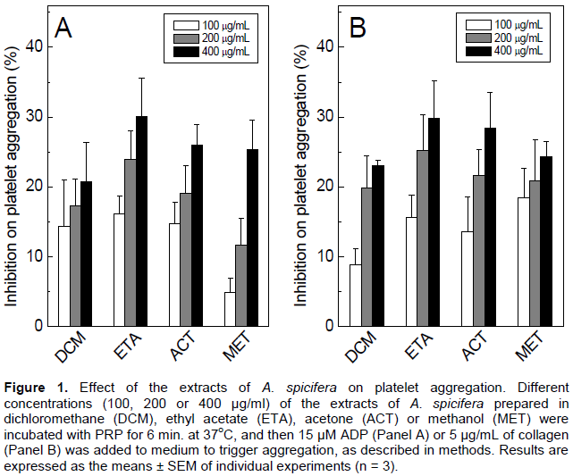
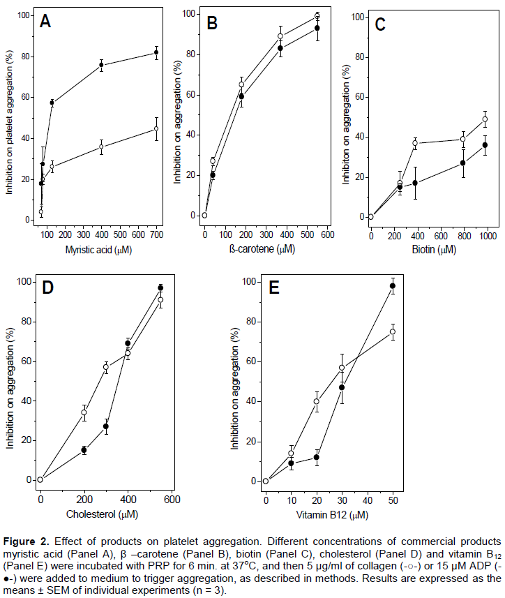
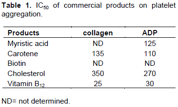
However, at the highest concentration, such difference disappeared, and both extracts inhibited around 25% the aggregation of platelets. The ETA, ACT, MET extracts achieved similar inhibitory percentages on collagen-induced aggregation. Because of the low inhibitory percentages, IC50 values of extracts were not possible to determine. Commercial molecules, which are present in A. spicifera seaweed (Ganesan et al., 2008), were tested on platelet aggregation. As seen in Figure 2, these products inhibited ADP- or collagen-induced platelet aggregation in a concentration-dependent manner; and, different half-maximal inhibitory concentration (IC50) values for the products were achieved (Table 1), ranging from 25 to 350 µM. On the other hand, IC50 of biotin was not able to determine, regardless the inducer tested (Table 1).
It is noteworthy that ADP or collagen does not share the same receptor and signaling pathways (Clemetson, 2012), so a difference at the percentage of inhibition can be achieved. After incubation of the extracts of A. spicifera (600 µg/ml) or products (2 mM) with platelets or erythrocytes, no trace of lactate dehydrogenase activity or release of hemoglobin was detected, respectively (data not shown). Thus, at such concentrations, the extracts of A. spicifera or products were devoid of toxicity. In general, products derived from seaweed have low cytotoxicity (Wang et al., 2007), and such assays are an important task that should be considered when the aim is to develop new drugs. Based on these results, the extracts of A. spicifera inhibited platelet aggregation and, therefore, could be considered to aid the development of new classes of antiplatelet agents.
Effect of the extracts of A. spicifera and products on coagulation
The effect of the extracts of A. spicifera on coagulation was investigated through the in vitro methods:
Prothrombin Time (PT), activated Partial Thromboplastin Time (aPTT), thrombin time (TT) and Fibrinogen Coagulation (FC), and results are shown in Table 2. These methods are traditionally employed to investigate bleeding or procoagulant disorders in patients as well as to look for molecules with procoagulant or anticoagulant effect. The PT and APPT tests measure the participation of the extrinsic and intrinsic pathway of the coagulation cascade, respectively. Both tests are inexpensive, easily performed and give accurate results over possible coagulation defects.
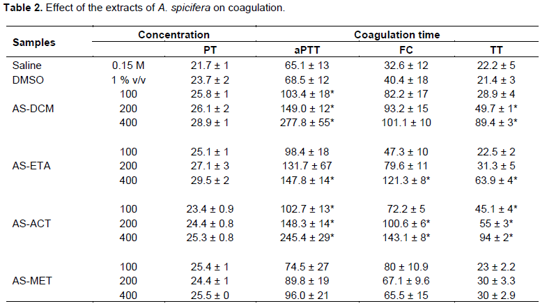
However, the division of the coagulation cascade into intrinsic and extrinsic pathways is only a useful concept for interpreting the results of such investigation, and does not have any in vivo validity. The TT and FC tests do not have a clinical signification by themselves, but they are important in combination with PT or aPTT test. As seen in Table 2, most of the extracts had anticoagulant action in such tests; and, none of them had a procoagulant activity. The extracts prepared in ACT or MET did not have any effect on coagulation time in the PT test, and at the highest concentration (400 µg/ml), the DCM or ETA extracts delayed coagulation time. The extract prepared in MET did not interfere in the coagulation time at any of all methods. On the other hand, the extracts in DCM, ETA or ACT delayed the plasma coagulation time with different potencies.
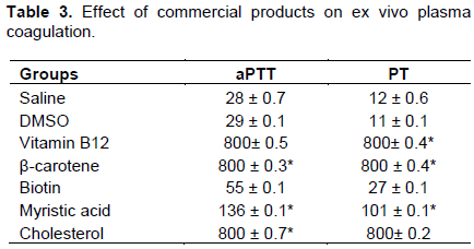
Overall, it is not possible to postulate a more efficient extract to inhibit coagulation. The extract in DCM or ACT inhibited the conversion of fibrinogen into fibrin through FC or TT tests. Thus, the extracts of A. spicifera prolonged coagulation by interfering at the intrinsic and extrinsic pathways of the coagulation cascade, and at the activity of thrombin, as well. Based on these results, one may speculate an interaction between molecules of extracts with thrombin, leading to the inhibition of the activity of enzyme. The commercial products were tested on ex vivo coagulation through the PT and aPTT tests, as well. As seen in Table 3, vitamin B12, b-carotene, myristic acid, and cholesterol delayed the plasma coagulation time of mice at both tests, when compared to vehicle (saline or DMSO).
For PT assay, the extracts of A. spicifera (100, 200 or 400 µg/ml) were incubated with plasma for 10 min at 37°C, and then thromboplastin was added to induce coagulation. For aPTT assay, the extracts of A. spicifera were incubated with plasma plus cephalin for 10 min at 37°C, and then, CaCl2 (8.3 mM) was added to induce coagulation. For FC assay, the extracts of A. spicifera were incubated for 10 min at 37°C with fibrinogen (2 mg/ml), and then, thrombin (10 nM) was added to induce coagulation.
For TT assay, the extracts of A. spicifera were incubated with plasma, and thrombin (1 nM) was added to induce coagulation. Results are expressed as the means ± SEM of individual experiments (n = 4). *, p < 0.05 when compared to saline or DMSO. AS-DCM, A. spicifera in dichloromethane, AS-ETA, A. spicifera in ethyl acetate, AS-ACT A. spicifera in acetone, AS-MET, A. spicifera in methanol. Mice received a single i.v. injection of 100 µL of vehicle (saline or DMSO) or 300 µg/mice of commercial products (vitamin B12, β-carotene, biotin, myristic acid, and cholesterol). After 2 h, blood was collected from mice, centrifuged and plasma was subjected to aPTT or PT tests, according to methods; *, p < 0.05 when compared to saline or DMSO.
Effect of the extracts of A. spicifera on hydrolysis of S-2238
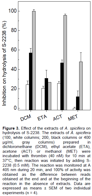
S-2238 is a specific chromogenic substrate, usually employed to test the enzymatic activity of thrombin, that it is a serine protease that cleaves fibrinogen (a soluble protein) into fibrin (an insoluble protein), resulting in the formation of a firm and stable clot. Besides, thrombin can trigger platelet aggregation and activate some coagulation factors of the cascade (factors V, VIII and XI), promotes vasoconstriction, and has mitogenic effects (Coughlin, 2000). Apart from the coagulant action, thrombin has anticoagulant effect through the activation of protein C, inactivating factors V and VIII (Crawley et al., 2007; Della-Valle et al., 2007). Some of these functions of thrombin depend on its catalytic site and two other sites (the anion-binding exosites I and II), in which are positively charged, and may interact with negative surfaces, promoting the pharmacological functions of thrombin (Crawley et al., 2007; Della-Valle et al., 2007). Anticoagulants, as hirudin and analogs have been designed to allow the binding to thrombin through these charged sites (Nutescu, 2016; Huntington, 2014). As seen in Figure 3, at all concentrations of extracts (100, 200 or 400 µg/ml), inhibition of the enzymatic activity of thrombin, measured by the hydrolysis of S-2238 was achieved. Again, the methanolic extract (MET) inhibited the activity of thrombin less, and the extracts in DCM and ACT achieved the highest inhibitory percentage, close to 100%. Surprisingly, these extracts (DCM and ACT) had the highest inhibitory effect at all the coagulation tests, as well. The extract in ETA inhibited around 20% of the thrombin activity, at any of the tested concentration.
CONCLUSION
Taken together, the extracts of different polarity of A. spicifera and commercial products inhibited aggregation of platelets and coagulation of plasma, and, thus, they have antihemostatic properties. However, the mechanism of action on aggregation should be investigated further. This work also shows the importance of bioprospecting studies for discovering molecules of natural sources without toxicity and easily cultivated, that could be used to develop drugs to improve antithrombotic therapy.
CONFLICT OF INTERESTS
The authors have not declared any conflict of interests.
ACKNOWLEDGEMENTS
The authors appreciate the support rendered by the International Foundation for Science (IFS grant F/4571-1), Conselho Nacional de Desenvolvimento Científico e Tecnológico (VLT 304070/2014-9 and 443930/2014-7; ALF 304719/2012-9), Fundação de Amparo à Pesquisa do Estado do Rio de Janeiro (VLT E-26/110.205/2013 and E-26/CNE-2014; ALF E-26/201.163/2014 and E-26/01.001918/2015), Coordenação de Aperfeiçoamento de Pessoal de Nível Superior and Universidade Federal Fluminense/Pró-reitoria de Pesquisa, Pós-graduação e Inovação.
REFERENCES
|
Agnelli G (2005). Current issues in anticoagulation. Pathophysiology of Haemostasis and Thrombosis 34(Suppl. 1):2-9. |
|
|
Aihara MS, Yamamoto HY (1968). Occurrence of antheraxanthin in two rhodophyceae Acanthophora spicifera and Gracilaria lichenoides. Phytochemistry 7:497-499. |
|
|
Alwan A, Maclean DR, Riley LM, d'Espaignet ET, Mathers CD, Stevens GA, Bettcher D (2010). Monitoring and surveillance of chronic non-communicable diseases: progress and capacity in high-burden countries. The Lancet 376:1861–1868. |
|
|
Ávila E, Méndez-Trejo MC, Riosmena-Rodríguez R, López-Vivas JM, Sentíes A (2012). Epibiotic traits of the invasive red seaweed Acanthophora spicifera in La Paz Bay, South Baja California (Eastern Pacific). Marine Ecology 33:470–480. |
|
|
Bauer M, Lautenschlaeger C, Kempe K, Tauhardt L, Schubert, US, Fischer D (2012). Poly(2-ethyl-2-oxazoline) as alternative for the stealth polymer poly(ethylene glycol): comparison of in vitro cytotoxicity and hemocompatibility. Macromolecular Bioscience 12:986–998. |
|
|
Choi JT, Shin KA, Kim YK (2013). Prevalence of aspirin resistance and clinical characteristics in patients with cerebral infarction. Journal Experimental Biomedicine Science 19:233–238. |
|
|
Cirne-Santos CC, de Souza Barros C, Nogueira CCR, Amorim LC, Campos RM, Ratcliffe NA, Teixeira VL, Ferreira DF, Paixão ICN (2018). Antiviral effect of the seaweed Osmundaria obtusiloba against the Zika virus. Journal of Medicinal Plants Research 12:387–395. Provide a valid link. |
|
|
Clemetson KJ (2012). Platelets and Primary Haemostasis. Thrombosis Research 129:220–224. |
|
|
Collins KG, Fitzgerald GF, Stanton C, Ross RP (2016). Looking beyond the terrestrial: the potential of seaweed derived bioactives to treat non-communicable diseases. Marine Drugs 14:60–91. |
|
|
Coughlin SR (2000). Thrombin signalling and protease-activated receptors. Nature 14:258–264. |
|
|
Crawley JT, Zanardelli S, Chion CK, Lane DA (2007). The central role of thrombin in hemostasis. Journal of Thrombosis Haemostasis 5:95–101. |
|
|
Dayong SHI, Shuju GUO, Xiao F (2011). A new ketosteroid from red alga Acanthophora spicifera. Chinese Journal of Oceanology Limnology 29:674–678. |
|
|
de Andrade Moura L, Marqui AC, Domingos TF, Ortiz-Ramirez F, Cavalcanti DN, Teixeira VL, Fuly AL (2014). Antiplatelet and anticoagulant effects of diterpenes isolated from the marine alga, Dictyota menstrualis. Marine Drugs 30:2471-2478. |
|
|
de Andrade Moura L, Ortiz-Ramirez F, Cavalcanti DN, Ribeiro SM, Muricy G, Teixeira VL, Fuly AL (2011). Evaluation of marine brown algae and sponges from Brazil as anticoagulant and antiplatelet products. Marine Drugs 9:1346-1358. |
|
|
Della-Valle P, Pavani G, D'Angelo A, Crawley JT, Zanardelli S, Chion CK, Lane DA (2007). The protein C pathway and sepsis. The central role of thrombin in hemostasis. Journal of Thrombosis Haemostasis 5:95–101. Provide a valid link. |
|
|
DeWald TA, Becker RC (2014). The pharmacology of novel oral anticoagulants. Journal of Thrombosis Thrombolysis 37:217–233. |
|
|
Duarte ME, Cauduro JP, Noseda DG, Noseda MD, Gonçalves AG, Pujol CA, Damonte EB, Cerezo AS (2004). The structure of the agaran sulfate from Acanthophora spicifera (Rhodomelaceae, Ceramiales) and its antiviral activity. Relation between structure and antiviral activity in agarans. Carbohydrate Research 22:335–347. |
|
|
Duraikannu K, Shameem RK, Anithajothi R, Umagowsalya G, Ramakritinan CM (2014). In-vivo anticancer activity of red algae (Gelidiela acerosa and Acanthophora spicifera). International Journal Pharmaceutical Sciences and Research 35:3347–3352. |
|
|
Flora G, Rani MS (2013). GC-MS analysis of Acanthophora spicifera. International Journal Pharma and Bio Sciences 4:649–653. |
|
|
Fuly AL, Machado OL, Alves EW, Carlini CR (1997). Mechanism of inhibitory action on platelet activation of a phospholipase A2 isolated from Lachesis muta (Bushmaster) snake venom. Thrombosis and Haemostasis 78:1372–1380. |
|
|
Furie B, Furie BC (2008). Mechanisms of thrombus formation. The New England Journal of Medicine 359(9):938-949 |
|
|
Ganesan P, Kumar CS, Bhaskar N (2008). Antioxidant properties of methanol extract and its solvent fractions obtained from selected Indian red seaweeds. Bioresearch Technology 99(8):2717–2723. |
|
|
Huntington JA (2014). Natural inhibitors of thrombin. Thrombosis and Haemostasis 112(04):583-589. |
|
|
Kijjoa A, Sawangwong P (2004). Drugs and Cosmetics from the Sea. Marine Drugs 2(2):73-82. |
|
|
Kilar JA, McLachlan J (1986). Ecological studies of the alga, Acanthophora spicifera (Vahl) Bøerg. (Ceramiales: Rhodophyta): Vegetative fragmentation. Journal of Experimental Marine Biololgy and Ecology 104(1-3):1-21. |
|
|
Kumari P, Reddy R, Jha B (2014). Quantification of Selected Endogenous Hydroxy-oxylipins from Tropical Marine Macroalgae. Marine Biotechnology 16:74-87. |
|
|
Kwak JY (2014). Fucoidan as a marine anticancer agent in preclinical development. Marine Drugs 12(2):851-870. |
|
|
Lorigooini Z, Ayatollahi SA, Amidi S, Kobarfard F (2015). Evaluation of anti-platelet aggregation Effect of some allium species. Iranian Journal of Pharmaceutical Research 14(4):1225. |
|
|
Mourão PA (2015). Perspective on the use of sulfated polysaccharides from marine organisms as a source of new antithrombotic drugs. Marine Drugs 13(5):2770-2784. |
|
|
Newman DJ, Cragg GM (2016). Natural products as sources of new drugs from 1981 to 2014. Journal of Natural Products 79(3):629-661. |
|
|
Nogueira CCR, Paixão ICN, Cirne-Santos CC, Stephens PRS, Villaça R C, Pereira HS, Teixeira VL (2016). Anti-HIV-1 activity in human primary cells and Anti-HIV1 RT inhibitory activity of extracts from the red seaweed Acanthophora spicifera. Journal of Medicinal Plants Research 10:621–625. |
|
|
Nutescu EA, Burnett A, Fanikos J, Spinler, S, Wittkowsky A (2016). Pharmacology of anticoagulants used in the treatment of venous thromboembolism. Journal of Thrombosis Thrombolysis 42(2): 296-311. |
|
|
Nutescu EA, Shapiro NL, Chevalier A, Amin AN (2005). A pharmacological overview of current and emerging anticoagulants. Cleveland Clinic Journal of Medicine 72:2–6. |
|
|
Pimentel SM, Bojo ZP, Roberto AV, Lazaro JE, Mangalindan GC, Florentino LM, Lim-Navarro P, Tasdemir D, Ireland CM (2003). Concepcion GP. Platelet aggregation inhibitors from Philippine marine invertebrate samples screened in a new microplate assay. Marine Biotechnology (NY) 5(4):395-400. |
|
|
Polat S, Ozogul Y (2008). Biochemical composition of some red and brown macro algae from the Northeastern Mediterranean Sea. International Journal of Food Science Nutrition 59(7-8):566-572. |
|
|
Qiu Y, Ciciliano J, Myers DR, Tran R, Lam WA (2015). Platelets and physics: How platelets "feel" and respond to their mechanical microenvironment. Blood Review 29(6):377-386. |
|
|
Roth GA, Huffman MD, Moran AE, Feigin V, Mensah GA, Naghavi M, Murray CJ (2015). Global and regional patterns in cardiovascular mortality from 1990 to 2013. Circulation 132(17):1667-1678. |
|
|
Ruggeri ZM (2002). Platelets in atherothrombosis. Nature Medicine 8(11):1227 |
|
|
Wang SC, Bligh SWA, Shi SS, Wang ZT, Hu ZB, Crowder J, Branford-White C, Vella C (2007). Structural features and anti-HIV- 1 activity of novel polysaccharides from red algae Grateloupia longifolia and Grateloupia filicina. International Journal of Biological Macromolecules 41(4):369-375. |
|
|
Yoon EK, Ku SK, Lee W, Kwak S, Kang H, Jung B, Bae JS (2015). Antitcoagulant and antiplatelet activities of scolymoside. BMB Reports 48(10):577. |
|
|
Zhao J, Yang D, Zheng D (2017). Coagulation is more affected by quick than slow bleeding in patients with massive blood loss. Blood Coagulation Fibrinolysis 28(2):121-125. |
|
Copyright © 2024 Author(s) retain the copyright of this article.
This article is published under the terms of the Creative Commons Attribution License 4.0


