Full Length Research Paper

ABSTRACT
Acute oral toxicity of Mystroxylon aethiopicum root bark aqueous was evaluated in albino mice of either sex. In this study, five groups of mice were orally treated with doses of 1000, 2000, 3000, 4000 and 5000 mg/kg body weight of crude extract. The mortality, signs of toxicity and body weights were observed individually for two weeks. At the end of the two weeks study, all animals were sacrificed and the hematological and biochemical parameters as well as organ weights relative to body weight of each animal were determined. No mortality, signs of toxicity and abnormalities in vital organs were observed in the entire period of study for both treated and control groups of mice. Additionally, there were no significant changes (p>0.05) in the blood hematology and biochemical analysis. However, the body weights of all mice increased significantly. The M. aethiopicum root bark aqueous extract were found to have a high safe margin when administered orally. Hence, the extract can be utilized for pharmaceutical formulations.
Key words: Mystroxylon aethiopicum, acute oral toxicity, albino mice.
INTRODUCTION
The use of medicinal plants has received great attention in the world as an alternative to conventional drugs and the demand for these remedies has recently increased (Phani and Kumar, 2014). Plant based medicines have been used by traditional healthcare in most parts of the world for thousands of years (Newman et al., 2000). According to the World Health Organization, 80% of the populations in developing countries rely on traditional medicines for their healthcare (WHO, 2007). Some of these traditional medicines involve the use of crude plant extracts in the form of infusion, decoction or tincture which may contain an extensive diversity of molecules, often with indefinite biological effects (Olowa and Nuñeza, 2013).
The Mystroxylon aethiopicum (Celastraceae) is a small to medium sized evergreen tree that grow up to 12 m high (Pope, 1995). The plant grows in a wide range of habitats including the forest margins, evergreen forests, open woodland, riverine fringes and also on termite mounds and rocky ridges (Burrows and Willis, 2005). In Africa, the plant is widely distributed in Ethiopia, Sudan, South Africa, Namibia, Angola, Cameroon, Madagascar, Seychelles and Comoro (Curtis and Mannheimer, 2005). In Tanzania, the species is found in highlands of Arusha and Kilimanjaro regions, where it locally known as “Oldonyanangui” in Maasai language (Kokwaro, 1993). Traditionally, the plant is consumed by many ethnic groups in Africa for the management of hemorrhagic diarrhea, stomach, respiratory tract infections, coughs and anemia (Boera et al., 2005: Iwu, 2014). In Kenya, root bark extract of this plant is reportedly to be used in making a kind of tea which is drank as a stomach medicine, particularly by children (Kokwaro, 1993). Despite the wide use of M. aethiopicum in the management of different diseases, there is lack of scientific studies regarding the prevalence of the toxicological profile of this plant. This study hypothesize that extracts of M. aethiopicum is safe for usage in traditional medicine since it has been used by many communities in Africa as an alternative to conventional drugs. The aim of this paper therefore was to determine the safety profile of the aqueous root bark extracts of M. aethiopicum in albino mice.
MATERIALS AND METHODS
Plant materials and preparation of extracts
The plant materials were collected from Imbibya village in Arusha rural district, Tanzania. Plant species were identified by Mr. Gabriel Laizer, a botanist from the Tropical Pesticide Research Institute (TPRI) and voucher specimen coded MA-001 was kept at Nelson Mandela African Institution of Science and Technology (NM-AIST). Root barks were harvested without affecting the plant, air dried under the shade and pulverized into fine particles using electric blender. Pulverized materials (250 g) were added to a 1 L of distilled water maintained at 30°C for 4 h and allowed to cool to room temperature. The extracts were sieved and centrifuged at 5000 rpm for 10 min. The supernatant was collected and filtered using Whatman No. 1 filter paper and dried by freezing to eliminate water by sublimation. The extracts were stored in a deep freezer at -20°C for further activities.
Experimental animals
Albino mice of both sexes, weighing between 19 and 20 g and aged 3 to 4 weeks were randomly obtained from the Plant Protection Division of the Tropical Pesticides Research Institute (TPRI) Arusha, Tanzania. The animals were allowed to stay in cages with sawdust litters in a controlled temperature environment of about 23°C. Lighting was controlled to supply 12 h of light and 12 h of darkness for each 24 h period.
Ethical consideration
Prior to the experimental work, an ethical clearance with notification number NIMR/HQ/R.8a/Vol. IX/2145 was given by the National Health Research Ethics Sub-Committee (NatHREC) of the National Institute for Medical Research (NIMR) in Tanzania.
Experimental design
Acute oral toxicity test was done according to OECD guideline number 425 of 2001 (OECD, 2001). The mice were acclimatized for 7 days before experimentation. Before dosing with extract, the mice were starved for 4 h with access to adequate drinking water only. The mice were divided into a control group and five experimental groups with six mice each (3 males and 3 females). Body weights of the mice were determined and the dose was calculated in accordance with their body weights. The control group received 1% tween 80 in normal saline only by the oral route (5 ml/kg body weight), whereas the animals in the treatment group were administered oral doses of 1000, 2000, 3000, 4000 and 5000 mg/kg body weight, respectively, of crude extract that was dissolved in 1% Tween 80 in normal saline. Food was withheld for 1 h after administration of the extract but not water. The mice were observed regularly for mortality and any sign of toxicity such as change in skin and fur, eyes, respiratory effects, mucus membrane, diarrhea and sleep.
By the 14th day, all mice were weighed and blood samples were collected by cardiac puncture into two vacutainer tubes for each animal. The first vacutainer tube contained anti-coagulant substance (EDTA) and the second vacutainer tube was plain. Hematological parameters including red blood cell (RBC), mean cell volume (MCV), mean cell hemoglobin (MCH), mean cell hemoglobin concentration (MCHC), white blood cell (WBC), lymphocyte and hemoglobin concentration (Hb) were determined using the blood samples contained in the EDTA tubes. The blood samples contained in plain vacutainer tubes were centrifuged at 4000 rpm for 10 min and the serum obtained were subjected to biochemical parameters which included cholesterol, protein, bilirubin and alkaline phosphate (ALP). After blood collection, all mice were sacrificed and dissected for macroscopic organ analysis. The internal organs such as liver, heart, lungs, spleen and kidneys were carefully removed and weighed.
Statistical analysis
The student’s t-test was employed to compare mean body weight between day zero and day fourteen for both control and treated groups of mice while one way analysis of variance (ANOVA) was used in multiple comparisons of the means for organ weight, hematological and biochemical data between the control and treated groups of animals. All statistical analysis was performed using STATISTICA software version 8 (StatSoft, 2007) with the level of significance set at pË‚ 0.05
RESULTS
Findings from this study indicate that there were no signs of toxicity in mice of both control and treated groups up to a dose level of 5000 mg/kg body weight. All animals were normal throughout the study period and they survived until the end of the 14th day of experimentation.
Body weight changes
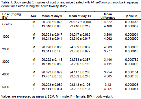
The body weights of the control and treated mice with aqueous extract of root barks of the plant are shown in Table 1. Results from this study revealed that there were a gradual increase in body weights of both control and treated groups of mice.
Hematological parameters
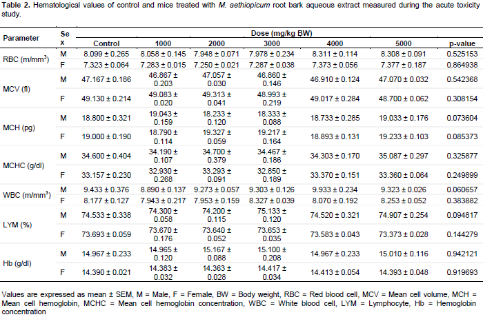
The results of hematological parameters of control and treated mice are shown in Table 2. These results show that there were no significant changes (p>0.05) in hematological parameters, both in the control and treated groups of mice after 14 days of treatment with M. aethiopicum root bark aqueous extract. All values of hematological parameters such as red blood cell, mean cell volume, mean cell hemoglobin, mean cell hemoglobin concentration, white blood cell, lymphocyte and hemoglobin concentration remained within normal limits throughout the experimental period.
Biochemical parameters
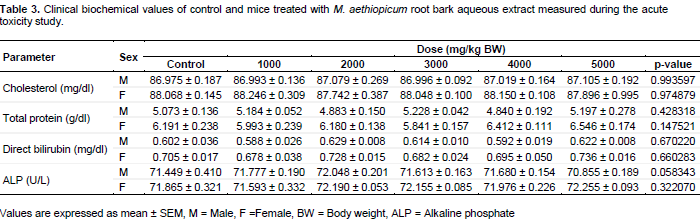
In this study, no significant changes (p>0.05) were observed at all doses in alkaline phosphate (ALP) and cholesterol levels between the control and treated group of mice as shown in Table 3. Furthermore, the values of protein and bilirubin did not differ significantly (p>0.05) in the treated mice as compared to the control group.
Macroscopic examination
The macroscopic examination of the internal organs of animals revealed no difference between the control and treated mice groups after administration even with higher dose of 5000 mg/kg body weight.
Organ weight
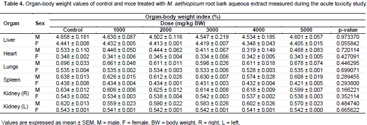
The organ weights relative to body weights of the animals were determined and results are summarized in Table 4. Findings from this study indicated that there were no significant differences (p>0.05) in weight changes of each organ between the control and treated mice at all doses.
DISCUSSION
Medicinal plants have been used worldwide for thousands of years in the form of crude drugs such as tinctures, teas, poultices, powders and other herbal formulations (Gurib, 2006). M. aethiopicum is among the medicinal plant which is known for many traditional applications in humans (Schmidt et al., 2002). The root bark extract has been commonly used by many populations in Africa for medicinal purposes (Burkil, 2004). Despite the usage of this plant as traditional medicine, there are few studies on the safety evaluations. Therefore, experimental screening methods using animal models are essential to ascertain the safety of this plant.
In this study, the M. aethiopicum root bark aqueous extract did not affect the body weight of the treatment mice as compared to the control mice. The gradual body weight gain shown by animals against the extract, provide circumstance evidence that the administration of the crude extract has negligibly level of toxicity on the growth of the animals. According to Raza et al. (2002) and Teo et al. (2002), the reduction in gain body weight is a sensitive indicator of toxicity after exposing the animals to the toxic substances and it is usually significant if such losses are more than 10% of the initial weight. Findings from this study are in agreement with previous study conducted by Ndukui et al. (2014) who reported weight gain in albino rats treated with aqueous leaf extract of M. aethiopicum in Uganda.
Blood parameters analysis is relevant to risk evaluation as the hematological system has a higher predictive value for toxicity in humans when assay involves animals (Olson et al., 2000). Blood is an important index of physiological and pathological status in both animals and humans and the parameters usually measured are red study has shown that acute oral ingestion of M. aethiopicum root bark aqueous extract did not lymphocyte and hemoglobin concentration (Vaghasiya et al., 2011). The normal range of these parameters can be altered by the intake of some toxic plants (Ajagbonna et al., 1999). This
study has shown that acute oral ingestion of M. aethiopicum root bark aqueous extract did not cause any change in hematological parameters for both control and treated mice verifying the nontoxic nature of the extract. Findings from this study validate the safety nature of the extract through oral administration. This explains why there is no reported case of intoxication due to the use of this plant among the Maasai communities in Tanzania (Kokwaro, 1993).
Disease or response to toxic substances is indicated by alterations in the biochemical parameters which are the sensitive indicators of organ function or metabolic defects (Reddy et al., 2013). Liver plays a major role in the metabolism and detoxification of compounds that reach the liver and hence it serves as a prime target organ for drugs and toxic substances (Reddy et al., 2013). A liver function test such as alkaline phosphate (ALP) is therefore useful in determining the extent of damage (Shah et al., 2011). In the same perspective, liver is the major site for cholesterol synthesis or disposal in mammals (Gautam and Goel, 2014). In this study, there were no significant changes in ALP and cholesterol levels for both control and treated mice, suggesting that M. aethiopicum root bark aqueous extract had no effects on the liver function and cholesterol metabolism of the mice and therefore strengthen the safety utilization of the plant in traditional medicine. Safety nature of the root bark extract of the plant is also indicated by other biochemical parameters such as protein and bilirubin which did not differ significantly as compared to control group of mice. These results support the reported use of M. aethiopicum by traditional healers in Uganda for treating various diseases with no severe adverse effect (Ndukui et al., 2014).
Macroscopic examination of internal organs of the experimental animals in this study did not reveal any abnormalities, presence of lesions or changes in the color for both control and treated group and therefore suggest that M. aethiopicum root bark aqueous extract is potentially safe for human consumption. In toxicological studies, internal organs such as liver, hearts, lungs, spleen and kidneys are primarily affected by metabolic reactions caused by the toxicants (Dybing et al., 2002).
Organ weight is an important index to diagnose whether the organ was exposed to the injury or not (Jothy et al., 2011). In this study, the weights of internal organs were not statistically significantly in both control and treated group of mice, indicating that the extract is virtually nontoxic. The non-toxicity shown by M. aethiopicum root bark aqueous extract towards albino mice, ratify the safety profile of the aqueous root bark extract of the plant. Results from this study collaborate with the previous cytotoxicity investigation study of the same plant growing in Uganda, which did not show significant changes in organ weight of albino rats (Ndukui et al., 2014).
CONCLUSION
This study presents evidence of nontoxic effects of M. aethiopicum root bark aqueous extract in animal models. No mortality or any sign of toxicity was observed in mice treated with the extract and therefore establishing its safety in use. The hematological and biochemical analysis showed no adverse effects between control and treated groups of mice. Furthermore, the plant extract did not induce any damage to the vital body organs and therefore considered as relatively safe for utilization especially in rural communities where conventional drugs are unaffordable due to their high costs. However, a detailed experimental analysis of sub-acute toxicity remains unveiled to complete the safety profile of this plant.
CONFLICT OF INTERESTS
The authors have not declared any conflict of interests.
ACKNOWLEDGEMENTS
The authors are grateful to the Government of Tanzania through the Commission for Science and Technology (COSTECH) for financial support. Herbalists from Imbibya village are appreciated for availing us ethnomedical information of medicinal plants. Last but not the least, the authors wish to thank Mr. Alfred Mwanyika of the Department of Physiology, Pharmacology and Toxicology at the Sokoine University of Agriculture for his technical support.
REFERENCES
|
Ajagbonna OP, Onifade KI, Suleiman U (1999). Hematological and biochemical changes in rats given extract of Calotropis procera. Sok. J. Vet. Sci. 1(1):36-42. |
|
|
Boera HJ, Koola A, Broberg A, William R, Mziray WR, Hedberg I, Levenfors JJ (2005). Anti-fungal and anti-bacterial activity of some herbal remedies from Tanzania. J. Ethnopharmacol. 96:461-469. |
|
|
Burkil HM (2004). The useful plants of west tropical Africa. Publisher Royal Botanic Gardens; Kew. pp. 25-28. |
|
|
Burrows JE, Willis CK (2005). Plants of the Nyika Plateau: an account of the Nyika National Parks of Malawi and Zambia. Southern African Botanical Diversity Network Report No. 31. SABONET, Pretoria. |
|
|
Curtis BA, Mannheimer CA (2005). Tree Atlas of Namibia National Botanic Research Institute. Windhoek. pp. 406-407. |
|
|
Dybing E, Doe J, Groten J, Kleiner JO, Brien J (2002). Hazard characterization of chemicals in food and diet: dose response, mechanism and extrapolation issues. J. Food Chem. Toxicol. 40(2):237-282. |
|
|
Ndukui JG, Mugisha K, Owiny D, Waako P (2014). Effects of crude aqueous leaf extracts of Citropsis articulata and Mystroxylon aethiopicum on sex hormone levels in male albino rats. Int. J. Pharm. Sci. Inven. 3(1):5-17. |
|
|
Gautam MK, Goel RK (2014). Toxicological study of Ocimum sanctum Linn Leaves: Hematological, biochemical and histopathological studies. J. Toxicol. 9:312-342. |
|
|
Gurib-Fakim A (2006). Medicinal plants: traditions of yesterday and drugs of tomorrow. Mol. Aspect. Med. 27(1):1-93. |
|
|
Iwu MM (2014). Handbook of African medicinal plants. Second edition: CRC Press, Taylor & Francis Group. pp. 28-29. |
|
|
Jothy LS, Zakaria Z, Chen Y, Ling Lau Y, Yoga Latha L, Sasidharan S (2011). Acute oral toxicity of methanolic seed extract of Cassia fistula in mice. Mol. 16(6):5268-5282. |
|
|
Kokwaro JO (1993). Medicinal plants of East Africa. Second edition: Published and printed by Kenya literature bureau, Nairobi Kenya. pp. 176-177. |
|
|
Newman DJ, Cragg GM, Snader KM (2000). The influence of natural products upon drug discovery. Nat. Prod. Rep. 17(3):215-234. |
|
|
Olowa LF, Nu-eza OM (2013). Brine shrimp lethality assay of the ethanolic extracts of three selected species of medicinal plants from Iligan City, Philippines. Int. Res. J. Biol. Sci. 2(11):74-77. |
|
|
Olson HG, Betton D, Robinson D, Thomas K, Monro A, Kolaja G, Lilly P, Sanders J, Sipes G, Bracken W, Dorato M, Van Deun K, Smith P, Berger B, Heller A (2000). Concordance of the toxicity of pharmaceuticals in humans and in animals. Regul. Toxicol. Pharm. 32(1):56-67. |
|
|
Organization for Economic Cooperation and Development (OECD) (2001). Guidelines for testing of chemicals. Health effects - Test No. 425: Acute oral toxicity: Up-and-Down procedure. |
|
|
Phani K, Kumar AR (2014). Toxicity studies of combined extracts of Vitex leucoxylon, Vitex negundo and Vitex trifolia. J. Chem. Pharm Sci. 7(1):54-58. |
|
|
Pope GV (1995). Flora Zambesiaca: Mozambique, Malawi, Zambia, Zimbabwe, Botswana. Flora Zambesiaca Management Committee. |
|
|
Raza M, Al-Shabanah OA, El-Hadiyah TM, Al-Majed AA (2002). Effect of prolonged vigabatrin treatment on hematological and biochemical parameters in plasma, liver and kidney of Swiss albino mice. J. Sci. Pharm. 70(2):135-145. |
|
|
Reddy RR, Lokanatha O, Ratnam K, Reddy CS, Raju IN, Reddy CD (2013). Acute and sub-acute toxicity of Moringa oleifera stem bark extract in Swiss albino mice. Int. J. Sci. Biotech. Pharm. Res. 2(4):74-82. |
|
|
Schmidt E, Lotter M, McCleland W (2002). Trees and shrubs of Mpumalanga and Kruger national park. Jacana Media. pp. 356-359. |
|
|
Shah R, Parmar S, Bhatt P, Chandra S (2011). Evaluation of hepatoprotective activity of ethyl acetate fraction of Tephrosia purpurea. Pharmacology 3:188-194. |
|
|
StatSoft Inc. STATISTICA (data analysis software system), version 8.0 (2007). |
|
|
Teo SD, Stirling S, Thomas A, Kiorpes A, Vikram K (2002). A 90-day oral gavage toxicity study of D-methylphenidate and D, L methylphenidate in Sprague-dawley rats. Toxicology 179(3):183-196. |
|
|
Vaghasiya YK, Shukla VJ, Chanda SV (2011). Acute oral toxicity study of Pluchea arguta boiss extract in mice. J. Pharmacol. Toxicol. 6(2)113-123. |
|
|
World Health Organization (WHO) (2007). Department of technical cooperation for essential drugs and traditional medicine. Guidelines for assessing quality of herbal medicines with reference to contaminants and residues, Geneva, Switzerland. |
|
Copyright © 2024 Author(s) retain the copyright of this article.
This article is published under the terms of the Creative Commons Attribution License 4.0


