In the body, the imbalance between oxidant/antioxidant in favor of the oxidation promotes the oxidative stress that causes proteins, lipids and DNA damages, as well as induces a variety of cellular responses through the generation of reactive oxygen species (ROS) that may be originated from cellular metabolism or environmental sources (Nita and Grzybowski, 2016). ROS have become a concern among researchers because they have been associated with a significant number of diseases involving inflammatory and painful processes that affect humans and animals. In inflammation, for example, ROS cause vascular damage which allows the permeability of macromolecules and inflammatory cells from the blood to tissue (Mittal et al., 2014). This permeability is controlled by vasoactive and chemotactic mediators, which make the inflammatory process active, including the pain (Silva, 2015). Among the mediators that regulate the events of inflammation, vasoactive amines, lipid-derived eicosanoids, cytokines, chemokines and adhesion molecules have been highlighted (Silva, 2015). On the other hand, the inflammatory pain has been treated with non-steroidal anti-inflammatory agents, but present high prevalence among adverse reactions to drugs (Derle et al., 2006). However, natural products, as flavonoids, have been investigated for their mechanisms against painful, inflammatory and oxidative processes (Agrawal, 2011; Iwalewa et al., 2007).
Palicourea is a plant genus in the family
Rubiaceae and contains about 200 species (Rosa et al., 2010). Plants of this genus, as
Palicourea rigida Kunth, are distributed in the Tropics of the New World, particularly in the South America’ Brazilian Cerrado region (Morel et al., 2011).
P. rigida, commonly known as “douradinha”, “bate-caixa” and “douradão” and the medicinal uses as antifungal, analgesic, diuretic, hypotensive, antiulcerogenic, cicatrizing and anti-inflammatory have been made by traditional communities (Rosa et al., 2010). From the chemical view-point, triterpenes, flavonoids, iridoids, alkaloids and peptides have been identified in this medicinal plant (Rosa et al., 2010; Morel et al., 2011; Pinto et al., 2012; Soares et al., 2012). Antioxidant (Rosa et al., 2010), antibacterial and insecticide (Pinto et al., 2012) and antiproliferative (Soares et al., 2012) activities have been related to these compounds.
Additionally, rutin is a flavone glycoside widely known and its hydrolysis produces quercetin and rutinose by the action of glucosidase (Chua, 2013). Both rutin and quercetin are found in medicinal plants, and anti-inflammatory (Chua, 2013; Choi et al., 2012), anti-tumor (Ren et al., 2003), anti-asthma (Jung et al., 2007), and antioxidant (Yang et al., 2008) activities are attributed to these compounds. Beside these data, quercetin 3-O-D-glucoside, quercetin 3-O-sophoroside and isorhamnetin 3-glucoside were identified in P. rigida (Rosa et al., 2010), which can justify the biological properties of this plant.
Considering that the scientific evidences of medicinal plants are fundamental for the therapeutic use, this article was described to investigate the antioxidant, antinociceptive and anti-inflammatory activities of EEPR using in vitro, in vivo and in silico tools. In addition, due to the chemical characterization and phenolic and flavonoid contents of EEPR, two markers (rutin and quercetin) were evaluated in order to establish possible bioactive compounds and mechanism of action.
Plant material and extraction
P. rigida leaves were collected in the city of São João del-Rei, Minas Gerais State, Southeast region of Brazil, in May 2010. The species was identified by Dr. Glauciemar Del-Vechio Vieira and registered in the Herbarium of the Department of Botany, Federal University of Juiz de Fora, Brazil, under number CESJ 42.677. After drying, 850 g of powdered leaves were subjected to extraction by static maceration in 95% ethanol (2.5 L) to obtain the ethanol extract (EEPR) through filtration. Extractive solution was evaporated (rotary evaporator, R-215 Büchi Labortechnik AG, Flawil, Switzerland) at 50 to 60°C. After removal of the water and solvent in a desiccator, the yield was of 66.68 g.
Chemicals
In this study, the following chemicals were used: Acetic acid, acetylsalicylic acid and aluminum chloride (Vetec Química Fina Ltda, Sigma-Aldrich Corporation, Rio de Janeiro, RJ, Brazil), formaldehyde (Quimibrás Indústria Química S/A, Rio de Janeiro, RJ, Brazil), Folin-Ciocalteu reagent, trichloroacetic acid, and ascorbic acid (Cromoline Química Fina, Diadema, SP, Brazil), potassium ferrocyanide, ferric chloride, methanol, ethanol, pyridine and sodium carbonate (Labsynth, Diadema, SP, Brazil), morphine hydrochloride (Merck Inc., Whitehouse Station, NJ, USA), naloxone and indomethacin (Sigma Chemical Co, St Louis, MO, USA) and DPPH, linoleic acid, β-carotene, tween® 40, galic acid, BHT, rutin, quercetin, kaempferol, luteolin, luteolin 7-O-β-D-glucoside, apigenin and apigenin 7-O-β-D-glucoside (Sigma-Aldrich Chemie, Buchs, SG, Switzerland).
Animals
In this experiment, Mus musculus L. (male Swiss albino mice, 50-70 days; 25-30 g) and Rattus norvegicus albinus (male Wistar rats, 90-110 days; 200-240 g) were supplied by the Central Biotery of the Federal University of Juiz de Fora (UFJF). Groups of animals were maintained in plastic cages (47 × 34 × 18 cm3) under a 12 h light/12 h dark cycle at room temperature (22 ± 2°C), with free access to rations (Nuvilab Rodents - Nuvital Nutrients, Colombo, Brazil) and water. The protocols (047/2012 and 049/2012) were approved by the ethical committee of UFJF, which are in accordance with the guidelines recommended by the Brazilian College of Animal Experimentation (COBEA).
Phytochemical screening
Preliminary phytochemical analysis of EEPR was determined by the following procedures (Tiwari et al., 2011).
Tannins
Gelatin test - To EEPR, 1% gelatin solution containing sodium chloride was added. Formation of white precipitate indicates the presence of tannins.
Flavonoids
Alkaline reagent test: EEPR was treated with few drops of sodium hydroxide solution. Formation of intense yellow colour, which becomes colourless on addition of dilute acid, indicates the presence of flavonoids.
Lead acetate test: EEPR was treated with few drops of lead acetate solution. Formation of yellow colour precipitate indicates the presence of flavonoids.
Diterpenes
Copper acetate test: EEPR was dissolved in water and treated with 3 to 4 drops of copper acetate solution. Formation of emerald green colour indicates the presence of diterpenes.
Phytosterols
Libermann Burchard’s test: EEPR was treated with chloroform and filtered. The filtrate was treated with few drops of acetic anhydride followed by boiled and cooled with addition of sulphuric acid. The positive reaction was observed through a brown ring at the junction.
Saponins
Foam test: 0.5 mg of EEPR with 2 ml of water was agitated in test tubes. Foaming for 10 min, it indicates positive reaction to saponins.
Coumarins
5 ml of EEPR was evaporated; the residue was dissolved in 1 to 2 ml of hot distilled water and the volume was divided into two parts. Half of the volume was taken as a witness and another volume of 0.5 ml 10% NH4OH was added. Two spots were placed on filter paper and examined under UV light. Intense fluorescence indicates the presence of coumarins.
Anthraquinones
Modified Borntrager’s test: EEPR was treated with ferric chloride solution and immersed in boiling water for around 5 min. The mixture was cooled and extracted with equal volume of benzene. The benzene layer was separated and treated with ammonia solution. Formation of rose-pink colour in the ammonical layer indicates the presence of anthranol glycosides.
Alkaloids
EEPR was dissolved in diluted hydrochloric acid and filtered.
Mayer’s test: Filtrated was treated with Mayer’s reagent (potassium mercuric iodide). Formation of a yellow coloured precipitate indicates the presence of alkaloids.
Dragendroff’s test: Filtrated was treated with Dragendroff’s reagent (potassium bismuth iodide solution). Formation of red precipitate indicates the presence of alkaloids.
Total phenolic determination
To quantify the total phenolic, spectrophotometric method was applied using the Folin-Ciocalteu reagent (Sousa et al., 2007). Concentrations of EEPR (400, 800 and 1200 µg/mL) were prepared for this determination. The calibration curve was established with gallic acid (200 to 760 µg/mL) and the obtained absorbances were submitted to linear regression analysis using the least squares method to acquire the equation of the line and the correlation coefficient (r). In this reaction, the Folin-Ciocalteu reagent is capable of oxidising phenolic constituents and the neutralization is done by sodium carbonate with generation of a blue staining. After 60 min of reaction, the absorbance was recorded at 765 nm in spectrophotometer (Shimadzu®, UV-1800, Tokyo, Japan). All analysis were carried out in triplicate and the average shown as gram of gallic acid equivalent (g/100 g).
Total flavonoids determination
Spectrophotometric method was performed for total flavonoid determination using rutin as standard (Sobrinho et al., 2008). For this quantification, concentrations of EEPR (400, 800 and 1200 µg/mL) were prepared. The calibration curve was elaborated with rutin (2 to 60 µg/mL) in AlCl3 (8% in ethanol) and the obtained absorbances were submitted to linear regression analysis using the least squares method to acquire the equation of the line and the correlation coefficient (r). In this procedure, aluminum chloride reacts with flavonoids of EEPR in the presence of acetic acid, pyridine:ethanol (2:8) and distilled water at room temperature for 30 min. After this time, the absorbances, in triplicate, were determined at 420 nm using a spectrophotometer (Shimadzu®, UV-1800, Tokyo, Japan). The results were demonstrated as gram of rutin equivalent (g/100 g).
High pressure liquid chromatography (HPLC) analysis
The methodology used in this analysis was described by Silva et al. (2013) previously validated by the Laboratory of Natural Products/Institute of Biological Sciences/UFJF. The HPLC system consisted of an Agilent Technologies 1200 Series with a PDA detector and an automatic injector. The column employed was a Zorbax SB-18; 250 × 4.6 mm, 5 μm particle size. Solvents that constituted the mobile phase were A (water pH adjusted to 4.0 with H3PO4) and B (acetonitrile). The elution conditions applied were: 0-30 min, 20% B isocratic. The mobile phase was returned to the original composition over the course of 30 min, and an additional 5 min were allowed for the column to re-equilibrate before injection of the next sample. The sample volume was 50 μl at a concentration of 1 mg/mL, the flow rate of 0.6 mL/min and the temperature was maintained at 25°C during the analysis. Detection was performed at 254 nm. Gallic acid, rutin, quercetin, kaempferol, luteolin, luteolin 7-O-β-D-glucoside, apigenin and apigenin 7-O-β-D-glucoside were also used as possible markers.
DPPH radical scavenging activity
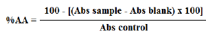
In this experiment, using DPPH method, the antioxidant activity was measured (Mensor et al., 2001). EEPR solutions (1 to 100 µg/mL) were prepared and mixed with methanol solution of DPPH (0.03 mM). Using a spectrophotometer (Shimadzu®, UV-1800, Tokyo, Japan), the absorbance values were measured at 518 nm after 60 min kept at 22 ± 2°C. The experiment was performed in triplicate. Rutin, quercetin and ascorbic acid were used as references. After obtaining the absorbances (Abs) of the samples, blank and control, the percentage of antioxidant activity (%AA) was determined using the following equation:

The 50% effective concentration (EC50) of EEPR was obtained by linear regression analysis using the least squares method to acquire the equation of the line and the correlation coefficient (r). Half maximum effective concentration (EC50) denotes the concentration (μg/mL) of EEPR required to reduce 50% of DPPH.
Antioxidant activity by reducing power
According to the method recommended by Oyaizu (1986), the antioxidant activity by reducing power was evaluated. EEPR solutions (750 to 100 μg/mL) reacted with 1% potassium ferrocyanide (in 0.2 mM phosphate buffer, pH 6.6) and kept at 50ºC for twenty minutes. After this time, 10% TCA (trichloroacetic acid) was mixed and centrifuged (3000 g over 10 minutes) to separate the supernatant. Distilled water containing 1% ferric chloride was mixed with the supernatant and the absorbance values, in triplicate, were recorded at 700 nm by spectrophotometry (Shimadzu®, UV-1800, Tokyo, Japan). Rutin, quercetin and ascorbic acid were used as references. EC50 was calculated from the graph of sample concentrations (X axis) and absorbances (Y axis) submitted to linear regression analysis using the least squares method to acquire the equation of the line and the correlation coefficient (r). The effective concentration (EC50) was determined in the absorbance of 0.5.
Antioxidant activity by lipid peroxidation method
Using the spectrophotometric method described by Miller (1971) with some modifications, the ð›½-carotene bleaching test was performed with 0.2 mg/mL ð›½-carotene (1 mL diluted in chloroform), linoleic acid (0.02 mL) and Tween 20 (0.2 mL). Then, the chloroform was evaporated (rotary evaporator, R-215 Büchi Labortechnik AG, Flawil, Switzerland), the mixture was previously oxygenated for 30 min with addition of distilled water to produce an emulsion. EEPR, rutin and quercetin (38.46 to 1.20 μg/mL) were placed in test tubes containing 5 mL of emulsion, which were inserted in water bath (50°C for 2 h). After this procedure, absorbances were determined spectrophotometrically (spectro-photometer Shimadzu®, UV-1800, Tokyo, Japan) in zero, 15, 30, 45, 60, 75, 90 and 105 min at 470 nm, in triplicate. BHT was used as standard. The percentage of inhibition of lipid peroxidation (%) was calculated.
Acute toxicity
To define the doses that were administered in male mice in the study of antinociceptive activity described below, the acute toxicity was evaluated using this animal gender. To perform this procedure, mice (n = 10) were orally (per oral route, p.o.) treated with doses of EEPR (0.5 to 3 g/kg) and saline (control group). The toxicity was also investigated by signs and symptoms and the number of death was totalized for 48 h. The probit test proposed by Litchfield and Wilcoxon (1949) was used to determine the LD50 (50% lethal dose). Based on the description of the pharmacological activity studies, the highest dose (400 mg/kg) is less than 500 mg/kg of acute toxicity, which may justify the doses chosen. Additionally, because rutin and quercetin are pharmaceutical and nutraceutical used by the population, their acute toxicity was not investigated.
Acetic acid-induced chemical nociception
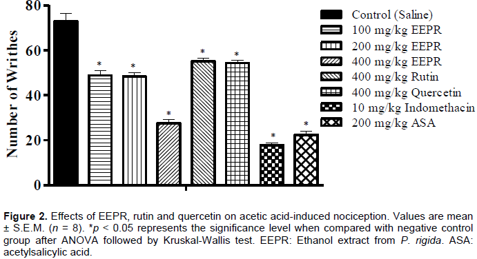
Considering the description on acute toxicity assay, animal groups (n = 8) were treated with EEPR (100 to 400 mg/kg, p.o.), rutin (400 mg/kg, p.o.), quercetin (400 mg/kg, p.o.), acetylsalicylic acid (200 mg/kg, p.o.), indomethacin (10 mg/kg, p.o.) and saline orally (10 mL/kg, p.o.) (Collier et al., 1968). One hour after treatment, 0.6% acetic acid (0.1 mL/10 g, i.p.) was applied intraperitoneally (i.p.). The abdominal writhes were measured between 10 and 30 min after application of acetic acid.
Formalin-induced chemical nociception
The experiment was conducted according to Hunskaar and Hole´s (1987) method using animal groups (n = 8). The treatment of the mice was performed with saline (10 mL/kg, p.o., negative control), EEPR (100 to 400 mg/kg, p.o.), rutin (400 mg/kg, p.o.), quercetin (400 mg/kg, p.o.), indomethacin (10 mg/kg, p.o., positive control) or morphine (5 mg/kg, s.c., positive control), one hour before formalin injection. After injection of 2.5% formalin (20 μL, in sterile saline) in the subplantar right hind paw region, the licking times of the neurogenic (0–5 min) and inflammatory phases (15-30 min) were evaluated.
Hot plate-induced thermal nociception
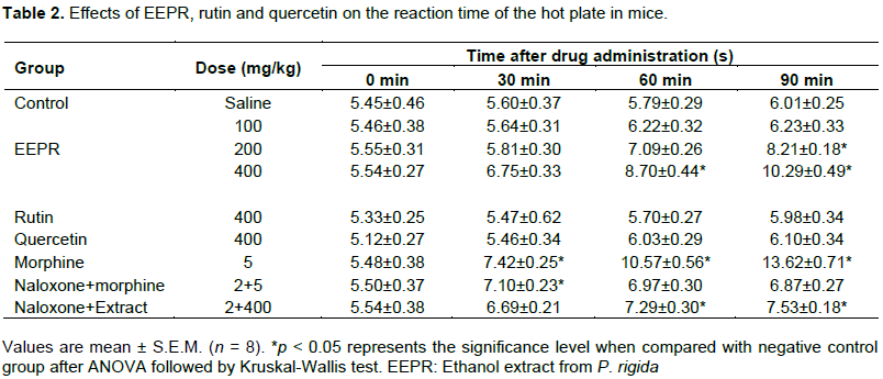
As recommended by Eddy and Leimbach (1953), mice (n = 8) were previously treated with EEPR (100 to 400 mg/kg, p.o.), rutin (400 mg/kg, p.o.), quercetin (400 mg/kg, p.o.), saline (10 mL/kg, p.o., negative control) and morphine (5 mg/kg, s.c., positive control). Then, the animals were placed on a hot plate (Model LE 7406, Letica Scientific Instruments, Barcelona, Spain) at 55 ± 1°C and the reaction time (s) was determined at 0, 30, 60 and 90 min. The maximum permanence time on the hot plate was 30 s. In separate groups, naloxone (2 mg/kg, s.c.), an opioid antagonist, was used in presence of EEPR (400 mg/kg) and morphine (5 mg/kg, s.c.) to investigate the central action mechanism.
Carrageenan-induced paw edema
Anti-inflammatory activity was evaluated by carrageenan-induced paw edema method in Wistar rats (Winter et al., 1962). In this assay, the animals were treated with EEPR (100 to 400 mg/kg, p.o.), rutin (400 mg/kg, p.o.), quercetin (400 mg/kg, p.o.), indomethacin (10 mg/kg, p.o., positive control) and saline (10 mL/kg, p.o., negative control). After 1 h of treatment, the rats were previously anesthetized with ketamine (60 mg/kg, i.p.) and xylazine (8 mg/kg, i.p.) solution and 1% carrageenan (0.1 mL) was administered into the right paw, while the left paw was injected with saline (0.1 mL). Using a plethysmometer (model LE 7500, Letica Scientific Instruments, Barcelona, Spain), the paw edema was measured at 1, 2, 3 and 4 h after carrageenan injection by differences between the paws (right and left).
Carrageenan-induced pleurisy
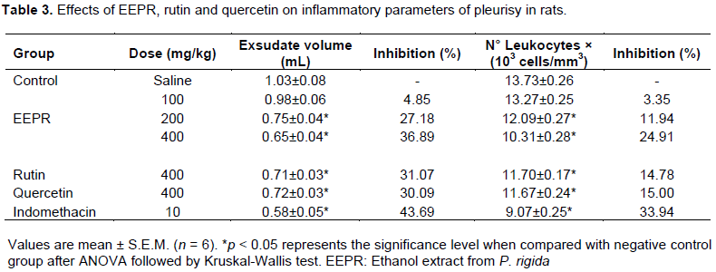
According to Vinegar et al. (1973) with minor modifications, rats (n= 6) were treated with EEPR (100 to 400 mg/kg, p.o.), rutin (400 mg/kg, p.o.), quercetin (400 mg/kg, p.o.), saline (10 mL/kg, p.o.) and indomethacin (10 mg/kg, p.o.). One hour after treatment, the animals were intraperitoneally anesthetized with ketamine and xylazine (60 and 8 mg/kg, respectively), and a suspension of saline containing 1% carrageenan (0.4 mL) was injected into the pleural cavity. After 4 h of pleurisy, overdose of anesthetic solution (120 mg/kg of ketamine and 16 mg/kg of xylazine 16 mg/kg, i.p.) was applied to induce the euthanasia of the animals. The pleural exudate was collected in the lung cavity to determine the volume and total leucocyte.
Evaluation of the molecular interaction profiles
The ligands (rutin, quercetin, acetylsalicylic acid and indomethacin) were generated in Marvin Sketch Program and refined by the semi-empirical method PM7 presents in the MOPAC2012 Program. The inflammatory enzymes were obtained from Protein Data Bank under the 1EQG (COX-1) and 5IKT (COX-2) codes. Subsequently, the Gasteiger’s loads were determined for all ligands and receptors through MGLTools Program. The molecular recognition was defined using the Discovery Studio v 4.5 2016 Program.
Statistical calculations
Mean and standard error of mean (S.E.M.) were obtained by statistical analysis. The level of significance (p < 0.05) was determined by one-way analysis of variance (ANOVA) followed by the Kruskal-Wallis or Tukey tests. For a better statistical determination, the Graph Pad® Prism 5.0. software was used.
Chemical determination and antioxidant activity
Using specific reagents, tannins, flavonoids, coumarins, terpenoids and steroids, saponins, anthraquinones and alkaloids were detected in EEPR.
EEPR revealed total phenolic and flavonoid contents and antioxidant activity (Table 1). In this extract, the total phenolic content was of 5.78±0.05 g/100 g, while the flavonoid reached 3.95±0.02 g/100 g. EEPR produced EC50 equal to 65.37±0.16 μg/mL (DPPH) and 697.36±2.48 μg/mL (reducing power). Using ð›½-carotene bleaching assay, the inhibition of lipid peroxidation of EEPR was 64.46%. As expected, rutin and quercetin showed antioxidant effect in these methods. In addition, quercetin was more active than rutin (a glycosylated flavonoid) in lipid peroxidation, since such assay is related to the oxidative stress in biological membranes.
High pressure liquid chromatography (HPLC) analysis
Considering the UV spectrum and the retention time of the main peaks, rutin (peak 13, tR = 30.72 min) was identified (Figure 1). However, rutin content was not quantified. The peak 12 represented a quercetin derivative. Although gallic acid, kaempferol, luteolin, luteolin 7-O-β-D-glucoside, apigenin and apigenin 7-O-β-D-glucoside had been reported in other species of Palicourea, using the conditions described above, these markers were not detected in EEPR. It is possible that flavonoids, together with other compounds, have a synergistic action that may justify the pharmacological effects of P. rigida.
Assessment of the acute toxicity
EEPR revealed no toxicity to treated animals, since produced LD50 up to 3000 mg/kg. After 48 h of treatment, the animals showed no signs or symptoms of toxicity. The LD50 value was important in defining the pharmacologic doses.
Effect on acetic acid-induced chemical nociception in mice
When compared to the control group, 100, 200 and 400 mg/kg of EEPR significantly (p < 0.05) reduced the writhes in 32.92, 33.62 and 62.26%, respectively. Rutin (24.36%) and quercetin (25.39%) also diminished the abdominal contortions (p < 0.05). Indomethacin (75.64%) and acetylsalicylic acid (69.22%) proved to be efficient as analgesic agents, since were able to inhibit the abdominal contortions (Figure 2).
Effect on formalin-induced chemical nociception in mice
The duration of paw licking for the control group was 74.25 ± 2.60 s (first phase) and 86.75 ± 2.40 s (second phase). EEPR (200 and 400 mg/kg) and morphine (5 mg/kg) significantly reduced the paw licking time in the nociceptive phase (Figure 3). In this phase, rutin and quercetin were not actives. The licking time was reduced after treatment with EEPR (100 to 400 mg/kg), rutin and quercetin in the second phase (p < 0.05). Even by different mechanism, indomethacin (cyclooxygenase inhibitor) and morphine (opioid agonist) were highly effective in decreasing the late phase.
Effects on hot plate-induced thermal nociception in mice
To evaluate the possible central effect indicated in the formalin test, the hot plate-induced thermal nociception method was used in this investigation. After one hour of treatment, EEPR (400 mg/kg) increased the reaction time of the thermal stimulation when compared to the control (p < 0.05). Doses of 200 (p < 0.05) and 400 mg/kg (p < 0.05) also increased the reaction time after 90 min (Table 2). In this experiment, rutin and quercetin produced no significant activity. As expected, morphine increased the reaction time and naloxone blocked this effect. Naloxone was not able to completely antagonize the effect of EEPR.
Effects on paw edema model
EEPR (400 mg/kg, 19.70%; p < 0.05) reduced the paw edema from 2 h after carrageenan application (Figure 4). This effect was observed after 3 h at the doses of 200 and 400 mg/kg of EEPR, as well as rutin and quercetin (p < 0.05). After 4 h carrageenan application, 200 and 400 mg/kg of EEPR, rutin and quercetin (p < 0.05) also showed significant effect. At this time, indomethacin reduced the paw edema by 25.92%.
Effects on pleurisy model
The results of the paw edema were confirmed by the pleurisy model (Table 3). In the Table 3, considering the control group, doses of 200 (27.18%) and 400 mg/kg (36.89%) of EEPR significantly (p < 0.05) reduced the exudate volume. These doses (200 and 400 mg/kg) also decreased the number of total leukocytes (p < 0.05) in 11.94 and 24.91%, respectively (Table 3). Rutin, quercetin and indomethacin (positive control) reduced the inflammatory parameters.
Molecular interaction profiles by docking
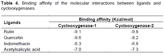
The molecular docking study showed that the ligands (rutin and quercetin) are capable of complexing with cyclooxygenase-1 (rutin = -9.1 Kcalmol-1; quercetin = -9.6 Kcalmol-1) and cyclooxygenase-2 (rutin = -9.8 Kcalmol-1; quercetin = -8.2 Kcalmol-1) more favorably than acetylsalicylic acid (-7.0 and -7.3 kcalmol-1 to COX-1 and COX-2, respectively) and indomethacin (-6.3 and -6.8 kcalmol-1 to COX-1 and COX-2, respectively) (Table 4). These data indicated that rutin and quercetin have higher affinity for the site of action of these enzymes than the reference compounds. In addition, van der Waals, dipole-dipole and hydrogen bonding interactions were recognized by the inflammatory enzymes (COX-1 and COX-2) (Figures 5 and 6).
Plants containing flavonoids have been associated with different medicinal uses such as diuretic, laxative, antispasmodic, anti-hypertensive, antinociceptive, and anti-inflammatory (Agrawal, 2011). The HPLC analysis of EEPR identified rutin, which has been reported as antioxidant, anti-inflammatory, antiasthmatic, and analgesic agents (Azevedo et al., 2013), and our results are in concordance with this description. In addition, flavonoids (quercetin 3-O-D-glucoside, quercetin 3-O-sophoroside and isorhamnetin 3-glucoside) were also isolated and identified in P. rigida (Rosa et al., 2010) and these compounds are well known for their ability to inhibit oxidative, painful and inflammatory mechanisms (Agrawal, 2011; Azevedo et al., 2013). Prostaglandins, for example, are related to generation of immunological mediators and are produced by the cyclooxygenase pathway, which can be inhibited by natural substances as flavonoids (Agrawal, 2011). Thus, based on our data, EEPR flavonoids, mainly rutin and quercetin tested in the current investigation, contributed for the antioxidant, antinociceptive and anti-inflammatory activities, since these compounds were effective in the applied tests.
Based on the literature, the flavonoid content has not been previously reported for P. rigida and the total phenolic content seems to be higher than that described by Rosa et al. (2010). Phenolic compounds and other natural products are known for their antioxidant action, since they are capable of donate electrons to free radicals. Using DPPH method, Rosa et al. (2001) showed an EC50 value eight times smaller than our result and associated the antioxidant effect with to the presence of phenolic compounds (total phenolic) in P. rigida that exhibit mechanism against this radical. In addition, the antioxidant effect of EEPR by the reducing power of iron has as mechanism to the donation of a hydrogen atom to break the free radical chain through the conversion of Fe3+ to Fe2+. (Cushnie and Lamb, 2005; Oyaizu, 1986). Considering the results showed in Table 1, EEPR transformed Fe+3 to Fe+2, demonstrating a reducing potential of P. rigida and confirmed the response observed by DPPH method.
On the other hand, the lipid peroxidation assay with the β-carotene/linoleic acid co-oxidation system is an in vitro test that reproduces physiological situations of oxidative stress, which can lead to cell death in extreme cases by compounds that act on lipid environment (Alam et al., 2013; Stutz et al., 2015). Our data revealed that EEPR had an antioxidant potential against lipid peroxidation by inhibiting oxidative process and was more effective than rutin and quercetin. The lipidic environment favors the action of compounds belonging to terpenoids and steroids that were detected in the phytochemical screening of EEPR.
The acute toxicity test on mice showed that the EEPR was no toxic for the animals. The signs and symptoms of toxicity were also not revealed demonstrating an important finding to the traditional use of P. rigida, since the population has used this plant for various medicinal purposes.
The application of acetic acid intraperitoneally produces a painful response characterized by writhe and body stretching. Acetic acid induces the opening of ion channels and transient receptor potential vanilloid 1 (TRPV1) in nociceptive afferent neurons (Ikeda et al., 2001; Julius and Basbaum, 2001). From the viewpoint of inflammatory metabolites, acetic acid promotes the release of TNF-α, interleukin 1β and interleukin 8, prostanoids and bradykinin (Ribeiro et al., 2000). Our results showed a dose-related antinociceptive activity of EEPR (Figure 2), which could be related to inhibition of peripheral mediators, such as prostaglandins. Moreover, this effect may be related to the synergism of different compounds, such as flavonoids, identified in P. rigida.
According to Figure 3, EEPR produced significant inhibition in the first and second phases. As clinical pain model, this assay is characterized by a local tissue injury distinguishing two phases of pain (neurogenic and inflammatory). The neurogenic phase (0-5 min after injection of formalin) causes a direct stimulation of nociceptors, while the inflammatory phase (15-30 min after injection of formalin) is due to the generation of inflammatory mediators (Hunskaar and Hole, 1987). Opioids, as morphine, are able to inhibit both phases, and anti-inflammatory agents (aspirin, indomethacin and dexamethasone) only block the second phase (Hunskaar and Hole, 1987; Le Bars et al., 2001). In this assay, substance P and bradykinin are generated in the neurogenic phase, while other chemical mediators, such as histamine, serotonin and prostaglandin, are involved in the nociceptive response of the second stage (Martins et al., 2006). Considering our results, EEPR inhibited both phases, but rutin and quercetin were active only in the second stage. In this sense, EEPR possess compounds that may exert a central action similar to morphine and peripheral action by the inhibition of inflammation mediators. In addition, rutin and quercetin showed peripheral effect only.
The antinociceptive effect mediated by central mechanisms can be evaluated by the hot plate model. In this model, the thermal stimulus actives TRPV1 and induces paw licking and jumping (Carter, 1991). Once activated, TRPV1 promotes Ca2+ influx, actives voltage-dependent Na+ channel, depolarizes nociceptive sensory fibers, and propagates the action potential (Julius and Basbaum, 2001). According to the presented data (Table 2), EEPR promoted a central effect that confirms the results of the neurogenic phase, and suggests at least a supraspinal modulatory effect. Furthermore, anti-nociceptive action induced by EEPR was not exclusively opioid-dependent system, because naloxone did not totally block this action (Table 2). In this experiment, rutin and quercetin did not contribute with the central effect.
Considering the formalin test, the anti-inflammatory activity of EEPR, rutin and quercetin was corroborated using the carrageenan-induced paw edema model. This model of acute inflammation is most commonly used to test compounds with anti-inflammatory potential, and involves different phases and inflammatory markers (Winter et al., 1962; Fereidonia et al., 2001). After carrageenan application, serotonin and histamine are mediators found in the first phase (1 to 2 h); kinins are present in the middle phase; the generation of prostaglandins occurs in the late phase (3 to 5 h) (Fereidonia et al., 2001). According to this report, our results showed that EEPR (400 mg/kg) was active on the first phase (2 h) indicating an inhibition of early mediators (as histamine and serotonin), while the effect of EEPR found in the second phase may be justified by a reduction of prostaglandins. Therefore, the present results indicate that EEPR, rutin, quercetin and indomethacin are able to protect the body against acute inflammation.
The anti-inflammatory effect was also confirmed using carrageenan-induced pleurisy model in rats. This model is able to quantify the pleural exudate and inflammatory cells related to inflammation. Anti-inflammatory agents, as indomethacin, reduce the volume of exudate and leukocytes migration between 3 and 6 h after induction of pleurisy (Vinegar et al., 1973; Mikami and Miyasaka, 1982). The tests performed with EEPR, rutin and quercetin in the pleurisy model showed that such products behave as inhibitors of leukocyte migration and pleural exudates formation when given orally.
To test the hypothesis of a possible action mechanism of quercetin derivatives, we evaluated the molecular interaction profile by docking tools. Our results showed that the ligands (rutin and quercetin) are capable of complexing with the inflammatory enzymes (cyclooxygenase-1 and cyclooxygenase-2) more efficient than reference substances (acetylsalicylic acid and indomethacin), since they produced lower free energy between the molecular interactions observed by affinity binding (Table 3 and Figures 5 and 6). These findings may corroborate the anti-inflammatory action of EEPR observed in the in vivo tests and they are in agreement with literature data (Pany et al., 2013).
In summary, the present study showed that EEPR possesses antioxidant, antinociceptive and anti-inflammatory effects that must involve peripheral and central components and could be associated with the synergism of substances found in P. rigida. In addition, rutin and quercetin derivative found in EEPR appear to contribute for these effects, possibly through at least an inhibitory action on signalling pathways of inflammation mediators. The results also corroborate the medicinal uses of P. rigida, but new scientific evidences are necessary for a better knowledge of their therapeutic applications.