Full Length Research Paper

ABSTRACT
A battery of short-term in vitro assays for apoptosis and/or cell cycle arrest, cytotoxicity, genotoxicity and antigenotoxicity are used to screen and establish the efficacy of medicinal plants. This study evaluated three concentrations (0.1, 0.2 or 0.4 mg/ml) of methanolic leaf extracts of Artemisia afra and Leucosidea sericea and their individual mixtures with Ethyl methanesulfonate (EMS) (0.15 mg/ml) for induction of those end points using the in vivo Allium cepa assay. Cytotoxicity was measured by the mitotic index, genotoxicity was expressed as the number of aberrant mitotic cells per 100 mitotic cells and modulatory effect (ME) was calculated as: ME = (B - C) - (A - C) / (A - C) and the value, positive or negative, indicated the number of units of the mutagen-induced genotoxicity (A) that equaled the mixture-induced genotoxicity (B). The three concentrations of A. afra extract tested did not induce cell cycle arrest and were not cytotoxic. The 0.4 mg/ml concentration and its mixture with EMS were genotoxic. The concentrations of L. sericea extract tested did not induce cell cycle arrest, were not cytotoxic nor genotoxic to the A. cepa root tip cells. The mixture of either 0.2 or 0.4 mg/ml L. sericea extract with EMS was genotoxic. The mixture of 0.4 mg/ml L. sericea extract with EMS was significantly more (ME = 4.40>2) genotoxic than EMS alone. Leaf extracts of A. afra and L. sericea lacked cell-cycle arrest activity, were non-toxic but lacked antigenotoxic activity against EMS-induced genotoxicity. High concentrations of A. afra were genotoxic whereas high concentrations of L. sericea interacted synergistically with EMS. Chromosomal abnormalities observed included sticky chromosomes, c-mitosis, chromosome largards, chromosome fragments, anaphase and telophase bridges.
Key words: Anti-genotoxicity, cell cycle arrest, safety of medicinal plants of Lesotho.
INTRODUCTION
Different human civilizations have depended for many centuries on plants and plant products for their medicinal (Balandrin et al., 1985) needs. The scientific basis for the use of plants in traditional medicine, in many ancient communities has been attributed largely to secondary metabolites which have been shown to possess various biological activities (Bourgaud et al., 2001). Advances in analytical techniques such as chromatography allowed the recovery of more and more of these molecules, which eventually formed the basis of the discipline of phytochemistry (Hussein and El-Anssary, 2018). Some of the phytochemicals to which the protective and therapeutic benefits of plants have been attributed include alkaloids, terpenoids, tannins and phenolics among others (Hertog et al., 1993; Harborne, 1998; Zhang et al., 2001). Herbal traditional medicines are therefore, in most cases, concoctions of crude extracts of plant materials in water, alcohol or distillates or essential oils, which contain many secondary metabolites (SMs) from various structural groups; so that the therapeutic activity of the concoction is often due to synergistic interactions of the SMs present (Mulyaningsih et al., 2010; Eid et al., 2012). The synergistic interactions of the SMs, such phenolic compounds and polysaccharides, present in the concoctions have also been credited with the apparent broad-spectrum activity of the concoctions used in traditional medicine (Wink, 2015). At high concentrations, SMs change membrane fluidity and increase permeability. Therefore, many lipophilic SMs (especially those in essential oils) exhibit antimicrobial and cytotoxic activities (van Wyk and Wink, 2015).
In Lesotho, as in many other countries in the world, a system of traditional medicine based on the use of plants, birds, animals, their products and their combinations to treat a broad spectrum of communicable and noncommunicable diseases is still being practiced, especially in areas with limited access to clinics or other health facilities (Shale et al., 1999; Padmanabhan and Sujana, 2008). Lesotho has approximately 2000 recorded species of flowering plants which have been used for medicinal purposes for many generations and the knowledge of such plants has been carried from generation to generation through oral tradition (Moteetee and Van Wyk, 2011). Two examples of such plants used for traditional medicinal purposes in Lesotho are Artemisia afra (Asteraceae) and Leucosidea sericea (Rosaceae). A. afra Jacq. Ex. Willd‘, also called “African wormwood”, is a well-known medicinal plant in South Africa, where it is known as “wilde als.” (van Wyk, 2008; Abad et al., 2012) and in Lesotho as “Lengana” in Sesotho. The roots, stems, and leaves of A. afra are used to treat numerous ailments including colds, coughs, diabetes, heartburn, bronchitis, asthma acne, boils, headaches, stomach disorders, sprains and rheumatic swellings. It is widely used for numerous ailments including colds, coughs, diabetes, heartburn, bronchitis, and asthma (van Wyk, 2008; Abad et al., 2012). The various uses (broad-spectrum effects) reveal that it possesses antiviral, antibacterial, and anti-inflammatory activities (Thring and Wertz, 2006; Liu et al., 2009; Moteetee and Van Wyk, 2011). The leaf extracts of the plant species L. sericea (known as “Cheche” in Sesotho) are used by indigenous peoples of Southern Africa as a vermifuge, astringent and anti-inflammatory agent and a paste made from the crushed leaves is used to treat ophthalmia (Van Wyk and Van Wyk, 1997; Nair et al., 2012).
The therapeutic benefits of plants notwithstanding, investigations have revealed that many plants which are used in traditional medicine have mutagenic, cytotoxic and genotoxic effects in in vitro and in vivo assays (Higashimoto et al., 1993; Schimmer et al., 1994; Kassie et al., 1996; Fennell et al., 2004; Çelik and Aslantürk, 2007; Kuete, 2014) and contain mutagenic and/or carcinogenic substances (Ames, 1986; de Sá Ferreira and Ferrão Vargas, 1999) and their use has been correlated with high rate of tumor formation in some human populations (Wynder et al., 1983; Ames, 1986; Nagao et al., 1986; Nguyen et al., 1989; Brito et al., 1990).
Medicinal herbs have also been shown to cause adverse effects or have the potential to interact with other medications (Zink and Chaffin, 1998; Markowitz et al., 2003), modify the mutagenic and carcinogenic effects of environmental contaminants and induce both mutagenic and antimutagenic effects on known mutagens in different test systems (Debisri et al., 1996; Gimmler-Luz et al., 1999). These observations raise concern about the potential mutagenic or genotoxic hazards that could result from the long-term use of such plants as food or medicine, especially because not enough information is available on the potential risk to health of many of these plants (Basaran et al., 1996).
The interest in plant-based pharmaceuticals intensified due to the development of methods of screening for anticarcinogenic drugs determined by their cytotoxic effects represented by inhibitory concentration (IC50) and Selectivity index (SI), using a panel of different independent cancer cell-lines as well as normal cell-lines (Hartwell, 1971, Cassady and Douros, 1980; Mbaveng et al., 2017) and through a battery of in vitro and in vivo short-term genotoxicity tests, and long-term rodent carcinogenicity assays (Moreira et al., 2014). The development resulted in the extension of the survey to include plant extracts and plant products able to modify the process of mutagenesis, because of the observation of a correlation between carcinogenesis and mutagenesis (McCann et al., 1975; Debisri et al., 1996) and medicinal plants usage gradually abandoned the anecdotal framework and became founded on empirical and explicatory facts (Petrovska, 2012). Whereas many in vivo tests on aqueous extracts largely support the safety of herbal medicines, most in vitro tests on isolated single cells mostly with extracts other than aqueous ones however, show contrary results and thus continue the debate on herbal medicine safety (Mensah et al., 2019).
The relationship between apoptosis and cell cycle arrest has been a recent focus and many chemotherapeutic agents exert their anticancer effects by inducing apoptosis and/or cell cycle arrest in cancer cells (Schuchmann and Galle, 2004; Belayachi et al., 2017). In in vitro studies, plant extracts can be evaluated for cell cycle arrest potential where, cancer cell lines are treated with various concentrations of plant extracts for 24 h, stained with propidium iodide (PI), and subsequently analyzed by flow cytometry to detect and quantify the amount of cells undergoing apoptosis as measured by the level of DNA fragmentation as well as the increase in the percentages of cells in the sub G1 phase compared to the untreated or solvent-treated cells (Pumiputavon et al., 2017). A decrease in the proportion of dividing cells in anaphase (A) and telophase (T) is an indication of metaphase arrest due to the poisoning of the spindle fibers, akin to the action of the well documented spindle poison, colcemid (Parry et al., 1999). The chemotherapeutic agents taxol, vincristine, vinblastine and nocodozole act in a similar manner (Alberts et al., 2008) to colcemid. These compounds act by binding to and stabilizing microtubules, inhibiting their dynamic instability and cuasing various genetic disruption, including the induction of cell cycle arrest (Alberts et al., 2008; Zhang et al., 2015). Drugs suppress microtubule dynamics by binding to different sites of tubulin heterodimer, disturb the assembly of the mitotic spindle apparatus and arrest cell cycle progression through M-phase, leading to eventual cell apoptosis (Chen et al., 2010). Therefore, cell cycle arrest is one of the targets for many anticancer drugs. Among them, taxanes, colchicines and vinca alkaloids are well-known examples that induce G2/M phase arrest leading to subsequent apoptosis (Kim et al., 2011). The anti-cancer activity of plant extracts and isolated compounds is often evaluated by assessing their ability to induce cell cycle arrest and apoptosis (Pumiputavon et al., 2017; Abu-Darwish and Efferth, 2018). Certain types of alkaloids, widely distributed in plants such as jerantinine B (Qazzaz, 2016), liriodenine (Nordin et al., 2015), and vinoreline (Rabbani-Chadegani et al., 2015), exhibit the ability to induce apoptosis and block the cell cycle in the G1 phase (Pumiputavon et al., 2017).
Screening medicinal plants for biological activity to harness their therapeutic potential involves cytotoxicity studies as a first step to evaluate the toxicity of plant extracts or plant-derived compounds and to establish their safety or lack thereof (McGaw et al., 2014; Mafole et al., 2017). Minimal to no toxicity is essential for the successful development of a pharmaceutical or cosmetic preparation and in this regard, cellular toxicity studies play a crucial role (McGaw et al., 2014). The toxicity of plant extracts has been attributed to the presence of cytoxic secondary plant metabolites in the solvent extracts (Asche, 2005; Hussein and El-Anssary, 2018; Priyanka et al., 2019). Since the observation of a high correlation between carcinogenicity and mutagenicity: 90% (156/ 174) of carcinogens are mutagenic (McCann et al., 1975) and the indication of epidemiological studies that many cancers are dependent on multiple mutational etiology, as well as on inherited mutator phenotype (Verschaeve and Van Staden, 2008), carcinogenic potential of environmental chemicals and plant extracts is assessed through a battery of in vitro and in vivo short-term genotoxicity tests, and long-term rodent carcinogenicity assays during research and development (R&D) of conventional drugs and to screen and establish the efficacy of medicinal plants (Moreira et al., 2014; Atanasov et al., 2015). Researchers often use a battery of in vitro assays to screen and establish the efficacy of medicinal plants (Atanasov et al., 2015). The advantage of this approach is that it takes into consideration the often complex and interwoven nature of the disease state (Houghton et al., 2005). However, Rank and Nielsen (1994) found an 82% correlation between the Allium cepa test and the mouse carcinogenicity test, concluding that A. cepa was even more sensitive than the Ames test. Teixeira et al. (2003) found the same results in tests comparing meristematic cells of onion roots, bone marrow cells of rats, and human lymphocytes as bioindicators, validating the safety of A. cepa for cytogenetic studies.
Antimutagen is described as an agent that reduces the apparent yield of spontaneous and or induced mutations in organism genome (Bhattacharya, 2011). Seaweeds were found to contain naturally occurring antimutagens and antioxidants compounds which demonstrated great protective effect against oxidative damage caused by free radicals (Arumugama et al., 2017). Therefore the Antimutagenic properties of plant extracts are considered of particular interest as it may be assumed that these antimutagenic natural substances are able to lower the risk of occurrences of mutation and cancer risk from everyday exposures to environmental mutagens as well as to mutagenic pharmaceuticals (Verschaeve and Van Staden, 2008; Nagarathna et al., 2013) The mechanism by which plant extracts exhibit antigenotoxic effects is either by inducing or inhibiting enzyme such as glutathione- stransferase or CYP1A1 respectively as well as antioxidant and scavenging properties of polyphenolics contained in it. The most important mechanism in antimutagenesis and anticarcinogenesis is the scavenging of bio active molecule (Mersch-Sundermann et al., 2006)
During research and development (R&D) of conventional drugs, carcinogenic potential is assessed through a battery of in vitro and in vivo short-term genotoxicity tests, and long-term rodent carcinogenicity assays (Moreira et al., 2014).
Studies on agents that modulate carcinogen-induced genotoxic effects in experimental animals provide end points that can be used for assessing the antimutagenic or anticarcinogenic properties of putative chemopreventive compounds and for predicting their protective efficacy in humans (Khaidakov et al., 2001).
The A. cepa L assay is an in vivo assay and one of the established plant bioassays, validated by the international programme on chemical safety (IPCS, WHO), as an efficient and standard test for chemicals screening, in situ monitoring of the genotoxicity of environmental substances (Leme and Marin-Morales, 2009) and to evaluate the genotoxic potential of medicinal plants (Camparoto et al., 2002; Knoll et al., 2006; Fachinetto et al., 2007; Lubini et al., 2008; Fachinetto et al., 2009). The A. cepa L assay is also a short-term in vivo genotoxicity test that tests genotoxicity using chromosomes and therefore detects chromosome structural and numerical alterations (Tedesco and Laughinghouse, 2012; Bonciu et al., 2018). The assay is relatively inexpensive, fast, give reliable results and chemicals which cause chromosomal aberration (CA) in plant cells also produce CA in cultured animal cells that are frequently identical (Grant, 1978; Ma et al., 1994). Ethyl methanesulphonate (EMS) is a direct-acting mutagen, tetratogen, and brain carcinogen (Stubbs et al., 1997).
In view of the foregoing therefore, the aim of this study was to evaluate the methanol extracts of the leaves of A. afra and L. sericea for cytotoxicity, genotoxicity and the modulation of EMS- induced genotoxicity using the A. cepa chromosome aberration assay system.
MATERIALS AND METHODS
Test organism
Onion (A. cepa) seeds of the variety, Texas Grano 502 P.R.R., a product of Sakata seeds, Lanseria 1748, Republic of South Africa were purchased from Maseru Garden Centre, Lesotho, Southern Africa.
Mutagens and chemicals
EMS was a product of Fluka (Biochemika, Germany). Methanol (absolute) was a product of Associated Chemical Enterprises (Pty) Ltd (Johannesburg, South Africa); hydrochloric acid glacial and acetic acid were products of UNILAB (Krugerdorp, South Africa); acetocarmine stain was obtained from Carolina Biological Supply Company, Burlington, North Carolina, USA.
Plant material collection and crude extract preparation
The aerial parts of L. sericea and A. afra were collected from the outskirts of the National University of Lesotho, with the following geographical coordinates: Latitude: 29° 22? 49? S, Longitude: 27° 33? 13? E. with altitude of 1600 m. The two plant species were authenticated by the curator of the Herbarium at the Department of Biology of the university, where the voucher specimens of these plants have been deposited. The leaves were dried in a fanned Labcon oven at 37°C to a constant weight and brittle, about 48 h. Thereafter, the dried leaves were ground to fine powder using a pulveriser (Kenwood) and the powder was stored in sealed amber bottles in the refrigerator at 4°C until they were used. Extraction was carried out according to the method of Magama et al. (2013). Briefly, the ground leaf material was soaked in 95% methanol (v/v in distilled water) and the mixture was shaken on the orbital shaker for 24 h and filtered through Whatman No.1 filter paper using suction. The filtrates obtained were then concentrated under vacuum in a rotary evaporator and oven dried at 35°C for 12 h to a constant weight and were stored at 4°C in the refrigerator until they were used.
Genotoxicity experiments
The preliminary assay to select concentrations of EMS and leaf extracts to use and Genotoxicity assay (including the treatment of A. cepa seedlings with test agents, root harvest, slide preparation and scoring of slides) were conducted according to the methods of Asita et al. (2017). From the results of the preliminary assays to select the concentrations of EMS and plant extracts to use, the following concentrations of leaf extracts (in mg/ml) of A. afra (0.1, 0.2 and 0.4) and L. sericea, (0.1, 0.2 and 0.4) and EMS (0.15) were assessed for cytotoxicity that is, mitotic index (MI), genotoxicity (GT) and the modulatory effect (ME) of plant extracts on EMS-induced genotoxicity.
The aberrations observed and scored were: sticky chromosomes (S), C-metaphase (C-Mit), lagging chromosomes (L), chromosome bridges at anaphase and telophase (A.B) and chromosome fragment (F). For calculating the GT, only aberrant mitotic cells were considered.
In each assay, three root tips (triplicate) were assessed at each concentration. On each of three slides (n = 3) per treatment, a total of 2000 cells, classified into interphase or dividing cell, that is, prophase (normal, N or aberrant, ABN), metaphase (N or ABN), anaphase (N or ABN) or telophase (N or ABN) were scored; that is, a total of 6000 cells each for the control and treatment groups. The aberrations assessed were sticky chromosomes (S), C-metaphase (C-Mit), lagging chromosomes (L), chromosome bridges at anaphase and telophase (A.B) and chromosome fragment (F). For calculating the GT, only aberrant mitotic cells were considered.
Analysis of slide preparations
Cytotoxicity, as determined by the mitotic index (MI) was expressed as the number of dividing cells per 100 cells scored according to the formula:
MI = Number of dividing cells/Total number of cells scored x 100. (1)
The MI was used as a measure of cytotoxicity (CT). The MI of each treatment group was compared with that of the negative control group using t-test at a probability level of 0.05, using the SPSS for windows, version 11.0 software.
Genotoxicity (GT) was expressed as the number of aberrant mitotic cells (AMC) per 100 mitotic cells [i.e AMC + normal mitotic cells (NMC)] scored according to the formula:
Frequency of GT = AMC/ (AMC + NMC) x 100 (2)
The mean GT of each group of three slides per concentration of test agent was compared with that of the negative control group using t-test. P values less than 0.05 (P < 0.05) were considered as indicative of significance. The modulatory effect (ME) of plant extract on EMS-induced genotoxicity (GT) was calculated using the formula of Asita et al. (2017):
ME = (B – C) – (A – C) / (A – C) (3)
Where ‘A’ is the genotoxicity induced by the mutagen (CP or EMS) alone that is, mutagen-induced genotoxicity; ‘B’ is the genotoxicity induced by mixture of plant extract and mutagen, that is, mixture-induced genotoxicity and ‘C’ is the genotoxicity induced by negative control, such as tap water alone.
The modulatory effect (ME) was thus obtained by comparing the mutagen-induced genotoxicity (A) with the mixture-induced genotoxicity (B). The ME value indicated the number of units of the mutagen-induced genotoxicity (A) that equaled the mixture-induced genotoxicity (B). ME was significant only if ME was ≥ 2, that is, mixture was at least twice (200%) more (+) or less (-) genotoxic than mutagen alone.
A positive ME (+ME) indicated that the mixture was more genotoxic (increased GT) than the mutagen and if mixture is also more genotoxic than the genotoxic plant extract alone then a synergistic interaction is inferred. But mutagen-potentiation is inferred if mixture is less genotoxic than the non-genotoxic plant extract alone.
A negative ME (-ME) indicated that the mixture was less genotoxic (reduced GT) than the mutagen alone. If mixture is less genotoxic than the mutagen and the genotoxic plant extract then antagonism is infered. However, if mixture is less genotoxic than mutagen and also, more or less genotoxic than the non-genotoxic plant extract then it is antimutagenicity.
Data analysis
The mean value of each group of three slides per concentration of test agent was compared with that of the negative (solvent) control group using student’s t-test. P-values less than 0.05 (P < 0.05) were considered as significant.
RESULTS
RESULTS
Cytotoxicity and genotoxicity figures and tables
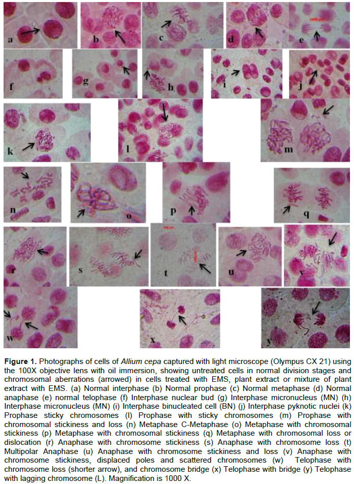
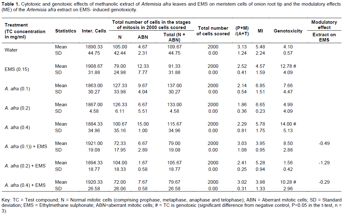
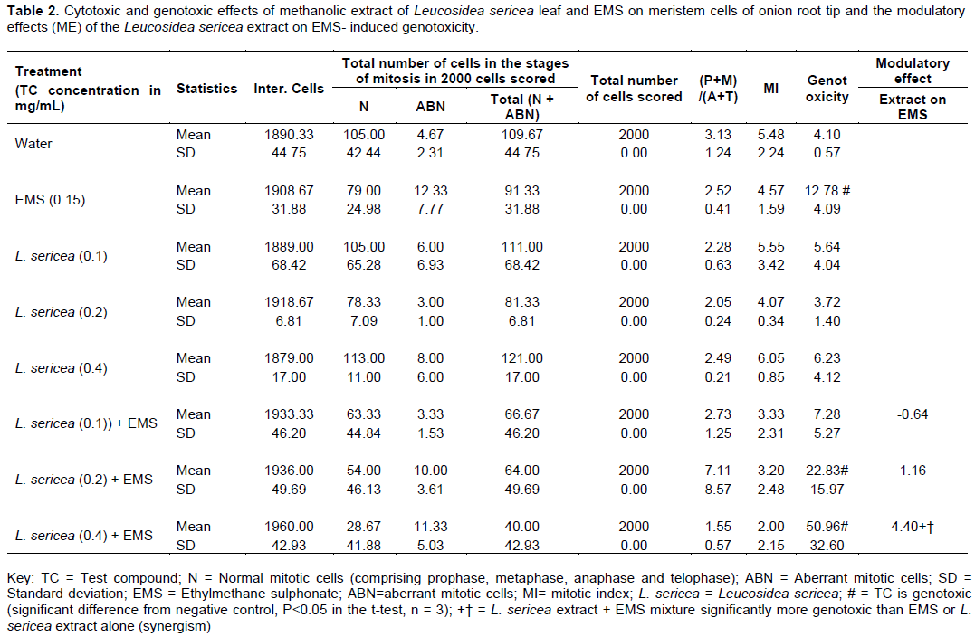
Photographs of the most representative pictures of normal mitotic cells and cells containing the different types of chromosome aberrations that were observed and scored are presented in Figure 1. The results of the cytotoxicity and genotoxicity experiments with methanol extract of leaves of A. afra and L. sericea are presented in Tables 1 and 2 respectively.
Results of experiments with A. afra leaf extract
(P+M)/ (A+T) Ratio
Examination of the (P+M)/(A+T) ratio in column 8 of Table 1 shows that the treatment with EMS (0.15 mg/ml), the different concentrations of A. afra leaf extract and the mixtures of AA extract with EMS did not induced a significant change in the (P+M)/(A+T) ratio, when compared with water treatment negative control group (P < 0.05).
Cytotoxicity
Examination of the MI in column 9 of Table 1 shows that the concentration of EMS (0.15 mg/ml) used was not toxic, none of the three concentrations of A. afra leaf extract (0.1, 0.2 or 0.4 mg/ml) tested was toxic and the mixtures of A. afra leaf extract with EMS were also not toxic to the A. cepa root meristem cells, when compared to the water treated negative control (P<0.05).
Genotoxicity (GT)
Examination of induction of genotoxicity in column 10 of Table 1 shows that only EMS, highest concentration (0.400 mg/ml) of A. afra leaf extract and the mixture of EMS and the 0.40 mg/ml A. afra leaf extract was genotoxic when compared to the water treatment negative control (P<0.05).
Modulatory effect (ME) of A. afra leaf extract on EMS-induced Genotoxicity (GT)
Examination of the modulatory effect (ME) in column 11 of Table 1 shows that the mixture of each of the three concentrations of A. afra leaf extract (0.1, 0.2 and 0.4 mg/ml) with EMS was insignificantly (< twofold or 200%) less genotoxic than EMS alone.
Results of experiments with Leucosidea sericea leaf extract
(P+M)/ (A+T) Ratio
Examination of the (P+M)/(A+T) ratio in column 8 of Table 2 shows that the treatment with EMS (0.15 mg/ml), the different concentrations of L. sericea leaf extract and the mixtures L. sericea leaf extract with EMS did not induced a significant change in the (P+M)/(A+T) ratio, when compared with water treatment negative control group (P < 0.05).
Cytotoxicity
Examination of the MI in column 9 of Table 2 shows that the concentration of EMS (0.15 mg/ml) used was not toxic, non of the three concentrations of L. sericea leaf extract (0.1, 0.2 or 0.4 mg/ml) tested was toxic and the mixtures of L. sericea leaf extract with EMS were also not toxic to the A. cepa root meristem cells, when compared to the water treated negative control (P<0.05).
Genotoxicity (GT)
Examination of induction of genotoxicity in column 10 of Table 2 shows that only three treatments, EMS, and the mixture of EMS with each of the two highest concentrations (0.2 and 0.4 mg/ml) of L. sericea leaf extract, were genotoxic when compared to the water treatment negative control (P<0.05).
Modulatory effect (ME) of leaf extract of L. sericea on EMS-induced genotoxicity (GT)
Examination of the modulatory effect (ME) in column 11 of Table 2 shows that only the mixture of the top concentration (0.4 mg/ml) of L. sericea leaf extract with EMS was significantly (> twofold or 200%) more genotoxic than EMS or L. sericea leaf extract alone (synergism), with a positive ME value of 4.40. This observation suggested a synergistic interaction between L. sericea leaf extract and EMS at high concentration of L. sericea leaf extract.
DISCUSSION
In this study, methanolic extracts of the leaves of A. afra and L. sericea used in traditional medicine in Lesotho to treat various diseases were evaluated for cytotoxicity, genotoxicity and modulatory effects on, the direct acting mutagen, EMS-induced genotoxicity using the in vivo onion (A. cepa L.) root tip meristem chromosome aberration assay system. The results of genotoxicity tests are presented in Tables 1 and 2.
As shown in Tables 1 and 2, the concentration of EMS (0.15 mg/ml) used in the present study did not reduce the mitotic index (MI) of meristem cells of the treated roots of A. cepa compared with the negative control and was adjudged not cytotoxic. The concentration of EMS used however induced genotoxicity in the root meristem cells of A. cepa. In a study by Çelik and Aslantürk (2010), EMS at a concentration of 2×10⁻² M (0.2484 mg/ml) was both toxic and mutagenic to root meristem cells of A. cepa.
The results of the assessments of the cytotoxic and genotoxic effects of the methanolic extracts of A. afra leaves are presented in Table 1. The concentrations tested did not induce any changes in the (P+M)/(A+T) ratio. In the present in vivo study, no such observations were made and it can be concluded that methanolic extract of A. afra leaves, at the concentrations (0.1, 0.2 or 0.4 mg/ml) used, did not induce or demonstrate any cell cycle arrest capabilities. However, in an in vitro experiment, ethanol extract of A. afra was cytotoxic against U937 and HeLa cancer cells via the decrease in mitochondrial membrane potential after G2/M arrest (Spies et al., 2013; Venables et al., 2016). Some Artemisia species and their active components that have been shown to arrest cells in in vitro experiments with cell lines include, A. sphaerocephala (Wang et al., 2017), A. princeps, cell cycle arrest at G2/M (Lee et al., 2013), A. monosperma, cell cycle arrest at S+G2/Mphase (Whelan and Ryan, 2004) and A. annua (Mizushina et al., 2010).
All three concentrations (0.1, 0.2 or 0.4 mg/ml) of the methanol extract of A. afra leaves and their individual mixtures with EMS (Table 1) tested in the present in vivo study were not cytotoxic to the A. cepa root meristem cells as determined by the reduction of mitotic index (MI) compared to the negative control treatment. But the methanol extract of another Artemisia species, A. annua showed cytotoxic effect on the root meristem cells of Allium cepa at >450 mg/ml (Karaismailoglu, 2014). The concentrations tested in that experiment were however, much higher than the ones tested here and the solvent for extraction was also methanol. In an in vitro experiment with U937 and HeLa cells, ethanol extract of A. afra was cytotoxic (Spies et al., 2013; Venables et al., 2016). Some Artemisia species and their active components that were cytotoxic in in vitro experiments with cell lines include, A. sphaerocephala (Wang et al., 2017), A. princeps (Lee et al., 2013) and A. monosperma (Whelan and Ryan, 2004).
Only the highest concentration (0.4 mg/ml) of the methanol extract of Artemisia afra leaves and its mixture with EMS (Table 1) were genotoxic. Dichloromethane and methanol extracts of A. afra leaves were not mutagenic in the in vitro Ames test with and without metabolic activation, using strains TA98 and TA100 (Elgorashi et al., 2003; Aremu et al., 2013). Dichloromethane and methanol extracts of leaves of A. afra were not toxic nor mutagenic when tested for genotoxic activity in human peripheral blood lymphocytes using the micronucleus test and the Ames test with TA98 and TA100 (Taylor et al., 2003; Verschaevea and Van Staden, 2008). However, some Artemisia species and their active components that have been shown to induce DNA damage and/or genotoxic effects in in vitro experiments with mammalian cell lines include, A. turanica (Tayarani-Najaran et al., 2013), A. indica (Zeng et al., 2015), A. capillaris (Masuda et al., 2015), A. annua (Mota et al., 2011; Alcântara et al., 2013) and A. armeniaca (Mojarrab et al., 2013). In a recent experiment with human peripheral blood lymphocytes (PBLs), methanol extracts of Artemisia vulgaris L. and Artemisia alba Turra significantly increased micronucleus (MN) frequency in the PBLs at concentrations of 50, 100 and 250 mg/ml (Jakovljevi? et al., 2020).
The mixtures of all three concentrations (0.1, 0.2 or 0.4 mg/ml) of the methanol extract of A. afra leaves with EMS (Table 1) tested in the present in vivo study were insignificantly less genotoxic than the EMS alone to the A. cepa root meristem cells. With the modulatory effects (ME) below 2 (-0.49, -1.29, -0.29) it was concluded that the leaf extract, at the concentrations tested (0.1, 0.2 or 0.4 mg/ml), did not intereact in any significant way with the mutagen to reduce the EMS-induced genotoxicity, that is, the extract was not antigenotoxic. In tests of dichloromethane and 90% methanol extracts of leaves of A. afra for antimutagenicity using the Ames test with TA 100 and TA 98, with or without metabolic activation against the mutagenic responses of the well-known mutagens sodium azide (SA), 4-Nitroquinoline-N-oxide (4-NQO) or 2-amino-anthraceen (2-AA), the extracts did not indicate any antimutagenic activity (Verschaevea and Van Staden, 2008). The lack of antimutagenic activity obtained in the present in vivo test is in agreement with those of the in vitro Ames tests with bacteria, against the direct-acting mutagen-induced mutagenicity.
In treatment against known mutagen (mitomycin C, MMC), methanol extracts of two other Artemisia species, A. vulgaris L. and A. alba Turra dose-dependently reduced MMC-induced micronucleus (MN) frequencies in human peripheral blood lymphocytes (PBLs) in comparison with the positive control (Jakovljevi? et al., 2020). The results of the assessments of the cytotoxic and genotoxic effects of the methanolic extracts of L. sericea leaves are presented in Table 2. The concentrations tested did not induce any significant changes in the (P+M)/(A+T) ratio. It can be concluded that methanolic extract of L. sericea leaves, at the concentrations of (0.1, 0.2 or 0.4 mg/ml) used in the present in vivo experiments, did not induce or demonstrate any cell cycle arrest effects. We are not aware of studies conducted to determine cell cycle arrest activities of the extract of these plants in the Allium cepa assay.
Whereas we could not find any study on cell cycle arrest by extracts of L. sericea but, in in vitro studies dichloromethane (DCM)/methanol (MeOH) (ratio 1:1) extract of L. sericea leaves, showed weak anti-cancer activity when assayed in a three-cell line panel consisting of renal TK10, breast MCF7 and melanoma UACC62 at the concentrations of (0.1, 0.2 or 0.4 mg/ml) (Fouche et al., 2008). However, hexane and methanol extract of flowers of another plant of the Rosaceae family, Rosa damascena Mill (Damask rose), induce apoptosis and blocked (arrested) the cell cycle in the G1 phase in cervical (HeLa) cancer cells (Al-Oqail et al., 2021). The crude extract of R. canina, another Rosaceae also induced cell cycle arrest at the G1 phase, apoptosis and exhibited a selective cytotoxic effect in treated human lung (A549) and prostate (PC-3) cancer cells compared to normal fibroblast cells (Kilinc et al., 2020). Crude methanolic extracts of leaves of four genera (four species) of the Annonaceae family namely Uvaria longipes Craib, Dasymaschalon sp., Artabotrys burmanicus A.DC., and Marsypopetalum modestum Pierre B., exhibited the ability to induce apoptosis and block (arrest) the cell cycle in the G1 phase when subjected to this cell cycle analysis by PI staining using three groups of cancer cell lines, cervical carcinoma, human hepatocellular carcinoma, and human hematopoietic cell lines (Pumiputavon et al., 2017)
All three concentrations (0.1, 0.2 or 0.4 mg/ml) of the methanol extract of L. sericea leaves and their individual mixtures with EMS (Table 2) tested in the present in vivo study were not cytotoxic to the A. cepa root meristem cells as determined by the reduction of mitotic index (MI) compared to the negative control treatment. However, in in vitro studies, ethyl acetate and methanol crude extracts and fractions obtained from L. sericea leaves exhibited weak antiproliferative or cytotoxicity activity, measured in IC50 (in µg/ml) against di?erent cancer cell lines as well as demonstrating cytotoxicity towards the normal human ?broblasts (BJ) cell line (Pendota et al., 2018). Ethanol extracts of leaves of L. sericea exhibited moderate cytotoxicity on B16-F10 mouse melanocytes cells (EC50 mg/ml/(mM) of 55.51) and comparatively higher toxicity on U937 human macrophage cells (EC50 mg/ml/(mM) of 26.03) (Sharma et al, 2014). Also, in the in vitro Cytotoxicity assay that used the tetrazolium-based colorimetric MTT (3-5-dimethyl thiazol-2-yl-2, 5-diphenyl tetrazolium bromide) assay with Vero monkey kidney cells, acetone extract of L. sericea leaves was toxic with LC50 (in mg/ml) of 0.0515, (Adamu et al., 2013). Based on the ?ndings by Sharma et al., (2014), L. sericea ethanol extract demonstrated moderate toxic e?ects on B16-F10 cells and comparatively higher toxicity on U937 cells based on MTT cytotoxicity assay. The crude methanol extracts of flowers of Rosa damascena Mill, another Rosaceae reduced the cell viability (was toxic) of three carcinoma cell lines, MCF-7, A-549, and HeLa, when assessed by MTT assay at 0- 1000 μg/ml for 24 h (Al-Oqail et al., 2021).
All three concentrations (0.1, 0.2 or 0.4 mg/ml) of the methanol extract of L. sericea leaves (Table 2) tested in the present in vivo study were not genotoxic to the A. cepa root meristem cells compared to the solvent negative control treatment. However, only the mixture of the 0.2 or 0.4 mg/ml concentration of the L. sericea leaf extract with EMS, were genotoxic. In vitro mutagenic evaluation of organic solvent extracts of stem and leaves of L. sericea, in terms of the number of histidine independent (His+) revertants per plate using Salmonella typhimurium tester strains (TA98, TA100, TA102 and TA1535) with and without S9 metabolic activation, the extracts did not exert mutagenic effects (Aremu et al., 2011). On the basis of the non-mutagenic effects of the extracts, in the Ames test Aremu et al. (2011) concluded that it indicated that L. sericea extracts are safe but suggested that, it might be appropriate to use some other in vivo and in vitro tests to further con?rm the safety of these extracts. In the present in vivo test, the L. sericea extracts were not genotoxic. Pitso and Ashafa (2015) reported the presence of β-thujone, a compound known for its genotoxicity, neurotoxicity, reproductive toxicity and chronic toxicity, in essential oil obtained from oven-dried leaves of L. sericea. However, methanol extracts of fruit of Crataegus oxyacantha (Rosaceae) exerted genotoxic and clastogenic/aneugenic effects in cultured human cells, and in the Ames, test using TA98 with metabolic activation, +S9 (de Quadros et al., 2017)
The mixture of the lowest concentration (0.1 mg/ml) of the methanol extract of L. sericea leaves with EMS (Table 2) tested in the present in vivo study was insignificantly less (ME = -0.64<2) genotoxic than the EMS alone to the A. cepa root meristem cells. The mixture of the middle concentration (0.2 mg/ml) of the methanol extract of L. sericea leaves with EMS was insignificantly more (ME = 1.16<2) genotoxic than the EMS alone. The mixture of the highest concentration (0.4 mg/ml) of the methanol extract of L. sericea leaves with EMS was significantly more (ME = 4.40>2) genotoxic than the EMS alone. It can be concluded that low concentrations of the extract did not have appreciable interaction with the EMS and did not modulate EMS-induced genotoxicity. High concentrations of L. sericea extract interacted with EMS in a mixture of the two in a synergistic manner, such that the mixture was more genotoxic than either EMS or extract alone. It can be concluded that at high concentrations, the methanol extract of L. sericea leaves interacted synergistically with EMS and was not antigenotoxic to EMS-induced genotoxicity in the in vivo, A. cepa assay. The methanol extracts of the leaves of another Rosaceae, Prunus africana lacked any antimutagenic effect against mutagen-induced mutagenicity in the Ames test (Verschaeve and Van Staden, 2008). However, methanol extracts of the fruit and bark of Cotoneaster integerrimus (Rosaceae) were reported to exhibit antimutagenic potential against sodium azide (SAZ), 2-aminoanthracene (2-AA), 2-amino?uorene (2-AF) and 4-nitro-o-phenylenediamine (4-NODP) induced mutations in the Ames test using TA98, TA100 with and without metabolic activation at 500, 1,000, 5,000 mg/plate (Uysal et al., 2016).
The chromosomal abnormalities observed following treatment of the root tip cells of A. cepa with the methanolic leaf extract of A. afra and L. sericea alone or in mixture with EMS included sticky chromosomes, c-mitosis, chromosome largards, Chromosome fragments, and anaphase and telophase bridges. Genotoxic effects of different medicinal herbs in the A. cepa root meristematic cells assay have been demonstrated (Soliman, 2001; Bidau et al., 2004; C?elik and Aslantürk, 2007; Akinboro and Bakare, 2007; Akintonwa et al., 2009; Oyedare et al., 2009).
CONCLUSION
In treated A. cepa root meristem cells, leaf extract (0.1, 0.2 or 0.4 mg/ml) of A. afra did not induce cell cycle arrest and was noncytotoxic nor antigenotoxic against EMS-induced genotoxicity. High concentration (0.4 mg/ml) of extract of A. afra alone or its mixture with EMS was genotoxic. In treated A. cepa root meristem cells, leaf extract of Leucosidea sericea did not induce cell cycle arrest, was noncytotoxic and nongenotoxic. Mixtures of high concentrations (0.2 or 0.4 mg/ml) of L. sericea with EMS were genotoxic. The mixture of 0.4 mg/ml concentration of extract of L. sericea with EMS was significantly more (ME = 4.40>2) genotoxic than the EMS alone which indicated a synergistic interaction with EMS instead of antigenotoxicity.
CONFLICT OF INTERESTS
The authors have not declared any conflict of interests.
ACKNOWLEDGEMENT
This work was supported by a Research grant provided by the Research and conference committee, National University of Lesotho, 2018.
REFERENCES
|
Abad MJ, Bedoya LM, Apaza L, Bermejo P (2012). The Artemisia L. genus: a review of bioactive essential oils. Molecules 17(3):2542-2566. |
|
|
Abu-Darwish MS, Efferth T (2018). Medicinal Plants from Near East for Cancer Therapy. Frontiers in Pharmacology 9:56. |
|
|
Adamu M, Naidoo V, Eloff JN (2013). Efficacy and toxicity of thirteen plant leaf acetone extracts used in ethnoveterinary medicine in South Africa on egg hatching and larval development of Haemonchus contortus. BMC Veterinary Research 9(1):38. |
|
|
Akinboro A, Bakare AA (2007). Cytotoxic and genotoxic effects of aqueous extracts of five medicinal plants on Allium cepa Linn. Journal of Ethopharmacology 112(3):470-475. |
|
|
Akintonwa A, Awodele O, Afolayan G, Coker HA (2009). Mutagenic screening of some commonly used medicinal plants in Nigeria. Journal of Ethopharmacology 125(3):461-470. |
|
|
Alberts B, Johnson A, Lewis J, Raff M, Roberts K, Walter P (2008). Molecular Biology of the Cell, 1601, Garland Science, New York. |
|
|
Alcântara DDFÁ, Ribeiro HF, Cardoso PCdS, Araújo TMT, Burbano RR, Guimarães AC, Khayat AS, Oliveira Bahia M (2013). In vitro evaluation of the cytotoxic and genotoxic effects of artemether, an antimalarial drug, in a gastric cancer cell line (PG100). Journal of Applied Toxicology 33(2):151-156. |
|
|
Al-Oqail MM, Farshori NN, Al-Sheddi ES, Al-Massarani SM, Saquib Q, Siddiqui MA, Al-Khedhairy AA (2021). Oxidative Stress Mediated Cytotoxicity, Cell Cycle Arrest, and Apoptosis Induced by Rosa damascena in Human Cervical Cancer HeLa Cells. Oxidative Medicine and Cellular Longevity Volume 2021, Article ID 6695634, 11 pages. |
|
|
Ames BN (1986). "Food constituents as a source of mutagens, carcinogens and anticarcinogens," in Genetic Toxicology of the Diet, I. Knudsen, Ed., Alan R. Liss, New York, NY, USA. pp. 55-62. |
|
|
Aremu AO, Amoo SO, Ndhlala AR, Finnie JF, Van Staden J (2011). Antioxidant activity, acetylcholinesterase inhibition, iridoid content and mutagenic evaluation of Leucosidea sericea. Food and Chemical Toxicology 49(5):1122-1128. |
|
|
Aremu AO, Moyo M, Amoo SO, Van Staden J (2013). Mutagenic evaluation of 10 long-term stored medicinal plants commonly used in South Africa. South African Journal of Botany 87:95-98. |
|
|
Arumugama P, Murugan M, Kamalakannan S, Murugan K (2017). Determination of Various Bioactive Potential of Stoechospermum marginatum (C. Agardh) Kutzing In-Vitro. Journal of Analytical and Pharmaceutical Research 5(4):00145-00156. |
|
|
Asche C (2005). Antitumour quinones. Mini Reviews in Medicinal Chemistry 5(5):449-467. |
|
|
Asita AO, Moramang S, Rants'o T, Magama S (2017). Modulation of mutagen-induced genotoxicity by vitamic C and medicinal plants in Allium cepa, L. Caryologia 70(2):151-165. |
|
|
Atanasov AG, Waltenberger B, Pferschy-Wenzig E.-M, Linder T, Wawrosch C, Uhrin P, Temml V, Wang L, Schwaiger S, Heiss EH, Rollinger JM, Schuster D, Breuss JM, Bochkov V, Mihovilovic MD, Kopp B, Bauer R, Dirsch VM, Stuppner H (2015). Discovery and resupply of pharmacologically active plantderived natural products: a review. Biotechnology Advances 33(8):1582-1614. |
|
|
Balandrin MF, Klocke JA, Wurtele ES, Bollinger WH (1985). Natural plant chemicals: sources of industrial and medicinal materials. Science 228 (4704):1154-1160. |
|
|
Basaran AA, Yu T-W, Plewa MJ, Anderson D (1996). "An investigation of some Turkish herbal medicines in Salmonella typhimurium and in the COMET assay in human lymphocytes." Teratogenesis Carcinogenesis and Mutagenesis 16(2):125-138. |
|
|
Belayachi L, Aceves-Luquero C, Merghoub N, de Mattos SF, Amzazi S, Villalonga P, Bakri Y (2017). Induction of cell cycle arrest and apoptosis by Ormenis eriolepis a Morrocan endemic plant in various human cancer lell lines. African Journal of Traditional, Complementary and Alternative Medicine 14(2):356-373 |
|
|
Bhattacharya S (2011). Natural antimutagens: a review. Research Journal of Medicinal Plant 5(2):116-26. |
|
|
Bidau CJ, Amat AG, Yajia M, Marti DA, Riglos AG, Silvestroni A (2004). Evaluation of the genotoxicity of aqueous extracts of Ilex paraguariensis St. Hil. (Aquifoliaceae) using the Allium test. Cytologia 69(2):109-117. |
|
|
Bonciu E, Firbas P, Fontanetti CS, Wusheng J, Karaismailo?lu MC, Liu D, Menicucci F, Pesnya DS, Popescu A, Romanovsky AV, Schiff S, ?lusarczyk J, de Souza CP, Srivastava A, Sutan A, Papini A (2018). An evaluation for the standardization of the Allium cepa test as cytotoxicity and genotoxicity assay. Caryologia 71(3):191-209. |
|
|
Bourgaud F, Gravot A, Milesi S, Gontier E (2001). Production of plant secondary metabolites: A historical perspective. Plant Science 161(5):839-851. |
|
|
Brito MT, Martinez A, Cadavid NFC (1990). Mutagenic activity in regional foods and beverages from the Venezuelan Andean region. Mutation Research 243 (2):115-120. |
|
|
Camparoto ML, Teixeira RO, Mantovani MS, Vicentini VEP (2002). Effects of Maytenus ilicifolia Mart. and Bauhinia candicans Benth infusions on onion root-tip and rat bone-marrow cells. Genetics and Molecular Biology 25(1):85-89. |
|
|
Cassady JM, Douros JD (1980). Anticancer Agents Based on Natural Product Models. Academic Press, New York. |
|
|
Çelik TA, Aslantürk ?S (2007). Cytotoxic and genotoxic effects of Lavandula stoechas aqueous extracts. Biologia Bratislava 62(3):292-296. |
|
|
Çelik TA, Aslantürk ?S (2010). Evaluation of cytotoxicity and genotoxicity of Inula viscosa leaf extracts with Allium Test. BioMed Research International 2010(1):1-9. |
|
|
Chen SP, Dong M, Kita K, Shi QW, Cong B, Guo WZ, Sugaya S, Sugita K, Suzuki N (2010). Anti-proliferative and apoptosis-inducible activity of labdane and abietane diterpenoids from the pulp of Torreya nucifera in HeLa cells. Molecular Medicine Reports 3(4):673-678. |
|
|
de Quadros APO, Mazzeo DEC, Marin-Morales MA, Perazzo FF, Rosa PCP, Maistro EL (2017). Fruit extract of the medicinal plant Crataegus oxyacantha exerts genotoxic and mutagenic effects in cultured cells. Journal of Toxicology and Environmental Health: Part A 80(3):161-170. |
|
|
de Sá Ferreira ICF, Ferrão Vargas VMF (1999). Mutagenicity of medicinal plant extracts in Salmonella/microsome assay. Phytotherapy Research 13(5):397-400. |
|
|
Debisri S, Archana S, Geeta T (1996). Plant extracts as modulators of genotoxic effects. The Botanical Review 62(4):275-300. |
|
|
Eid SY, El-Readi MZ, Wink M (2012). Synergism of three-drug combinations of sanguinarine and other plant secondary metabolites with digitonin and doxorubicin in multi-drug resistant cancer cells. Phytomedicine 19(14):1288-1297. |
|
|
Elgorashi EE, Taylor JLS, Maes A, van Staden J, De Kimpe N, Verschaeve L (2003). Screening of medicinal plants used in South African traditional medicine for genotoxic effects. Toxicology Letters 143(2):195-207. |
|
|
Fachinetto JM, Bagatini MD, Silva ACF, Tedesco SB (2007). Efeito anti-proliferativo das infusões de Achyrocline satureioides DC (Asteraceae) sobre o ciclo celular de Allium cepa. Revista Brasileira de Farmacognosia 17(1):49-54. |
|
|
Fachinetto JM, Tedesco SB (2009). Atividade antiproliferativa e mutagênica dos extratos aquosos de Baccharis trimera (Less.) A. P. de Candolle e Baccharis articulate (Lam.) Pers. (Asteraceae) sobre o sistema teste de Allium cepa. Revista Brasileira de Plantas Medicinais 11(4):360-367. |
|
|
Fennell CW, Lindsey KL, McGAW LJ, Sparg SG, Stafford GI, Elgorashi EE, Grace OM, Van Staden J (2004). Assessing African medicinal plants for ef?cacy and safety: pharmacological screening and toxicology. Journal of Ethnopharmacology 94(2-3):205-217. |
|
|
Fouche G, Cragg GM, Pillay P, Kolesnikova N, Maharaj VJ, Senabe J (2008). In vitro anticancer screening of South African plants. Journal of Ethnopharmacology 119(3):455-461. |
|
|
Gimmler-Luz MC, Cardoso VV, Sardiglia CU, da Silva Widholzer D (1999). Transplacental inhibitory effect of carrot juice on the clastogenicity of cyclophosphamide in mice. Genetics and Molecular Biology 22(1):1-13. |
|
|
Grant WF (1978). Chromosome Aberration in plants as monitoring system. Environmental Health Perspectives 27:37-43. |
|
|
Harborne AJ (1998). Phytochemical Methods A Guide to Modern Techniques of Plant Analysis. Dordrecht: Springer: 1-302. |
|
|
Hartwell JL (1971). Plants used against cancer. A survey. Lloydia 34(4):386-425. |
|
|
Hertog MG, Feskens EJ, Hollman PC, Katan MB, Kromhout D (1993). Dietary antioxidant flavonoids and risk of coronary heart disease: the Zutphen elderly study. Lancet 342(8878):1007-1011. |
|
|
Higashimoto M, Purintrapiban J, Kataoka K, Kinouchi T, Vinitketkumnuen U, Akimoto S, Matsumoto H, Ohnishi Y (1993). Mutagenicity and antimutagenicity of extracts of three spices and a medicinal plant in Thailand. Mutation Research 303(3):135-142. |
|
|
Houghton PJ, Hylands PJ, Mensah AY, Hensel A, Deters AM (2005). In vitro tests and ethnopharmacological investigations: wound healing as an example. Journal of Ethnopharmacology 100(1-2):100-107. |
|
|
Hussein RA, El-Anssary AA (2018). Plants Secondary Metabolites: The Key Drivers of the Pharmacological Actions of Medicinal Plants, Herbal Medicine, Philip F. Builders, IntechOpen. |
|
|
Jakovljevi? MR, Gruji?i? D, Vukajlovi? JT, Markovi? A, Milutinovi? M, Stankovi? M, Vukovi? N, Vuki? M, Miloševi?-Djordjevi? O (2020). In vitro study of genotoxic and cytotoxic activities of methanol extracts of Artemisia vulgaris L. and Artemisia alba Turra. South African Journal of Botany 132:117-126. |
|
|
Karaismailoglu MC (2014). Investigation of the cytotoxic and genotoxic effects of Artemisia annua methanol extract with the Allium test. Ekoloji Dergisi 23:64-74. |
|
|
Kassie F, Parzefall W, Musk S, Johnson I, Lamprecht G, Sontag G, Knasmuller S (1996). Genotoxic effects of crude juices from Brassica vegetables and juices and extracts from phytopharmaceutical preparations and spices of cruciferous plants origin in bacterial and mammalian cells. Chemico-Biological Interactions 102(1):1-16. |
|
|
Khaidakov M, Bishop ME, Manjanatha MG, Lyn-Cook LE, Desai VG, Chen JJ, Aidoo A (2001). Influence of dietary antioxidants on the mutagenicity of 7,12-dimethylbenz[a]anthracene and bleomycin in female rats. Mutation Research 480-481(1):163-170. |
|
|
Kilinc K, Demir S, Turan I, Mentese A, Orem A, Sonmez M, Aliyazicioglu Y (2020). Rosa canina extract has antiproliferative and proapoptotic effects on human lung and prostate cancer cells. Nutrition and Cancer 72(2):273-282. |
|
|
Kim SY, An JM, Lee HG, Du SK, Cheong CU, Seo JT (2011). 1H-[1,2,4] oxadiazolo[4,3-a]quinoxalin-1-one induces cell cycle arrest and apoptosis in HeLa cells by preventing microtubule polymerization. Biochemical and Biophysical Research Communications 408(2):287-292. |
|
|
Knoll MF, Silva ACF, Tedesco SB, Canto-Dorow TS (2006). Effects of Pterocaulon polystachyum DC. (Asteraceae) on onion (Allium cepa) root-tip cells. Genetics and Molecular Biology 29(3):539-542. |
|
|
Kuete V (2014). Toxicological Survey of African Medicinal Plants. 1st ed. In: Kuete V, editor. London: Elsevier Inc.; 2014. pp. 1-742. |
|
|
Lee SJ, Yum YN, Kim SC, Kim Y, Lim J, Lee WJ, Koo KH, Kim JH, Kim JE, Lee WS, Sohn S, Park SN, Park JH, Lee J, Kwon SW (2013). Distinguishing between genotoxic and non-genotoxic hepatocarcinogens by gene expression profiling and bioinformatic pathway analysis. Scientific Reports 3:2783. |
|
|
Leme MD, Marin-Morales MA (2009). Allium cepa test in environmental monitoring: a review on its application. Mutation Research 682(1):71-81. |
|
|
Liu NQ, Van der Kooy F, Verpoorte R (2009). Artemisia afra: A potential flagship for African medicinal plants? South African Journal of Botany 75(2):185-195. |
|
|
Lubini G, Fachinetto J, Laughinghouse H, Paranhos J, Silva A, Tedesco S (2008). Extracts affecting mitotic division in root-tip meristematic cells. Biologia 63(5):647-651. |
|
|
Ma TH, Cabrera GL, Cebulska-Wasilewska A, Chen R, Loarca F, Vandenberg AL, Salamone MF (1994). Tradescantia stamen hair mutation bioassay. Mutation Research/Fundamental and Molecular Mechanisms of Mutagenesis 310(2):211-220. |
|
|
Mafole TC, Aremu AO, Mthethwa T, Moyo M (2017). An overview on Leucosidea sericea Eckl. & Zeyh.: A multi-purpose tree with potential as a phytomedicine, Journal of Ethnopharmacology, 203: 288-303. |
|
|
Magama S, Lieta MI, Asita AO (2013). Antioxidant and free radical scavenging properties of four plant species used in traditional medicine in Lesotho. International Journal of Medicinal Plant Research 2(3):170-178. |
|
|
Markowitz JS, Donovan JL, DeVane CL, Taylor RM, Ruan Y, Wang JS, Chavin KD (2003). Effect of St. John's Wort on Drug Metabolism by Induction of Cytochrome P450 3A4 Enzyme. Journal of the American Medical Association 290(11):1500-1504. |
|
|
Masuda Y, Asada K, Satoh R, Kitajima TK (2015). Capillin, a major constituent of Artemisia capillaris Thunb. flower essential oil, induces apoptosis through the mitochondrial pathway in human leukemia HL-60 cells. Phytomedicine 22(5): 545-552. |
|
|
Mbaveng AT, Kuete V, Efferth T (2017). Potential of Central, Eastern and Western Africa Medicinal Plants for Cancer Therapy: Spotlight on Resistant Cells and Molecular Targets. Frontiers in Pharmacology 8:343. |
|
|
McCann J, Choi E, Yamasaki E, Ames BN (1975). Detection of carcinogens as mutagens in the Salmonella/microsome test: Assay of 300 chemicals. Proceedings of the National Academy of Sciences of the United States of America 72(12):5135-5139. |
|
|
McGaw LJ, Elgorashi EE, Eloff JN (2014). 8 - Cytotoxicity of African Medicinal Plants Against Normal Animal and Human Cells, Editor(s): Victor Kuete, Toxicological Survey of African Medicinal Plants, Elsevier, 2014, pp. 181-233. |
|
|
Mensah MLK, Komlaga G, Forkuo AD, Firempong C, Anning AK, Dickson RA (2019). Toxicity and Safety Implications of Herbal Medicines Used in Africa. Herbal Medicine. http://dx.doi.org/10.5772/intechopen.72437. |
|
|
Mersch-Sundermann V, Bahorun T, Stahl T, Neergheen VS, Soobrattee MA, Wohlfarth R, Sobel R, Brunn HE, Schmeiser T, Lamy E, Aruoma OI (2006). Assessment of the DNA damaging potency and chemopreventive effects towards BaP-induced genotoxicity in human derived cells by Monimiastrum globosum, an endemic Mauritian plant. Toxicology in Vitro 20(8):1427-1434. |
|
|
Mizushina Y, Nishimura K, Takenaka Y, Takeuchi T, Sugawara F, Yoshida H, Tanahashi T (2010). Inhibitory effects of docosyl p-coumarate on DNA topoisomerase activity and human cancer cell growth. International Journal of Oncology 37(4):993-1000. |
|
|
Mojarrab M, Lagzian, MS, Emami SA, Asili J, Tayarani-Najaran Z (2013). In vitro antiproliferative and apoptotic activity of different fractions of Artemisia armeniaca. Revista Brasileira de Farmacognosia 23(5):783-788. |
|
|
Moreira DL, Teixeira SS, Monteiro MHD, De-Oliveira ACAX, Paumgartten FJR (2014). Traditional use and safety of herbal medicines. Revista Brasileira de Farmacognosia 24(2014): 248-257. |
|
|
Mota TC, Cardoso P, Gomes LM, Vieira P, Corrêa R, Santana PDPB, Miranda MS, Burbano RM, Bahia MO (2011). In vitro evaluation of the genotoxic and cytotoxic effects of artesunate, an antimalarial drug, in human lymphocytes. Environmental and Molecular Mutagenesis 52(7):590-594. |
|
|
Moteetee A, van Wyk BE (2011). The medical ethnobotany of Lesotho; a review. Bothalia 41(1):209-228. |
|
|
Mulyaningsih S, Sporer F, Zimmermann S, Reichling J, Wink M (2010). Synergistic properties of the terpenoids aromadendrene and 1, 8- cineole from the essential oil of Eucalyptus globulus against antibiotic-susceptible and antibiotic-resistant pathogens. Phytomedicine 17(13):1061-1066. |
|
|
Nagao M, Wakabayashi K, Fujita Y, Tahira T, Ochiaia T, Sugimura T (1986). Mutagenic compounds in soy sauce, Chinese cabbage, coffee and herbal teas, In: Genetic Toxicology of the Diet, I. Knudsen, Ed., pp. 55-62, Alan R. Liss, New York, NY, USA. |
|
|
Nagarathna PKM, Wesley MJ, Sriram Reddy P, Reena K (2013). Review on Genotoxicity, its Molecular Mechanisms and Prevention. International Journal of Pharmaceutical Sciences Review and Research 22(1, 43):236-243. |
|
|
Nair JJ, Aremu AO, Van Staden J (2012). Anti-inflammatory effects of Leucosidea sericea (Rosaceae) and identification of the active constituents. South African Journal of Botany 80:75-76. |
|
|
Nguyen T, Fluss L, Hodej R, Ginther G, Leighton T (1989). "The distribution of mutagenic activity in red rose and white wines. Mutation Research 223(2):205-212. |
|
|
Nordin N, Majid NA, Hashim NM, Rahman MA, Hassan Z, Ali HM (2015). Liriodenine, an aporphine alkaloid from Enicosanthellum pulchrum, inhibits proliferation of human ovarian cancer cells through induction of apoptosis via the mitochondrial signaling pathway and blocking cell cycle progression. Drug Design, Development and Therapy 9:1437-1448. |
|
|
Oyedare BM, Bakare AA, Akinboro A (2009). Genotoxicity assessment of water extracts of Ocimum gratissimum, Morinda lucida and Citrus medica using the Allium cepa assay. Boletín Latinoamericano y del Caribe de Plantas Medicinales y Aromáticas 8(2):97-103. |
|
|
Padmanabhan P, Sujana KA (2008). Animal products in traditional medicine from Attappady hills of Western Ghats. Indian Journal of Traditional Knowledge 7(2):326-329. |
|
|
Parry EM, Mumba LE, Asita A, Parry JM (1999). Mechanisms of Action of Aneuploidy Inducing Chemicals with Particular Reference to Spindle Inhibitors and Neurotoxins. In: Trends in Environmental Mutagenesis. Sobti RC, Obe G, Quillardet P. (Eds., Tausco Book distributors, New Delhi, India, pp. 101-110. |
|
|
Pendota SC, Aremu AO, Slav?tínská LP, Rárová L, Grúz J, Doležal K, Van Staden J (2018). Identi?cation and characterization of potential bioactive compounds from the leaves of Leucosidea sericea. Journal of Ethnopharmacology 220:169-176. |
|
|
Petrovska BB (2012). Historical review of medicinal plants' usage. Pharmacognosy reviews 6(11):1-5. |
|
|
Pitso TR, Ashafa AO (2015). Influence of different drying methods on the composition and antimicrobial activities of essential oil from Leucosidea sericea Eckl. & Zeyh. Journal of Essential Oil Bearing Plants 18(1):146-153. |
|
|
Priyanka MR, Kumari P, Kumar B (2019). Cytotoxicity Evaluation of Aqueous Extracts of Medicinal Plants on Allium cepa L. IOSR Journal of Biotechnology and Biochemistry 5(2):35-49. |
|
|
Pumiputavon K, Chaowasku T, Saenjum C, Osathanunkul M, Wungsintaweekul B, Chawansuntati K, Wipasa J, Lithanatudom P (2017). Cell cycle arrest and apoptosis induction by methanolic leaves extracts of four Annonaceae plants. BMC Complementary and Alternative Medicine 17:294-305. |
|
|
Qazzaz ME, Raja VJ, Lim KH, Kam TS, Lee JB, Gershkovich P, Bradshaw TD (2016). In vitro anticancer properties and biological evaluation of novel natural alkaloid jerantinine B. Cancer Letters, 370(2):185-197. |
|
|
Rabbani-Chadegani A, Paydar P, Amirshenava M, Aramvash A (2015). An in vitro study on the effect of vinca alkaloid, vinorelbine, on chromatin histone, HMGB proteins and induction of apoptosis in mice non-adherent bone marrow cells. Drug and Chemical Toxicology 38(2):220-226. |
|
|
Rank J, Nielsen MH (1994). Evaluation of the Allium anaphase-telophase test in relation to genotoxicity screening of industrial wastewater. Mutation Research 312:1724. |
|
|
Schimmer O, Kruger A, Paulini H, Haefele F (1994). An evaluation of 55 commercial plant extracts in the Ames mutagenicity test. Pharmazie 49(6):448-451. |
|
|
Schuchmann M, Galle PR (2004). Sensitizing to apoptosis-sharpening the medical sword. Journal of Hepatology 40:335-336. |
|
|
Shale TL, Stirk WA, Van Staden J (1999). Screening of medicinal plants used in Lesotho for anti-bacterial and anti-inflammatory activity. Journal of Ethnopharmacology 67:347-354. |
|
|
Sharma R, Kishore N, Hussein AA, Lall N (2014). The potential of Leucosidea sericea against Propionibacterium acne's. Phytochemistry Letters 7(1):124-129. |
|
|
Soliman MI (2001). Genotoxicity testing of neem plant (Azadirachta indica A. Juss.) using the Allium cepa chromosome aberration assay. Journal of Biological Sciences 1(11):1021-1027. |
|
|
Spies L, Koekemoer T, Sowemimo A, Goosen E, Van de Venter M (2013). Caspase-dependent apoptosis is induced by Artemisia afra Jacq. ex Willd. in a mitochondria-dependent manner after G2/M arrest. South African Journal of Botany 84:104-109. |
|
|
Stubbs L, Carver EA, Cacheiro NL, Shelby M, Generoso W (1997). Generation and characterization of heritable reciprocal translocations in mice. Methods 13(4):397-408. |
|
|
Tayarani-Najaran Z, Sareban M, Gholami A, Emami SA, Mojarrab M (2013). Cytotoxic and apoptotic effects of different extracts of Artemisia turanica Krasch. on K562 and HL-60 cell lines. The Scientific World Journal Volume 2013, Article ID 628073, P 6. |
|
|
Taylor JLS, Elgorashi EE, Maes A, Van Gorp U, De Kimpe N, Van Puyvelde L, Van Staden J, Verschaeve L (2003). Investigating the safety of plants used in South African traditional medicine-testing for genotoxicity in the micronucleus and alkaline Comet assays. Environmental and Molecular Mutagenesis 42:144-154. |
|
|
Tedesco SB, Laughinghouse IV HD (2012). Bioindicator of Genotoxicity: The Allium cepa Test. In: Srivastava JK (ed.) Environmental Contamination. Rijeka Croatia: InTech. pp. 137-156. |
|
|
Teixeira R O, Camparoto M L, Mantovani M S, Vicentini V E P (2003). Assessment of two medicinal plants, Psidium guajava L. and Achillea millefolium L., in in vitro and in vivo assays. Genetics and Molecular Biololy 26(4):551-555. |
|
|
Thring TSA, Weitz FM (2006). Medicinal plant use in the Bredasdorp/Elim region of the Southern Overberg in the Western Cape Province of South Africa. Journal of Ethnopharmacology 103(2):261-275. |
|
|
Uysal A, Zengin G, Mollica A, Gunes E, Locatelli M, Yilmaz T, Aktumsek A (2016). Chemical and biological insights on Cotoneaster integerrimus: A new (-)- epicatechin source for food and medicinal applications. Phytomedicine 23(10):979-988. |
|
|
Van Wyk B, Van Wyk P (1997). Field guide to trees of Southern Africa. 1st Ed. Cape-town: Struik publishers. |
|
|
van Wyk BE (2008). A broad review of commercially important southern African medicinal plants. Journal of Ethnopharmacology 119(3):342-355. |
|
|
van Wyk B-E, Wink M (2015). Phytomedicines, Herbal Drugs and Poisons. Briza, Kew Publishing, Cambridge University Press: Cambridge, UK. |
|
|
Venables L, Koekemoer T, Van de Venter M, Goosen E. (2016). Isoalantolactone, a sesquiterpene lactone from Artemisia afra Jacq. ex Willd. and its in vitro mechanism of induced cell death in HeLa cells. South African Journal of Botany 103:216-221. |
|
|
Verschaeve L, Van Staden J (2008). Mutagenic and antimutagenic properties of extracts from South African traditional medicinal plants. Journal of Ethnopharmacology 119(3):575-587. |
|
|
Wang J, Yang X, Bao A, Liu X, Zeng, J, Liu X, Yao J, Zhang J, Lei Z (2017). Microwave-assisted synthesis, structure and anti-tumor activity of selenized Artemisia sphaerocephala polysaccharide. International Journal of Biological Macromolecules 95:1108-1118. |
|
|
Whelan L, Ryan M (2004). Effects of the polyacetylene capillin on human tumour cell lines. Anticancer Research 24:2281-2286. |
|
|
Wink M (2015). Modes of Action of Herbal Medicines and Plant Secondary Metabolites. Medicines 2(3):251-286. |
|
|
Wynder EL, Hall NEL, Polansky M (1983) "Epidemiology of coffee and pancreatic cancer. Cancer Research 43(8):3900-3906. |
|
|
Zeng YT, Jiang JM, Lao HY, Guo JW, Lun YN, Yang M (2015). Antitumor and apoptotic activities of the chemical constituents from the ethyl acetate extract of Artemisia indica. Molecular Medicine Reports 11(3):2234-2240. |
|
|
Zhang CZ, Spektor A, Cornils H, Francis JM, Jackson EK, Liu S, Meyerson M, Pellman D (2015). Chromothripsis from DNA damage in micronuclei. Nature 522(7555):179-184. |
|
|
Zhang Z, Chang Q, Zhu M, Huang Y, Ho WKK, Chen ZY (2001). Characterization of antioxidants present in hawthorn fruits. The Journal of Nutritional Biochemistry 12(3):144-152. |
|
|
Zink T, Chaffin J (1998). Herbal health products: what family physicians need to know. American Family Physician 58(5):1133-1140. |
|
Copyright © 2024 Author(s) retain the copyright of this article.
This article is published under the terms of the Creative Commons Attribution License 4.0


